


Figure 3.
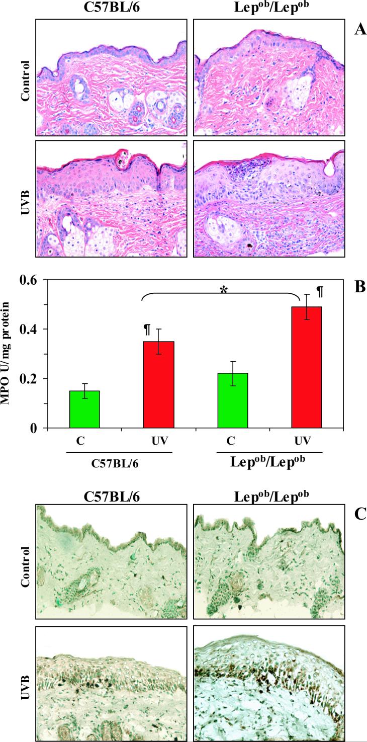
Chronic exposure of Lepob/Lepob mice to UVB results in greater leukocyte infiltration, and higher levels of MPO and hydrogen peroxide production than observed in UVB-irradiated wild-type (C57BL/6) mice. Mice were exposed to UVB on alternate days for one month and sacrificed 24 h after the last UVB exposure. Skin samples were collected and frozen in OCT under liquid nitrogen. A, UVB-induced edema and leukocyte infiltration was higher in Lepob/Lepob mice than wild-type mice. The paraffin-embedded skin samples (5 μm thick) were processed for routine H & E staining following a standard protocol. Magnification, x40; n=5. B, MPO was determined as a marker of UVB-induced tissue infiltration. The levels of UVB-induced MPO were greater in Lepob/Lepob mice compared to their wild-type counterparts. Data were reported as mean ± SD, n=5. Significant difference versus controls (non-UVB exposed), ¶p<0.01. Significant difference versus UVB-exposed wild-type, *p<0.05. C, The 5 μm thick sections of frozen skin samples were analyzed for UVB-induced H2O2 production following immunohistochemistry, as described in Materials and methods. The immunostaining intensity and the number of UVB-induced H2O2-positive cells appear dark brown. n=5, magnification, x40. Representative examples of micrographs of staining figures are shown from experiments conducted in skin samples from 5 mice that had identical patterns.