


Figure 5.
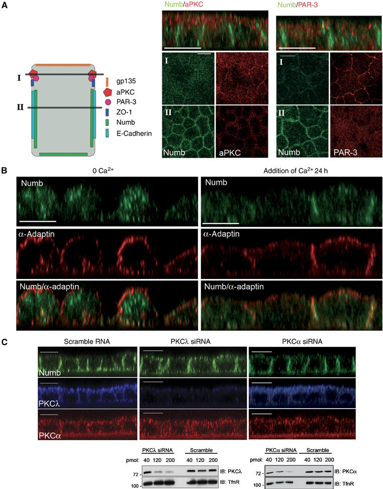
Cell polarity determines Numb localization in MDCK cells. (A) Polarized MDCK cells were fixed and co-stained with anti-NumbC (green) and either anti-aPKC or anti-Par3 (red). Shown are confocal X–Y sections taken at the subapical region of the cells (I) and 2–4 μm below at the basolateral region (II) as depicted in the diagramatic representation of the cells to the left of the images. Accumulation of Numb at the lateral membranes, below the subapical aPKC and Par3 staining, is shown in the Z sections shown above. (B) Polarized MDCK cells were grown in low calcium medium for 20 h (calcium switch) followed by re-addition of normal media for 24 h. Fixed cells were co-stained with anti-NumbC (green) and anti-α-adaptin (red). (C) MDCK cells were treated with 200 pmol aPKCλ or PKCα siRNA oligonucleotides and grown on filters for 72 h. Following fixation and permeabilization, the monolayers were co-stained with anti-aPKC (which also recognizes aPKCλ; blue), anti-PKCα (red), and anti-NumbC (green). Size bars indicate 10 μm. In lower panel MDCK cells were transfected with 40, 120, or 200 pmol of aPKCλ or PKCα siRNA oligonucleotides, or scrambled RNA control as indicated. In total, 20 μg of protein lysate for each condition was separated by SDS–PAGE and blotted with anti-aPKC or anti-PKCα. Membranes were reprobed with anti-Transferrin receptor as a loading control.