


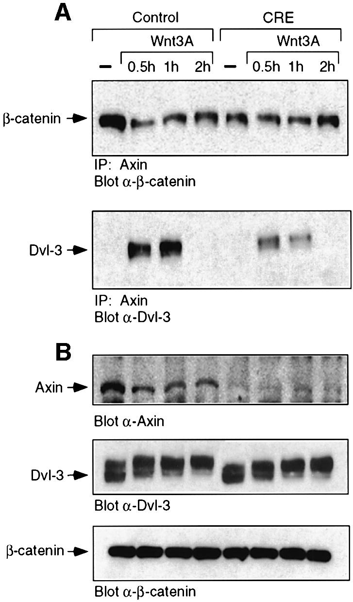
Fig. 8. Dab2 ablation leads to decreased axin and decreased axin– β-catenin–Dvl-3 interactions. (A) Control and Dab2–/– MEFs were treated with control (–) or Wnt-3A-conditioned medium for the times indicated. Cell lysates were prepared and subjected to immunoprecipitation with α-axin antibody. Following resolution by SDS–PAGE, the presence of β-catenin (top panel) or Dvl-3 (bottom panel) in the immunocomplex was detected by western blot analysis. The position of β-catenin and Dvl-3 is indicated by the arrow. (B) The expression of axin, Dvl-3 and β-catenin in the above lysates was determined by western analysis for each of the proteins as indicated.