


Figure 2.
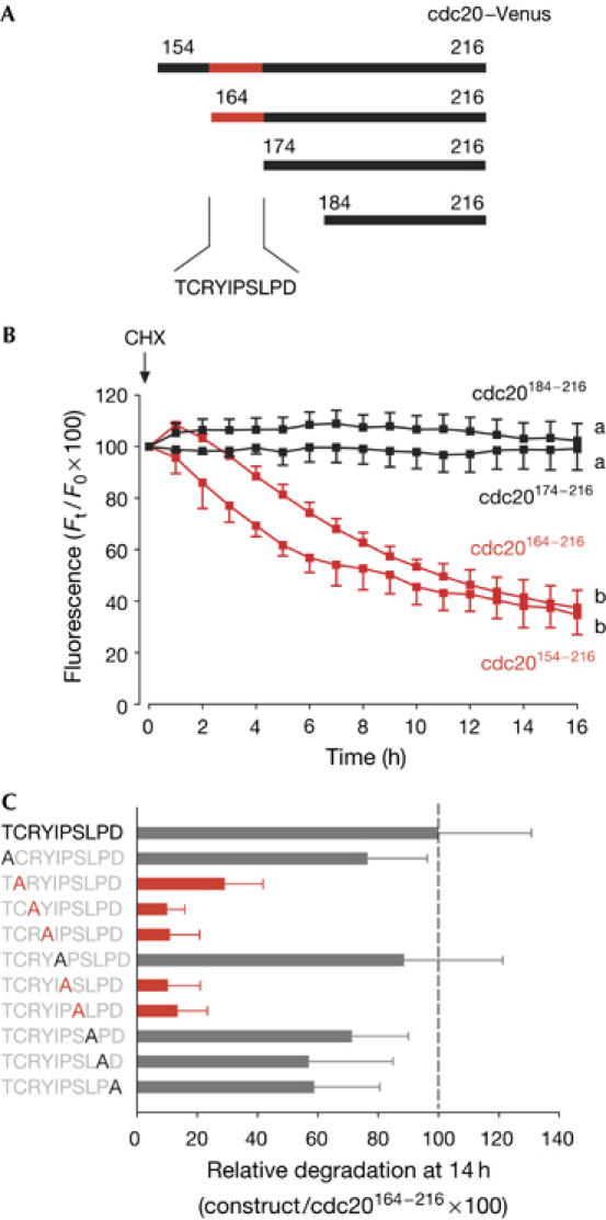
Characterization of the CRY degron in cdc20. (A,B) Cdc20–Venus complementary RNA constructs (A) were expressed in oocytes; cycloheximide (CHX) was then added to block further protein translation. (B) Cdc20174−216 (n=13) and cdc20184−216 (n=17; black traces) were stable, whereas cdc20154−216 (n=20) and cdc20164−216 (n=15; red traces) were degraded. The cdc20 sequence became stable when residues 164–174 were removed. a and b denote significantly different fluorescence at the 16 h time point (P<0.05 χ2 test). (C) To examine which residues were crucial for degradation in cdc20 residues 164–174, point mutations of each residue were made to alanine, as indicated. Mean loss of fluorescence of green fluorescent protein (GFP) in oocytes after 14 h was expressed as a quotient with respect to cdc20164−216–GFP, such that cdc20164−216 was assigned 100% degradation (n=15–20 per construct). Mutations that are defined as very stable are marked in red.