


Figure 4.
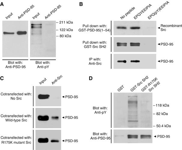
Src SH2 domain binding does not require tyrosine phosphorylation of the N-terminal region of PSD-95. (A) Immunoprecipitations from PSD fractions (100 μg) with anti-PSD-95. Immunoprecipitants were sequentially probed with a different anti-PSD-95 antibody (left) and anti-pY (right). A band corresponding to PSD-95 was not detectable with anti-pY even following longer exposures (data not shown). Input was 8 μg of PSD proteins. Molecular weight markers are shown on right. Similar results were observed in three separate experiments. (B) Upper: pull-down assays with GST-PSD-95(1–54) and recombinant Src preincubated with no peptide, EPQYEEIPIA or EPQ(pY)EEIPIA (1 mM). Src that remained bound to GST-PSD-95(1–54) was probed with anti-Src. Middle: pull-down assays from rat forebrain extracts (600 μg) with GST-Src SH2 preincubated with no peptide, EPQYEEIPIA or EPQ(pY)EEIPIA (1 mM). Proteins that remained associated with GST-Src SH2 were probed with anti-PSD-95. Lower: immunoprecipitations with anti-Src from lysates of HEK293 cells (300 μg) cotransfected with PSD-95 and Src, and incubated with no peptide, EPQYEEIPIA or EPQ(pY)EEIPIA (1 mM). Proteins were probed with anti-PSD-95. Similar results were found in each of three experiments. (C) Immunoprecipitations with anti-Src from lysates of HEK293 cells (300 μg) transfected with PSD-95 and no Src (upper), wild-type Src (middle) or R175K mutant Src (lower). Immunoprecipitants were probed with anti-PSD-95. Input was 30 μg of cell lysate proteins. Similar results were found in each of three experiments. (D) Pull-down assays from forebrain extracts (600 μg) with GST alone, GST-Src SH2 or GST-R175K Src SH2. Proteins were sequentially probed with anti-pY (upper) and anti-PSD-95 (lower). Molecular weight markers are indicated on right. Results are representative of three experiments.