



Abstract
GABAA receptors mediate the majority of fast synaptic inhibition in the brain. The accumulation of these ligand-gated ion channels at synaptic sites is a prerequisite for neuronal inhibition, but the molecular mechanisms underlying this phenomenon remain obscure. To further understand these processes, we have examined the cellular origins of synaptic GABAA receptors. To do so, we have created fluorescent GABAA receptors that are capable of binding α-bungarotoxin (Bgt), facilitating the visualization of receptor endocytosis, exocytosis and delivery to synaptic sites. Imaging with Bgt in hippocampal neurons revealed that GABAA receptor endocytosis occurred exclusively at extrasynaptic sites, consistent with the preferential colocalization of extrasynaptic receptors with the AP2 adaptin. Receptor insertion into the plasma membrane was also predominantly extrasynaptic, and pulse-chase analysis revealed that these newly inserted receptors were then able to access directly synaptic sites. Therefore, our results demonstrate that synaptic GABAA receptors are directly recruited from their extrasynaptic counterparts. Moreover, they illustrate a dynamic mechanism for neurons to modulate GABAA receptor number at inhibitory synapses by controlling the stability of extrasynaptic receptors.
Keywords: bungarotoxin binding site, endocytosis, exocytosis, GABAA receptor, inhibitory synaptic sites
Introduction
GABAA receptors are the major mediators of fast synaptic inhibition in the brain and the sites of action for both benzodiazepines and barbiturates. These receptors are pentameric, Cl− selective ligand-gated ion channels that can be assembled from seven subunit classes: α1–6, β1–3, γ1–3, γ, ɛ and π providing the structural basis for extensive heterogeneity of GABAA receptor structure (Rudolph et al, 2001; Sieghart and Sperk, 2002). However, a combination of molecular, biochemical and genetic approaches suggests that in the brain the majority of benzodiazepine receptor subtypes are composed of α, β and γ2 subunits (Rudolph et al, 2001; Sieghart and Sperk, 2002). γ2-containing receptors are highly enriched at synaptic sites in neurons, a process that is critical for efficient inhibitory neurotransmission and appropriate animal behavior (Essrich et al, 1998; Crestani et al, 1999; Kittler and Moss, 2003; Luscher and Keller, 2004; Alldred et al, 2005).
The mechanisms by which neurons facilitate the accumulation of GABAA receptors at synaptic sites remain poorly defined, but it is evident that synaptic GABAA receptors exhibit reduced rates of lateral mobility compared to their synaptic counterparts (Jacob et al, 2005; Thomas et al, 2005), consistent with studies on both glutamate and glycine receptors (Meier et al, 2001; Borgdorff and Choquet, 2002; Dahan et al, 2003). For GABAA receptors, this confinement is at least partially facilitated by the postsynaptic scaffold protein gephyrin (Kneussel and Betz, 2000; Keller et al, 2004), which has recently been shown to decrease the diffusion rates of GABAA receptors (Jacob et al, 2005), promoting their accumulation at synapses (Essrich et al, 1998; Kneussel et al, 1999). In addition to lateral movements, GABAA receptors also undergo significant rates of constitutive endocytosis and recycling, which can directly modulate the efficacy of synaptic inhibition (Kittler et al, 2000a, 2005; Chen et al, 2005).
To further understand how neurons construct inhibitory synapses, we have developed tools to visualize GABAA receptor endo- and exocytosis in addition to their accumulation at synaptic sites by creating receptor subunits modified with N-terminal pHluorin reporters that are also capable of binding α-bungarotoxin (Bgt) with high affinity. Using labelled Bgt derivatives, we then reveal that the principal sites of both GABAA receptor endo- and exocytosis are extrasynaptic in hippocampal neurons. In addition, our study demonstrates that newly inserted extrasynaptic receptors are capable of directly accessing synaptic sites. Our results thus reveal that synaptic GABAA receptors are derived directly from their extrasynaptic counterparts, and constitute a dynamic mechanism for neurons to rapidly modulate receptor number at inhibitory synapses by controlling the availability and stability of extrasynaptic receptors.
Results
Characterizing the properties of GABAA receptors engineered with the BBS and ecliptic pHluorin
Studies on the dynamics of cell-surface GABAA receptor populations have been compromised by the lack of suitable high-affinity probes. To measure GABAA receptor endo- and exocytosis directly, we have engineered the receptor β3 subunit with the bungarotoxin binding site (BBS) peptide WRYYESSLEPYPD. The BBS is derived from the α subunit of the muscle nicotinic acetylcholine receptor and has been established to bind Bgt with an affinity of approximately 3 nM (Scherf et al, 2001; Katchalski-Katzir et al, 2003). The BBS together with a pHluorin reporter separated by a 13-amino-acid alanine/proline linker was added between amino acids 4 and 5 of the mature β3 subunit (Figure 1A). The resulting construct, pHBBSβ3, was then expressed in HEK-293 cells and 48 h after transfection SDS-soluble lysates were subjected to immunoblotting with anti-β3 antibody. This revealed the presence of a major band of 87 kDa only in cells expressing pHBBSβ3, in agreement with the predicted molecular mass of this subunit (Figure 1B).
Figure 1.
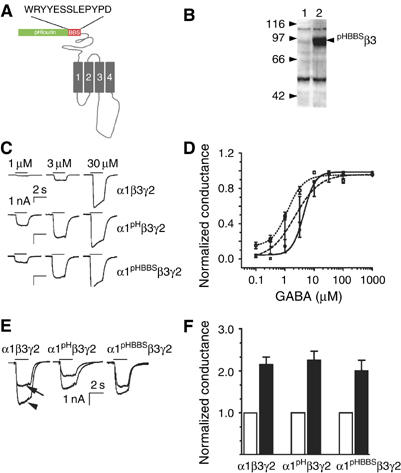
Functional expression of GABAA receptors incorporating pHBBSβ3 subunits. (A) The structure of the GABAA receptor pHBBSβ3 subunits. pHluorin and BBS were added between amino acids 4 and 5 of the mature β3 subunits and were separated by a 13-amino-acid alanine–proline linker. Tm domains 1–4 are shown in gray. (B) Analyzing expression of the pHBBSβ3 subunit by immunoblotting. Control (lane 1) or HEK-293 cells expressing GABAA receptor α1pHBBSβ3 and γ2 subunit cDNAs (lane 2) were immunoblotted with antibodies against the receptor β3 subunit. The migration of molecular mass standards is indicated. (C, D) Agonist sensitivity of GABAA receptors incorporating pHBBSβ3 subunits. HEK-293 cells were transfected with the respective murine cDNAs in a 1:1:1 ratio. At 48 h after transfection, IGABA was recorded under voltage clamp at −50 mV. Responses from individual cells are shown in panel C. Currents at individual agonist concentrations were then normalized to the maximal response observed and fitted with the Hill equation. I/Imax=1/(1+(EC50/AnH) are shown in panel D. EC50 values of 9.89±2.30, 2.38±0.4 and 6.12±1.94 were seen for GABAA receptors composed of α1β3 γ2 (□), α1pHβ3 γ2 (○) and α1pHBBSβ3γ2 (▴), respectively (mean±s.e.m.; n=6–8). (E, F) Benzodiazepine modulation of GABAA receptors incorporating pHBBSβ3 subunits. HEK-293 cells expressing GABAA receptors were voltage clamped at −50 mV. The magnitude of IGABA at EC20 agonist concentration was then measured in the absence and presence of 100 nM flurazepam as illustrated in panel E. Normalized conductances in the absence of flurazepam were given a value of 1. IGABA was potentiated by 2.15±0.18 (α1β3γ2), 2.26±0.21 (α1pHβ3γ2) and 2.09±0.25 (α1pHBBSβ3γ2) respectively (mean±s.e.m.; n=9–15) as shown in panel F.
We further assessed the ability of pHBBSβ3 subunits to assemble with receptor α1 and γ2 subunits to form functional heteromeric α1β3γ2 receptors using patch clamp recording (Kittler et al, 2000b; Bedford et al, 2001; Jacob et al, 2005). Fast application of GABA produced inward currents (IGABA) for cells expressing receptors composed of α1β3γ2, α1pHβ3γ2 or α1pHBBSβ3γ2 subunits (Figure 1C). Using dose–response analysis, it was evident that these differing receptors exhibited similar EC50 values for GABA of 9.89±2.30, 2.38±0.4 and 6.12±1.94 μM (mean±s.e.m.; n=6–8) respectively in addition to maximal currents, strongly suggesting similar receptor densities on the plasma membrane for these varying subunit combinations (Figure 1D). To measure the incorporation of the γ2 subunit into functional receptors, we analyzed modulation by benzodiazepines, a property critically dependent upon incorporation of this subunit into heteromeric αβγ2 receptors (Rudolph et al, 2001; Sieghart and Sperk, 2002). Flurazepam at a concentration of 100 nM produced similar robust enhancements of IGABA at EC20 GABA concentrations of approximately 220% for receptors composed of α1β3γ2, α1pHβ3γ2 or α1pHBBSβ3γ2 subunits (Figure 1E and F). Together, these observations suggest that the addition of the BBS and pHluorin reporters to the N-terminus of the β3 subunit does not compromise receptor functional expression, a finding consistent with prior studies using GABAA receptors engineered with N-terminal reporters GFP, pHluorin or myc reporters (Connolly et al, 1996; Kittler et al, 2000b; Taylor et al, 2000; Jacob et al, 2005).
GABAA receptors modified with BBS bind Bgt with high affinity
The ability of GABAA receptors incorporating pHBBSβ3 subunits to bind Bgt was analyzed by exposing live transfected HEK-293 cells to varying concentrations of 125I-Bgt at 37°C for 10 min. After extensive washing, the level of bound Bgt was measured (Figure 2A). This revealed that cells expressing GABAA receptors incorporating pHBBSβ3 subunits bound Bgt with a KD of 7.85±2.41 × 10⁻⁹ M (mean±s.e.m.; n=3), consistent with previous studies on the BBS or AMPA receptors modified with this sequence (Scherf et al, 2001; Katchalski-Katzir et al, 2003; Sekine-Aizawa and Huganir, 2004). No significant binding was observed in cells expressing GABAA receptors containing wild-type β3 subunits (Figure 2A).
Figure 2.

GABAA receptors engineered to incorporate pHBBSβ3 subunits bind Bgt with high affinity. (A) GABAA receptors containing pHBBSβ3 subunits bind Bgt with high affinity. HEK-293 cells were transfected with GABAA receptors α1pHBBSβ3 and γ2 subunit cDNAs. After 48 h, cells were labelled with varying concentrations of 125I-Bgtx (1000 Ci/mmol) at 37°C for 10 min before extensive washing. The level of bound Bgt after correction for nonspecific binding was then used to estimate the affinity of Bgt for receptors containing pH/BBSβ3 subunits (•) or pHβ3 (□) (calculated using Origin software; data represent mean±s.e.m.; n=4). (B–D) Bgt slowly dissociates from GABAA receptors containing pHBBSβ3 subunits. Transfected HEK-293 cells were labelled with 5 μg/ml Rd-Bgt for 5 min and washed extensively. Labelled cells were transferred to the stage of a confocal microscope maintained at 15°C and imaged over 180 min. Images of an individual cell at 5 and 180 min are shown in panels B and C, respectively (red=Rd-Bgt, green=pHluorin). (D) The intensity of Rd-Bgt in 2–5 μm2 areas of interest was then compared to the initial levels seen at zero time, which was given a value of 100% (each data point represents mean±s.e.m.; n=4–6).
To visualize cell-surface GABAA receptor populations, live expressing HEK-293 cells were stained with rhodamine-conjugated Bgt (Rd-Bgt) and then visualized using confocal microscopy. This imaging revealed robust labelling of the surface of cells exhibiting endogenous green fluorescence, indicating expression of GABAA receptors incorporating pHBBSβ3 subunits (Figure 2B). No binding was observed in non-transfected cells. To examine the stability of Rd-Bgt binding, cells expressing receptors incorporating pHBBSβ3 subunits were labelled for 10 min with Rd-Bgt, washed and incubated at 15°C to block endo- and exocytosis and subjected to live confocal imaging. The intensity of Bgt staining was then measured at the periphery of expressing cells over time (Figure 2B and C) and compared to the signal seen at zero time, which was given a value of 100%. Rd-Bgt labelling at the surface of HEK-293 cells remained unaltered over a 200 min time course, suggesting that Rd-Bgt complexes are stable (Figure 2D).
Next we analyzed the ability of Rd-Bgt to detect GABAA receptors incorporating pHBBSβ3 subunits when expressed in hippocampal neurons. 8–10-Div neurons were transfected with pHBBSβ3 and 4–7 days later live neurons were exposed to Rd-Bgt, fixed and subjected to confocal microscopy. Neurons expressing pHBBSβ3 subunits exhibited endogenous green fluorescence and also significant levels of cell-surface Rd-Bgt binding, whereas the background labelling to non-fluorescent untransfected neurons was negligible (Figure 3A). Rd-Bgt staining in transfected neurons was blocked by excess of unlabelled Bgt (Figure 3B). To further control for possible binding of Bgt to endogenous neuronal AChRs, we examined the effects of curare on Rd-Bgt staining in neurons expressing pHBBSβ3 subunits. Although curare blocks Bgt binding to neuronal AChRs containing α7 subunits, it does not block binding to the BBS (Schoepfer et al, 1988; Sekine-Aizawa and Huganir, 2004). In agreement with this, 100 μM curare did not modify Rd-Bgt binding to neurons expressing pHBBSβ3 subunits, suggesting that fluorescent Bgt binding sites in our experiments represent GABAA receptors and not endogenous AChRs (Figure 3E and F). We also examined the stability of Rd-Bgt/GABAA receptor complexes in hippocampal neurons by incubating Rd-Bgt-stained neurons at 15°C. The level of Rd-Bgt staining in 1–2 μm2 areas of interest was measured over a time course of 200 min (Figure 3E and F) with levels evident at zero time being given a value of 100%. No statistically significant loss of Rd-Bgt staining was seen over this time period (Figure 3G), suggesting that Rd-Bgt/GABAA receptor complexes are stable on the surface of neurons.
Figure 3.

Imaging GABAA receptors incorporating pHBBSβ3 subunits in hippocampal neurons. (A–D) Visualizing GABAA receptors incorporating pHBBSβ3 subunits in neurons. 14-Div hippocampal neurons expressing GABAA receptor pH/BBSβ3 subunits were labelled with 5 μg/ml Rd-Bgt in the absence (A) or presence (B) of 50 μg/ml of unlabelled Bgt. Cells were then fixed and images recorded by confocal microscopy (red=Rd-Bgt, green=pHluorin, yellow arrows=transfected neurons expressing pHBBSβ3, white=non-transfected neurons; scale bars=15 μm). Rd-Bgt staining was also performed on neurons expressing pHBBSβ3 in the absence (C) and presence of 100 μM curare (D). (E–H) Bgt slowly dissociates from neurons expressing pHBBSβ3 subunits. Transfected neurons were labelled with 5 μg/ml Rd-Bgt for 5 min, washed, transferred to the stage of a confocal microscope maintained at 15°C and imaged for 180 min (red=Rd-Bgt, green=pHluorin). Images of an individual transfected neuron stained with Rd-Bgt at 0 and 180 min are shown in panels E and F. The lower panels represent enlargements of the boxed areas in the upper panels. (G) The intensity of Rd-Bgt staining was measured in 1–2 μm2 areas of interest over the entire recording period with levels at zero time being given a value of 100% (data represent mean±s.e.m.; n=6). (H) GABAA receptors incorporating pHBBSβ3 subunits are targeted to synaptic sites. 14-Div hippocampal neurons expressing pHBBSβ3 subunits were stained with 5 μg/ml Rd-Bgt for 5 min, washed, fixed, permeabilized, stained with anti-synapsin antibodies and imaged using confocal microscopy (red=Rd-Bgt, green=pHluorin, blue=synapsin). The lower panel represents an enlargement of the boxed area in the upper panel; the white arrowheads indicate synaptic GABAA receptors, whereas the yellow arrowheads indicate extrasynaptic populations (scale bar=10 μm).
The accumulation of GABAA receptors incorporating pHBBSβ3 at synaptic sites in hippocampal neurons was also addressed. Expressing neurons were labelled with Rd-Bgt and fixed. Presynaptic terminals were then stained with antibodies against synapsin. Consistent with published studies analyzing the distribution of endogenous β3 subunits, Rd-Bgt staining on the surface of hippocampal neurons was evident as distinct clusters in addition to diffuse staining (Danglot et al, 2003; Jacob et al, 2005). We next calculated the number of these receptor clusters that were apposed to synapsin staining and therefore judged to be synaptic, as it has been documented that clusters containing the β3 subunit are found at both synaptic and extrasynaptic sites (Danglot et al, 2003; Jacob et al, 2005). A total of 24.6±6.4% of these clusters were apposed to synapsin staining and were therefore judged to be synaptic (Figure 3H; 250 clusters counted, mean±s.e.m.; n=5). Similar levels of synaptic targeting for endogenous GABAA receptors containing the β3 subunit have been previously reported in 12–14-Div hippocampal neurons (Danglot et al, 2003; Jacob et al, 2005).
Extrasynaptic GABAA receptors exhibit short residence times on the cell surface
We next compared the relative residence times on the cell surface of synaptic and extrasynaptic GABAA receptors. Neurons expressing receptor pHBBSβ3 subunits were labelled with Rd-Bgt and incubated at 37°C for up to 90 min in the presence of 50 nM unlabelled Bgt in the external media. At differing time points, neurons were fixed and then incubated with synapsin antibodies to differentiate synaptic and extrasynaptic GABAA receptors (Kittler et al, 2001; Jacob et al, 2005). At zero time, abundant diffuse extrasynaptic Rd-Bgt staining was seen in addition to labelled synaptic receptors (Figure 4, panels 1 and 2). However, a consistent finding after 90 min was an extensive loss of extrasynaptic Rd-Bgt labelling (Figure 4, panels 3 and 4).
Figure 4.

Extrasynaptic GABAA receptors have short residence times at the cell surface. 12–14-Div hippocampal neurons expressing GABAA receptors incorporating receptor pH/BBSβ3 subunits were labelled with 5 μg/ml of Rd-Bgt without (1–4) or with (5–8) 1 h preincubation with myristoylated p4 peptide. Neurons were then incubated at 37°C for between 0 and 90 min. At varying time intervals, samples were removed, permeabilized, stained with antibodies against synapsin and then visualized by confocal microscopy (red=Rd-Bgt, green=pHluorin, blue=synapsin). Images at zero time for an individual neuron are shown in 1, 2, 5 and 6, whereas those in 3, 4, 7 and 8 represent neurons incubated for 90 min at 37°C. The right-hand panels represent enlargements of the boxed areas in the left-hand panels. The yellow and white boxed areas indicate synaptic and extrasynaptic receptor populations respectively (scale bars=5 μm).
To quantify this apparent loss of extrasynaptic receptors, we measured Rd-Bgt staining in 1–2 μm2 areas of interest representing synaptic receptors and extrasynaptic receptors that were at least 2 μm from the nearest synapses and within the same neuron (Figure 4, panels 3 and 4). The ratio of extrasynaptic-to-synaptic Rd-Bgt staining (Bgt-ES/Bgt-S) was then determined and ratios at zero time were given an arbitrary value of 1.0. This ratio decreased rapidly with time of incubation at 37°C, reaching statistical significance after 10 min and steady state at 60 min (Figure 5A and B). At 90 min, this ratio had significantly decreased to 0.42±5.2% (Figure 5B; P<0.01; Student's t-test; n=6). Given the high stability of Bgt/GABAA receptors complexes (Figures 2 and 3), our results demonstrate that extrasynaptic GABAA receptors containing pHBBSβ3 have much shorter residence times on the cell surface as compared to their synaptic counterparts.
Figure 5.

Extrasynaptic GABAA receptors preferentially undergo clathrin-dependent endocytosis. (A, B) Cell-surface stability of extrasynaptic GABAA receptors is enhanced by inhibitors of clathrin-dependent endocytosis. The ratio of Rd-ES-Bgt:S-Bgt staining was measured in adjacent synaptic and extrasynaptic domains of RD-Bgt-stained neurons expressing pHBBSβ3 subunits over 90 min as shown in Figure 4 under control conditions (▪) or after 1 h pretreatment with M-p4 (▪) or M-Sc (▪). Data at varying time points were compared to ratios seen at zero time (which was given a value of 1). Each data point represents mean±s.e.m. from 44 to 90 synapses (n=6–8). Ratios at 90 min were also compared to the respective levels seen at zero time as shown in panel B (P4=1 h pretreatment with membrane-impermeable peptide; *significantly different from control; P<0.01; Student's t-test, n=6–8). (C–F) Internalization of Bgt-labelled GABAA receptors incorporating BBSβ3 subunits. 14-Div neurons expressing BBSβ3 subunits were labelled with 5 μg/ml Rd-Bgt for 5 min at 37°C, washed and incubated at 37°C for 120 min. After incubation, neurons were labelled with 5 μg/ml Alex-Bgt for 5 min at 37°C, washed, fixed and imaged by confocal microscopy; C=Rd-Bgt, D=Alx-Bgt, E=BBSβ3 fluorescence, F=merge. The white arrows represent internalized Rd-Bgt/GABAA receptors and yellow arrows represent clustered Rd-Bgt staining on the plasma membrane; scale bar=10 μm. (G, H) Extrasynaptic GABAA receptors show preferential colocalization with the AP2 adaptin. 14-Div hippocampal neurons expressing pHBBSβ3 were fixed, permeabilized, stained with antibodies against AP2 (red) and synapsin (blue) and visualized by confocal microscopy (green represents endogenous pHBBSβ3 fluorescence; scale bar=5 μm) The images in panel H are enlargements of the boxed area in panel G. Panel G shows a merged image and the individual channels with green representing pHluorin. The arrows represent extrasynaptic receptors that colocalize with AP2, whereas the arrowheads indicate synaptic GABAA receptors that colocalize with synapsin.
Extrasynaptic GABAA receptors undergo clathrin-dependent endocytosis
A number of studies have illustrated that GABAA receptors undergo constitutive clathrin-dependent endocytosis. We therefore sought to determine whether the time-dependent loss of extrasynaptic receptors seen in Figure 4 represents their preferential endocytosis. To do so, hippocampal neurons expressing pHBBSβ3 subunits were preincubated for 1 h with 50 μM myristoylated membrane-permeable p4 peptide (M-p4), a potent inhibitor of GABAA receptor endocytosis (Kittler et al, 2000a, 2005; Chen et al, 2005) before labelling with Bgt. Pretreatment of neurons with M-p4 appeared to block the loss of extrasynaptic receptors seen in untreated neurons (Figure 4, panels 5 and 6). After quantifying these results, it was evident that M-p4 blocked the time-dependent decrease in Bgt-ES/Bgt-S ratio seen in controls, an effect that was not replicated with a scrambled myristoylated version of p4 peptide (M-Sc) or non-myristoylated (non-cell permeable) p4 peptide (P4) (Figure 5A and B).
To further analyze the intracellular fate of internalized GABAA receptors, neurons were labelled for 5 min with Rd-Bgt, washed extensively and then incubated at 37°C for 120 min. After this time period, neurons were finally stained for 5 min with Alx-Bgt before fixation and analysis by confocal microscopy. The majority of the Rd-Bgt label over the incubation period at 37°C accumulated in perinuclear structures with endosomal-like morphology. These intracellular organelles also contained pHluorin fluorescence (Figure 5C–F). In contrast, Alx-Bgt staining was found extensively over the entire plasma membrane as diffuse staining in addition to clusters (Figure 5C–F). Remaining cell-surface Rd-Bgt staining was also found within these clusters, consistent with its stabilization at synaptic sites, as illustrated in Figure 4. Given the high stability of GABAA receptor Bgt complexes at 15°C, conditions at which both endo- and exocytosis (Figure 3) are blocked, our results suggest that internalized Bgt-GABAA receptors are specifically targeted to endosomal structures, where they are capable of rapid recycling back to the plasma membrane or are targeted for lysozomal degradation (Kittler et al, 2000a, 2004).
We further analyzed the selective endocytosis of extrasynaptic GABAA receptors by comparing their distribution with that of the clathrin AP2 adaptin, which directly binds to motifs within the intracellular domains of receptor subunits, facilitating their endocytosis (Kittler et al, 2000a, 2005; Chen et al, 2005). Neurons expressing pHBBSβ3 subunits were stained with antibodies against AP2 adaptin and presynaptic terminals were labelled with synapsin antibodies and then imaged using confocal microscopy. Punctate staining that incorporated both GABAA receptor pHBBSβ3 subunits and AP2 immunoreactivity was found on both neuronal cell bodies and processes. Synaptic GABAA receptors were also evident as measured by the colocalization of pHBBSβ3 fluorescence and synapsin staining (Figure 5C and D). We then calculated the number of synaptic GABAA receptors that also contained immunoreactivity for the AP2 adaptin. Only 19.2±1.2% of synaptic GABAA receptors exhibited detectable levels of AP2 staining (mean±s.e.m.; 165 synapses analyzed, n=4). This suggests that GABAA receptor AP2 complexes are found primarily at extrasynaptic sites, consistent with their preferential endocytosis.
Imaging newly inserted GABAA receptors using Bgt staining
To selectively measure GABAA receptor insertion into the plasma membrane, we first blocked constitutive receptor endocytosis. Previous biochemical experiments have revealed that in excess of 25% of the total cell-surface receptor population are internalized within 15 min at steady state (Kittler et al, 2004). To do so, neurons were first treated with M-p4 and incubated with Rd-Bgt at 37°C. At varying time points, neurons were fixed and stained with synapsin antibodies to differentiate synaptic and extrasynaptic GABAA receptor populations. Using this procedure, Rd-Bgt binding could be detected within 5 min of incubation and appeared to increase dramatically with further incubation at 37°C (Figure 6A–F). The specificity of this new Rd-Bgt staining was measured by coincubating neurons with Rd-Bgt and a large excess of unlabelled toxin. This treatment completely blocked the appearance of new Rd-Bgt staining compared to neurons incubated with Rd-Bgt alone (Figure 6G and H). Newly inserted GABAA receptors were evident at both synaptic and extrasynaptic sites at 5 min, suggesting that insertion can occur throughout the entire surface (Figure 6B). However, as inhibitory synaptic sites represent only a small fraction of the neuronal surface, the majority of newly inserted receptors will be delivered to extrasynaptic sites.
Figure 6.

Imaging the insertion of new GABAA receptors on the surface of hippocampal neurons. 14-Div hippocampal neurons expressing GABAA receptors incorporating pH/BBSβ3 subunits were pretreated with M-p4 peptide for 1 h. Neurons were then exposed to 5 μg/ml unlabelled Bgt to label existing cell-surface receptors. After washing, neurons were incubated for 30 min at 37°C in the presence of M-p4. Neurons were then labelled with 10 nM Bgt at various time points, fixed, permeabilized and stained with antibodies against synapsin and imaged using confocal microscopy. (A–D) Neurons incubated for 5 min. (E–H) Neurons incubated for 30 min. (I–L) A neuron incubated with Rd-Bgt, M-p4 and 100 nM unlabelled toxin. In all images, red=Rd-Bgt, blue=synapsin and green=pHluorin. Images C and D, G and H, K and L represent enlargements of the boxed areas in panels A, E and I, respectively. Panels B, F and J show the Rd-Bgt channel only, whereas panels D, H and L show both Rd-Bgt and synapsin staining. The arrowheads indicate synaptic receptor populations, whereas the arrows indicate extrasynaptic receptors. (M) The ratios of newly inserted GABAA receptors were measured for extrasynaptic and synaptic domains of neurons, as outlined in Figure 5. Each data point represents mean±s.e.m. from 60 to 80 synapses; *significantly different from control; P<0.01; Student's t-test; n=7–8.
To provide quantitative information on the delivery of newly inserted receptors to synaptic sites over time, we measured the ratio of newly inserted Rd-Bgt binding sites at synaptic and extrasynaptic sites (S-Bgt:ES-Bgt), with values evident at 5 min (zero time) given a value of 100%. In excess of 85% of all synapses analyzed in our experiments showed increased levels of Rd-Bgt staining, and these were chosen for further analysis, with the ratios seen at zero time being given a value of 1.0. This ratio increased slowly and reached a level of 1.41±0.08 at 30 min (Figure 6J), which was significantly different from zero time (P<0.001; n=4). This suggests that newly inserted receptors accumulate slowly at postsynaptic inhibitory synaptic sites.
Newly inserted extrasynaptic receptors are able to access synaptic sites
The slow accumulation of newly inserted GABAA receptors at synaptic sites may reflect either slower direct insertion of GABAA receptors at synaptic sites as compared to extrasynaptic sites or the diffusion of newly inserted extrasynaptic receptors to synaptic sites and their subsequent stabilization. To address these possibilities, we utilized pulse-chase analysis with Bgt conjugated to distinct fluorophores. Neurons were first pretreated with M-p4 peptide to block endocytosis and existing populations of receptors containing pHBBSβ3 subunits were blocked with unlabelled toxin. All GABAA receptors inserted into the plasma membrane over a 5 min time period were then labelled with Rd-Bgt toxin, washed extensively and then incubated at 37°C in toxin-free media. At differing time periods, neurons were fixed and stained with VIAAT antibodies to visualize presynaptic inhibitory terminals and imaged by confocal microscopy. Newly inserted receptors were clearly visible after 5 min incubation with Rd-Bgt (zero time) and also after incubation at 37°C, but there appeared to be increased labelling of synaptic sites at later time points (Figure 7C and D). To quantify these results, we compared the level of Rd-Bgt staining at synaptic and extrasynaptic sites over time as outlined in Figures 4, 5 and 6, with ratios evident at zero time being given a value of 1.0. This ratio significantly increased with time of incubation at 37°C (Figure 7D), suggesting that newly inserted GABAA receptors at extrasynaptic domains can over time move to synaptic sites.
Figure 7.

Analyzing the movement of newly inserted extrasynaptic receptors to synaptic sites. 14-Div hippocampal neurons expressing GABAA receptors were treated with 100 nM unlabelled Bgt for 10 min, washed and then subjected to a second labelling period of 5 min at 37°C with 10 nM Rd-Bgt. They were then incubated at 37°C for varying time periods before fixing followed by immunohistochemistry with anti-VIAAT antibodies. (A) shows a neuron fixed immediately after incubation with Rd-Bgt, whereas (D) shows a neuron incubated for 30 min after labelling with Rd-Bgt. In all images, red=Rd-Bgt, blue=VIAAT and green=pHluorin. Images in (B/C) and (E/F) represent enlargements of the panels in A and D, respectively. Panels C and D show only Rd-Bgt and VIAAT staining. The arrowheads indicate synaptic receptor populations, whereas the arrows indicate extrasynaptic receptors. (G) The ratio of synaptic Bgt to extrasynaptic staining was calculated from neurons expressing pH/BBSβ3 subunits over time, with the ratio evident at zero time (directly after labelling with Rd-Bgt) being given a value of 1.0. Each data point represents mean±s.e.m. from 44 to 90 synapses; *significantly different from control; P<0.001, n=4.
Discussion
The specific accumulation of GABAA receptors at postsynaptic sites is a critical determinant of fast synaptic inhibition. This process is regulated by the relative rates of receptor exocytosis, endocytosis, lateral mobility and receptor stabilization at postsynaptic specializations (Choquet and Triller, 2003; Kittler and Moss, 2003; Luscher and Keller, 2004). Although live imaging studies have provided evidence on the comparative diffusion rates of synaptic and extrasynaptic GABAA receptors (Jacob et al, 2005), measurements on the stability of these distinct receptor populations and their sites of exo- and endocytosis have been hampered by the lack of suitable probes.
To address these issues, we modified GABAA receptors within their N-terminal cytoplasmic domains with BBS and pHluorin reporters (Scherf et al, 2001; Katchalski-Katzir et al, 2003). These additions did not compromise the assembly or functional properties of physiologically relevant heteromeric GABAA receptors composed of α1pHBBSβ3 and γ2 subunits; however, these receptors were able to bind Bgt with a KD of approximately 7 nM and a very slow off-rate, consistent with previous studies on the affinity of the BBS for Bgt and AMPA receptors modified with this 13-amino-acid motif (Sekine-Aizawa and Huganir, 2004). Significantly, no binding was seen in cells expressing GABAA receptor β3 subunits modified with just pHluorin reporters. Recent studies have suggested that recombinant homomeric GABAA receptors composed of β3 subunits can bind Bgt with an approximate affinity of 50 nM when expressed in mammalian cells (McCann et al, 2006). However, this finding does not have an impact on our results, as the KD of Bgt for GABAA receptors composed of α1pHBBSβ3 and γ2 receptors was estimated at 7 nM, and our labelling studies used 10 nM Bgt. Furthermore, this low-affinity-binding Bgt site is abolished on coassembly of β3 subunits into heteromeric α1pHBBSβ3 and γ2 receptors (McCann et al, 2006).
In hippocampal neurons transfected with pHBBSβ3, fluorescent Bgt staining was seen only on the cell surface of neurons that exhibited endogenous green fluorescence, demonstrating the expression of GABAA receptors incorporating pHBBSβ3 subunits (Jacob et al, 2005). Moreover, Bgt staining in these neurons was insensitive to curare, a potent inhibitor of Bgt binding to AChRs. Together, these results demonstrate that the Bgt sites seen in neurons expressing pHBBSβ3 subunits originate from engineered GABAA receptors and not from endogenous AChRs.
We then utilized staining with Bgt derivatives to compare the relative residence times at the cell surface for extrasynaptic and synaptic GABAA receptors containing pHBBSβ3 subunits. This revealed a rapid loss of extrasynaptic Rd-Bgt staining over a 90 min time course at 37°C. Given the slow off-rate of Bgt for GABAA receptors incorporating pHBBSβ3 subunits, our results directly demonstrate that such extrasynaptic receptors have significantly shorter residence times on the cell surface as compared to synaptic receptors. To assess the mechanism underlying the distinct cell-surface stabilities of synaptic and extrasynaptic GABAA receptors, we focused on the role of dynamin-dependent endocytosis, a process key in determining receptor number at synaptic sites (Kittler et al, 2000a, 2005; Chen et al, 2005). Pretreatment of neurons with a membrane-permeable inhibitor of dynamin specifically blocked this loss of extrasynaptic GABAA receptors. Using immunohistochemistry, it was also evident that only extrasynaptic receptors exhibited high levels of colocalization with the AP2 adaptin, a critical regulator of GABAA receptor endocytosis. Our results thus strongly suggest that extrasynaptic GABAA receptors preferentially undergo clathrin-dependent endocytosis, which has important implications for the construction of inhibitory synapses. Specifically, decreasing the rate of GABAA receptor endocytosis using inhibitors of dynamin or by blocking the binding of GABAA receptors to the AP2 adaptin increases receptor number at synaptic sites over a time course of 15–30 min (Kittler et al, 2000a, 2005; Chen et al, 2005). Given our findings, these effects are likely to reflect the stabilization of extrasynaptic receptors and their subsequent accumulation at synaptic sites.
To further address this issue, we used imaging with Bgt to analyze the sites of GABAA receptor exocytosis. To do so, we first labelled existing cell-surface populations of receptors incorporating pHBBSβ3 subunits with unlabelled Bgt. Newly inserted receptors were then labelled with fluorescent Bgt in the presence of blockers of endocytosis. This revealed that newly inserted receptors were detected within a 5 min labelling period and these receptors were evident over the entire neuronal surface. However, the accumulation of these newly inserted receptors at synaptic sites was slow and did not reach statistical significance until 30 min later. These results may reflect either the movement of extrasynaptic receptors to synaptic sites or their slower direct insertion at synapses. To address these possibilities, we examined the fate of newly inserted receptors over a 5 min labelling period using pulse-chase analysis in the presence of blockers of endocytosis. This revealed that newly inserted receptors at extrasynaptic sites can directly access synaptic sites within a 15 min time period, a process that is facilitated by the high rates of diffusion of extrasynaptic compared to synaptic GABAA receptors (Choquet and Triller, 2003; Jacob et al, 2005).
Together, our results with GABAA receptors engineered to bind Bgt have provided novel insights into how neurons construct inhibitory synapses. Specifically, they have demonstrated that GABAA receptor endo- and exocytosis are localized to extrasynaptic membrane domains. Our studies also provide the first direct evidence that synaptic GABAA receptors are directly recruited from extrasynaptic receptors. Finally, our results reveal a dynamic mechanism for neurons to rapidly modulate both GABAA receptor number at inhibitory synapses and the efficacy of synaptic inhibition by controlling the availability and stability of extrasynaptic receptors.
Materials and methods
DNA constructs
The pH/BBSβ3 subunit construct was derived from pHβ3 (Jacob et al, 2005). Briefly, pHβ3 was subjected to two rounds of mutagenesis by PCR to introduce the following changes: to introduce the 13-amino-acid BBS tag directly after the pHluorin tag and to add 12 alanines and one proline to separate the BBS tag from the pHluorin tag. The final construct contains the pHluorin tag between amino acids 3 and 4 of the mature β3 subunit. The pHluorin tag is followed by 13-amino-acid alanine/proline spacer.
Cell culture and transfection
Hippocampal neuron cultures were prepared as described previously (Kittler et al, 2000a, 2000b). Neurons were transfected with Effectene according to the manufacturer's specifications (Qiagen, Valencia, CA, USA). Briefly, 1 μg of DNA was used for every two 35 mm cell culture dishes containing hippocampal neurons. DNA was added to 150 μl of EC buffer and 8 μl of Enhancer. The solution was incubated for 5 min at room temperature and 75 μl of Effectene was added. After 10 min incubation at room temperature, the final solution was added to hippocampal neurons. The medium was changed after 6 h. HEK-293 cells were transfected using electroporation, as detailed earlier (Connolly et al, 1996, 1999).
Electrophysiology
HEK-293 cells were transfected with equimolar mixes of the relevant expression construct. After 18–48 h, coverslips containing the transfected HEK-293 cells were transferred to a recording chamber mounted on the stage of an inverted microscope and transfected cells identified by their endogenous fluorescence (Kittler et al, 2000b; Jacob et al, 2005). The external solution contained (in mM) NaCl 140, KCl 4.7, HEPES 10, NaHCO3 23, glucose 11, MgCl2 1.2 and CaCl2 2.5 (adjusted to pH 7.4 with NaOH) continuously oxygenated with a mixture of 95%O2/5%CO2. The recording chamber was perfused at a rate of 5–10 ml/min and maintained at 32°C (Kittler et al, 2000a, 2005; Chen et al, 2005; Jacob et al, 2005). The internal solution contained (in mM) KCl 140, CaCl2 1.0, HEPES 10, MgCl2 2 and ATP (adjusted to pH 7.2 with KOH). Pipettes had a resistance of 2–4.5 MΩ when filled with this internal solution. Patch clamp experiments were performed in the whole cell configuration using an Axopatch 200B amplifier. Series resistance and membrane capacitance were partially compensated (70–80%) and current traces were low-pass filtered at 2 kHz using a four-pole Bessel filter. The holding potential in all experiments was −50 mV. Drugs were rapidly applied to single cells using a modified U-tube, placed 50–100 μm away from the cell of interest (Kittler et al, 2000a, 2005; Bedford et al, 2001; Chen et al, 2005; Jacob et al, 2005).
Radiolabelled Bgt binding
Heteromeric GABAA receptors containing pHBBSβ3 subunits were expressed in HEK-293 cells via electroporation (Connolly et al, 1996, 1999). After 48 h, cells were labelled with varying concentrations of 125I-Bgtx (1000 Ci/mmol) at 37°C for 10 min before extensive washing, as detailed previously (Wells et al, 1998). After correcting for nonspecific binding using HEK-293 cells expressing heteromeric receptors containing wild β3 subunits, the level of bound Bgt was measured in a γ counter and then analyzed using the Origin software package to estimate the affinity of Bgt for pH/BBSβ3 subunits.
Fluorescent Bgt labelling
HEK-293 cells or hippocampal neurons were incubated with Rd-Bgt at a final concentration of 10 nM for 5 min at 37°C. Nonspecific binding was evident in the presence of 100 nM/ml unlabelled Bgt. The cells were then washed extensively and used for immunocytochemistry or live imaging. Blocking of existing cell-surface receptors was performed using 50 μg/ml of unlabelled Bgt for 10 min at 37°C.
GABAA receptor endocytosis
14-Div hippocampal neurons expressing pH/BBSβ3 subunits were pretreated with 50 μM myristoylated p4 peptide, scrambled myristoylated p4 peptide, non-myristoylated p4 peptide or vehicle for 1 h. Neurons were then labelled with 10 nM Rd-Bgt for 5 min, washed extensively and placed in the fresh medium containing the peptides supplemented with 50 nM unlabelled Bgt. The neurons were then fixed at varying time points, subjected to immunocytochemistry with anti-synapsin antibodies and examined using confocal microscopy.
Visualizing GABAA receptor membrane insertion
14-Div hippocampal neurons expressing recombinant GABAA receptors were pretreated with M-p4 peptide for 1 h. All cell-surface pH/BBSβ3-containing GABAA receptors were blocked with unlabelled Bgt for 10 min. The cells were then washed extensively and left to recover in the medium containing M-p4 peptide for 5 min. Newly inserted receptors were labelled with Rd-Bgt and fixed immediately after the recovery period (time point 0) or 15 and 30 min after the recovery period. Fixed cells were stained with antibodies against synapsin.
Immunocytochemistry
HEK-293 cells and transfected hippocampal neurons were processed for immunocytochemistry under either permeabilized or non-permeabilized conditions as described previously (Jacob et al, 2005; Kittler et al, 2000a, 2000b). The following primary antibodies were used: anti-vesicular polyclonal anti-synapsin 1 at 1:500 (Affinity BioReagents) and anti-β-adaptin (BD Biosciences).
Image acquisition and data analysis
Confocal images of immunostained neurons were taken using a × 60 objective, acquired with Bio-Rad software and analyzed with MetaMorph. A receptor cluster was defined as being approximately 0.5–2 μm in length and approximately two- to three-fold more intense than background diffuse fluorescence. Synaptic clusters were colocalized with or directly apposed to ynspasin staining. Clusters further than 1 μm from presynaptic marker staining were considered extrasynaptic (Jacob et al, 2005). All channels of an image were first background subtracted, then thresholded and stacked to determine apposition of postsynaptic receptor subunits with a presynaptic marker (Jacob et al, 2005). The threshold value was determined for each culture and used for all images from that culture.
Acknowledgments
This work was supported by MRC (UK), the Wellcome Trust, NIH grants NS 046478, NS 048045 and NS 051195 (SJM), the Deutsche Forschungsgemeinschaft (MI 960/1-1) and the Koeln Fortune Program/Faculty of Medicine, University of Cologne. We thank Y Haydon and F Sinjin for their comments on the manuscript.
References
- Alldred MJ, Mulder-Rosi J, Lingenfelter SE, Chen G, Luscher B (2005) Distinct gamma2 subunit domains mediate clustering and synaptic function of postsynaptic GABAA receptors and gephyrin. J Neurosci 25: 594–603 [DOI] [PMC free article] [PubMed] [Google Scholar]
- Bedford FK, Kittler JT, Muller E, Thomas P, Uren JM, Merlo D, Wisden W, Triller A, Smart TG, Moss SJ (2001) GABA(A) receptor cell surface number and subunit stability are regulated by the ubiquitin-like protein Plic-1. Nat Neurosci 4: 908–916 [DOI] [PubMed] [Google Scholar]
- Borgdorff AJ, Choquet D (2002) Regulation of AMPA receptor lateral movements. Nature 417: 649–653 [DOI] [PubMed] [Google Scholar]
- Chen ZW, Chang CS, Leil TA, Olcese R, Olsen RW (2005) GABAA receptor-associated protein regulates GABAA receptor cell-surface number in Xenopus laevis oocytes. Mol Pharmacol 68: 152–159 [DOI] [PubMed] [Google Scholar]
- Choquet D, Triller A (2003) The role of receptor diffusion in the organization of the postsynaptic membrane. Nat Rev Neurosci 4: 251–265 [DOI] [PubMed] [Google Scholar]
- Connolly CN, Kittler JT, Thomas P, Uren JM, Brandon NJ, Smart TG, Moss SJ (1999) Cell surface stability of gamma-aminobutyric acid type A receptors. Dependence on protein kinase C activity and subunit composition. J Biol Chem 274: 36565–36572 [DOI] [PubMed] [Google Scholar]
- Connolly CN, Krishek BJ, McDonald BJ, Smart TG, Moss SJ (1996) Assembly and cell surface expression of heteromeric and homomeric gamma-aminobutyric acid type A receptors. J Biol Chem 271: 89–96 [DOI] [PubMed] [Google Scholar]
- Crestani F, Lorez M, Baer K, Essrich C, Benke D, Laurent JP, Belzung C, Fritschy JM, Luscher B, Mohler H (1999) Decreased GABAA-receptor clustering results in enhanced anxiety and a bias for threat cues. Nat Neurosci 2: 833–839 [DOI] [PubMed] [Google Scholar]
- Dahan M, Levi S, Luccardini C, Rostaing P, Riveau B, Triller A (2003) Diffusion dynamics of glycine receptors revealed by single-quantum dot tracking. Science 302: 442–445 [DOI] [PubMed] [Google Scholar]
- Danglot L, Triller A, Bessis A (2003) Association of gephyrin with synaptic and extrasynaptic GABAA receptors varies during development in cultured hippocampal neurons. Mol Cell Neurosci 23: 264–278 [DOI] [PubMed] [Google Scholar]
- Essrich C, Lorez M, Benson JA, Fritschy JM, Luscher B (1998) Postsynaptic clustering of major GABAA receptor subtypes requires the gamma 2 subunit and gephyrin. Nat Neurosci 1: 563–571 [DOI] [PubMed] [Google Scholar]
- Jacob TC, Bogdanov YD, Magnus C, Saliba RS, Kittler JT, Haydon PG, Moss SJ (2005) Gephyrin regulates the cell surface dynamics of synaptic GABAA receptors. J Neurosci 25: 10469–10478 [DOI] [PMC free article] [PubMed] [Google Scholar]
- Katchalski-Katzir E, Kasher R, Balass M, Scherf T, Harel M, Fridkin M, Sussman JL, Fuchs S (2003) Design and synthesis of peptides that bind alpha-bungarotoxin with high affinity and mimic the three-dimensional structure of the binding-site of acetylcholine receptor. Biophys Chem 100: 293–305 [DOI] [PubMed] [Google Scholar]
- Keller CA, Yuan X, Panzanelli P, Martin ML, Alldred M, Sassoe-Pognetto M, Luscher B (2004) The gamma2 subunit of GABA(A) receptors is a substrate for palmitoylation by GODZ. J Neurosci 24: 5881–5891 [DOI] [PMC free article] [PubMed] [Google Scholar]
- Kittler JT, Chen G, Honing S, Bogdanov Y, McAinsh K, Arancibia-Carcamo IL, Jovanovic JN, Pangalos MN, Haucke V, Yan Z, Moss SJ (2005) Phospho-dependent binding of the clathrin AP2 adaptor complex to GABAA receptors regulates the efficacy of inhibitory synaptic transmission. Proc Natl Acad Sci USA 102: 14871–14876 [DOI] [PMC free article] [PubMed] [Google Scholar]
- Kittler JT, Delmas P, Jovanovic JN, Brown DA, Smart TG, Moss SJ (2000a) Constitutive endocytosis of GABAA receptors by an association with the adaptin AP2 complex modulates inhibitory synaptic currents in hippocampal neurons. J Neurosci 20: 7972–7977 [DOI] [PMC free article] [PubMed] [Google Scholar]
- Kittler JT, Moss SJ (2003) Modulation of GABAA receptor activity by phosphorylation and receptor trafficking: implications for the efficacy of synaptic inhibition. Curr Opin Neurobiol 13: 341–347 [DOI] [PubMed] [Google Scholar]
- Kittler JT, Rostaing P, Schiavo G, Fritschy JM, Olsen R, Triller A, Moss SJ (2001) The subcellular distribution of GABARAP and its ability to interact with NSF suggest a role for this protein in the intracellular transport of GABA(A) receptors. Mol Cell Neurosci 18: 13–25 [DOI] [PubMed] [Google Scholar]
- Kittler JT, Thomas P, Tretter V, Bogdanov YD, Haucke V, Smart TG, Moss SJ (2004) Huntingtin-associated protein 1 regulates inhibitory synaptic transmission by modulating gamma-aminobutyric acid type A receptor membrane trafficking. Proc Natl Acad Sci USA 101: 12736–12741 [DOI] [PMC free article] [PubMed] [Google Scholar]
- Kittler JT, Wang J, Connolly CN, Vicini S, Smart TG, Moss SJ (2000b) Analysis of GABAA receptor assembly in mammalian cell lines and hippocampal neurons using gamma 2 subunit green fluorescent protein chimeras. Mol Cell Neurosci 16: 440–452 [DOI] [PubMed] [Google Scholar]
- Kneussel M, Betz H (2000) Clustering of inhibitory neurotransmitter receptors at developing postsynaptic sites: the membrane activation model. Trends Neurosci 23: 429–435 [DOI] [PubMed] [Google Scholar]
- Kneussel M, Brandstatter JH, Laube B, Stahl S, Muller U, Betz H (1999) Loss of postsynaptic GABA(A) receptor clustering in gephyrin-deficient mice. J Neurosci 19: 9289–9297 [DOI] [PMC free article] [PubMed] [Google Scholar]
- Luscher B, Keller CA (2004) Regulation of GABAA receptor trafficking, channel activity, and functional plasticity of inhibitory synapses. Pharmacol Ther 102: 195–221 [DOI] [PubMed] [Google Scholar]
- McCann CM, Bracamontes J, Steinbach JH, Sanes JR (2006) The cholinergic antagonist {alpha}-bungarotoxin also binds and blocks a subset of GABA receptors. Proc Natl Acad Sci USA 103: 5149–5154 [DOI] [PMC free article] [PubMed] [Google Scholar]
- Meier J, Vannier C, Serge A, Triller A, Choquet D (2001) Fast and reversible trapping of surface glycine receptors by gephyrin. Nat Neurosci 4: 253–260 [DOI] [PubMed] [Google Scholar]
- Rudolph U, Crestani F, Mohler H (2001) GABA(A) receptor subtypes: dissecting their pharmacological functions. Trends Pharmacol Sci 22: 188–194 [DOI] [PubMed] [Google Scholar]
- Scherf T, Kasher R, Balass M, Fridkin M, Fuchs S, Katchalski-Katzir E (2001) A beta-hairpin structure in a 13-mer peptide that binds alpha-bungarotoxin with high affinity and neutralizes its toxicity. Proc Natl Acad Sci USA 98: 6629–6634 [DOI] [PMC free article] [PubMed] [Google Scholar]
- Schoepfer R, Whiting P, Esch F, Blacher R, Shimasaki S, Lindstrom J (1988) cDNA clones coding for the structural subunit of a chicken brain nicotinic acetylcholine receptor. Neuron 1: 241–248 [DOI] [PubMed] [Google Scholar]
- Sekine-Aizawa Y, Huganir RL (2004) Imaging of receptor trafficking by using alpha-bungarotoxin-binding-site-tagged receptors. Proc Natl Acad Sci USA 101: 17114–17119 [DOI] [PMC free article] [PubMed] [Google Scholar]
- Sieghart W, Sperk G (2002) Subunit composition, distribution and function of GABA(A) receptor subtypes. Curr Top Med Chem 2: 795–816 [DOI] [PubMed] [Google Scholar]
- Taylor PM, Connolly CN, Kittler JT, Gorrie GH, Hosie A, Smart TG, Moss SJ (2000) Identification of residues within GABA(A) receptor alpha subunits that mediate specific assembly with receptor beta subunits. J Neurosci 20: 1297–1306 [DOI] [PMC free article] [PubMed] [Google Scholar]
- Thomas P, Mortensen M, Hosie AM, Smart TG (2005) Dynamic mobility of functional GABA(A) receptors at inhibitory synapses. Nat Neurosci 8: 889–897 [DOI] [PubMed] [Google Scholar]
- Wells GB, Anand R, Wang F, Lindstrom J (1998) Water-soluble nicotinic acetylcholine receptor formed by alpha7 subunit extracellular domains. J Biol Chem 273: 964–973 [DOI] [PubMed] [Google Scholar]