



Abstract
7,8-dihydro-8-oxoguanine (8-oxoG) is thought to be a major lesion formed in DNA by oxidative attack at the nucleobase guanine. Recent studies have shown that 8-oxoG has a lower reduction potential than the parent guanine and is a hot spot for further oxidation. Spiroiminodihydantoin (Sp) has been identified as one of these further oxidation products. Chromium(VI) is a human carcinogen that, when reduced by a cellular reductant such as ascorbate, can oxidize DNA. In this study, duplex DNA was reacted with Cr(VI) and ascorbate to identify and quantify the base lesions formed. Guanine bases were observed to be preferentially oxidized with 5′ guanines within purine repeats showing enhanced oxidation. Trapping of the guanine lesions by the base excision repair enzymes hOGG1 and mNEIL2 showed nearly exclusive trapping by mNEIL2, suggesting that 8-oxoG was not the major lesion but rather a lesion recognized by mNEIL2 such as Sp. Formation of the Sp lesion in the Cr(VI)/Asc oxidation reaction with DNA was confirmed by LC–ESI-MS detection. HPLC-ECD was used to identify and quantify any 8-oxoG arising from Cr(VI)/Asc oxidation of DNA. Concentrations of Cr(VI) (3.1–50 μM) with a corresponding 1:10 ratio of Asc oxidized between 0.3% and 1.5% of all guanines within the duplex DNA strand to Sp. 8-oxoG was also identified but with the highest Cr(VI) concentration converting ~0.1% of all guanines to 8-oxoG. These results show that Sp was present in concentrations ~20 times greater than that of 8-oxoG in this system. The results indicate that 8-oxoG, while present, was not the major product of Cr(VI)/Asc oxidation of DNA and that Sp predominates under these conditions. These results further imply that Sp may be the lesion that accounts for the carcinogenicity of this metal in cellular systems.
Introduction
Chromate, Cr(VI), has been established as a human carcinogen, although its mechanism of action has not been clearly defined (1, 2). The reaction of chromate with DNA creates a number of putative lesions in cellular systems including inter- and intrastrand cross-linked adducts, DNA–protein cross-links, DNA strand breaks, abasic sites, and oxidized nucleic acid bases (3–7). The tetrahedral anionic conformation of the +6 oxidation state of chromium facilitates active transport into cell systems through the phosphate and sulfate cellular transport systems (8). However, Cr(VI) is not the oxidation state that reacts with DNA, and cellular reduction of Cr(VI) to its stable +3 oxidation state forms a wide variety of intermediate high-valent (+4 and +5) oxidation states of chromium, as well as reductant-specific carbon-, oxygen-, and sulfur-based free radicals (9–12). Both the high-valent chromium intermediates and free radicals have the potential to cause oxidative DNA damage promoted by Cr(VI). The reduction of Cr(VI) to Cr(III) intracellularly is primarily nonenzymatic and promoted by endogenous reductants such as ascorbate (vitamin C) and the nonprotein thiols of cysteine and glutathione (13, 14). Ascorbate has been shown to be a kinetically efficient reductant of Cr(VI) (14) and has been identified as the major reducing component of Cr(VI) in rat liver and kidney (15). The nature of the oxidizing intermediates from Cr(VI) reduction by ascorbate is dependent on the Cr(VI)/ascorbate ratios with formation of Cr(IV) predominating in reactions with excess ascorbate. (12). The DNA damage that occurs during this reduction process remains in debate, specifically with regard to the type of lesion(s) that is being formed and the mechanism of its formation.
Oxidative damage and the formation of oxidized lesions in DNA is considered one of the critical steps in the induction of carcinogenesis by Cr(VI). Oxidation of DNA can occur at the deoxyribose sugar creating DNA strand breaks or at the nucleic acid bases creating oxidized base lesions (16, 17). Many oxidative lesions of nucleobases have been identified and quantified in vitro and in vivo, but to date, only 7,8-dihydro-8-oxoguanine (8-oxoG)1 has been identified in reactions with Cr(VI) (18, 19). The ubiquity of this lesion in many oxidizing systems arises from the lower reduction potential of guanine in comparison to the other nucleic acid bases. Oxidation of guanine has also been observed to be influenced by DNA sequence with the 5′ guanine in a run of purines showing enhanced levels of oxidation (20, 21). The 8-oxoG lesion is mildly mutagenic and has been shown to mispair with adenine during DNA replication giving rise to G:C → T:A transversion mutations (22). Recent studies have shown that 8-oxoG has a significantly lower reduction potential than the parent guanine itself, making it highly reactive toward further oxidation (23, 24). Oxidative hole migration can occur over long distances, (25) and certain lesions, such as 8-oxoG, can act as sinks for electron hole trapping leading to a hot spot for damage and mutation. Previously, the reaction of Cr(V) complexes with single-stranded DNA containing the 8-oxoG lesion has shown further oxidation exclusively at the 8-oxoG site (26, 27). The further-oxidized lesions formed in this reaction were determined to be spiroiminodihydantoin (Sp) and guani-dinohydantoin (Gh), Figure 1 (26).
Figure 1.

Further-oxidized lesions of guanine: spiroiminodi-hydantoin (Sp) and guanidinohydantoin (Gh).
Sp and Gh lesions have also been observed from the preferential oxidation of 8-oxoG in DNA by single-electron oxidants IrCl6, singlet oxygen, and ONOO− (24, 28, 29). Sp and Gh lesions produced the G:C → T:A mutations observed for 8-oxoG but at significantly greater levels. Sp and Gh have also been observed to produce significant levels of G:C → C:G transversion mutations and polymerase arrest (26, 30–33). Significantly, these G:C → T:A and G:C → C:G transversion mutations are the primary mutations observed in human lung tumors from chromate-exposed workers and in shuttle vector replication assays in Cr(VI)-treated mammalian cells (34, 35).
Recently, we have shown that the mammalian base excision repair (BER) glycosylases, NEIL1 and NEIL2, have a high affinity for recognition and cleavage of DNA containing Gh and Sp but almost no affinity for 8-oxoG in DNA (36). This result was the first to demonstrate the existence of a mammalian BER system for these further-oxidized lesions of guanine.
On the basis of our previous results, we have proposed that the further-oxidized products of guanine, Sp and Gh, are equally if not more common in Cr(VI) oxidation reactions with DNA than 8-oxoG itself and, being more mutagenic, may play a primary role in chromium-induced carcinogenesis. These lesions had yet to be observed by the direct oxidation of guanine in duplex DNA with any carcinogenic metal. In this study, we have reacted a duplex DNA oligonucleotide with concentrations of Cr(VI) normally used in cell culture studies and a molar excess of the reductant ascorbate. The site- and sequence-specificity of DNA oxidation in this reaction was monitored using PAGE sequencing, and lesion formation was determined by a combination of reductive trapping by BER enzymes, HPLC–ESI-MS and HPLC-ECD. Sequencing gels of the Cr(VI)/Asc oxidation showed preferential oxidation of guanine over the other nucleobases, with enhanced oxidation at 5′ guanines in purine runs. Reductive trapping of modified DNA with NaCNBH3 and SDS–PAGE using base excision repair glycosylases specific to 8-oxoG and Sp (hOGG1 and mNEIL2, respectively) showed that NEIL2 had a much greater affinity to the lesions being formed than hOGG1, suggesting that 8-oxoG was not the major lesion formed in this reaction. Digestion of Cr(VI)/Asc-oxidized DNA followed by HPLC–ESI-MS (selected ion monitoring, SIM) identified the formation of Sp in this reaction. An internal standard of isotopically labeled 18O-Sp was used for quantification of the Sp in these reactions. The highest concentrations of Cr(VI)/Asc (50 μM/500 μM) showed the conversion of ~1.5% of all guanines to Sp. The lowest concentrations of Cr(VI)/Asc (3.12 μM/31.2 μM) converted ~0.3% of all guanines to Sp. In comparison, HPLC-ECD clearly showed the presence of 8-oxoG (the putative intermediate in Sp formation) but at significantly lower concentrations. The highest concentration of Cr(VI)/Asc converted ~0.1% of all guanines to 8-oxoG, while the lowest concentration converted ~0.02% of all guanines to 8-oxoG. Thus, the further-oxidized lesion of guanine, Sp, was present in amounts at least 10–20 times greater than 8-oxoG, demonstrating that 8-oxoG is not the major lesion formed under these reaction conditions. These results suggest that further-oxidized lesions of guanine, such as Sp, may be the predominant lesion formed intracellularly and may explain the mutagenicity and carcinogenicity of this metal.
Materials and Methods
Deoxyribonucleotides
Oligonucleotides used in this study were purchased from Integrated DNA Technologies or Trilink Biotechnologies. The oligonucleotide sequences were d(5′-AGT-TGAGXGGACTTTCCCAGCC-3′), where X denotes G or 8-oxoG, and its complement d(5′-GGCTGGGAAAGTCCCCTCAACT-3′). Purification of the oligonucleotides prior to use was accomplished by HPLC using a Dionex Nucleopac PA-100, 4 mm × 250 mm anion-exchange column employing a linear gradient from 90% mobile phase A (10% aqueous acetonitrile) and 10% mobile phase B (1.5 M ammonium acetate, pH 6.0, in 10% acetonitrile) to 100% mobile phase B over 31 min. Eluting oligonucleotides were monitored at 268 nm. The fraction containing the oligomer was eluted as a single peak. The collected samples were lyophilized and purified into deionized water using a Bio-Rad Micro Bio Spin 6 Column. Pure oligonucleotides were stored at –20 °C until needed.
Synthesis of Spiroiminodihydantoin (Sp) Oligonucleotide Standards
Sp lesions were made at specific sites within the oligonucleotide by reaction of Cr(V) –salen with the 8-oxoG containing oligonucleotide. The Cr(V) –salen synthesis and reaction with DNA were carried out as described previously (26). Briefly, a 20 mM stock solution of the Cr(V) –salen complex in dry acetonitrile was prepared fresh, and a typical reaction mixture contained 10–100 μM DNA and 800 μM Cr(V) –salen. Reactions were allowed to proceed at room temperature for 15 min prior to HPLC purification of the oxidized product. Pure oligonucleotides containing specific lesions were identified by LC–ESI-MS as previously described (26).
Synthesis and Purification of 16O-Sp and 18O-Sp Nucleoside Standards
Na2IrCl6·6H2O, Ir(IV), was purchased from Acros Organics. 8-Oxo-2′-deoxyguanosine was purchased from Sigma-Aldrich. Water–18O (18O 95%) was purchased from Cambridge Isotope Laboratories, Inc. 18O-Sp was synthesized by oxidation of 8-oxoG in H218O using Ir(IV) as described previously (28). Briefly, a reaction volume containing 300 μL of 8-oxo-2′-deoxyguanosine (2.5 mM) was incubated for 1 h at 60 °C with Na2IrCl6 (5 mM) in 75 mM phosphate buffer (pH 7.4) followed by addition of 30 μL (200 mM; pH 4.8) of Na–Acetate buffer. Excess Ir(IV) was removed by microanion-exchange chromatography, using a column containing 20 mg of DEAE Sephadex A-25 anion-exchange resin. Samples were eluted with four 100 μL washes of deionized (DI) water. Samples were pooled and further purified by HPLC using a Zorbax C18 semipreparation 9.4 mm × 25 cm column with a gradient of 5%/min acetonitrile in water and a flow rate of 1.8 mL/min. Samples were collected and lyophilized to ~50 μL and quantified by UV–vis, ε230 = 4900 M−1 cm−1 (37).
DNA Oxidation Using Cr(VI) and Ascorbate
Ascorbate (>99%) and sodium dichromate dihydrate were purchased from Sigma-Aldrich. Purified duplex DNA oligonucleotides (2.5 μM) were oxidized by the addition of a 10:1 molar ratio of ascorbate to Cr(VI) in PBS buffer (pH 7.0). Concentrations of Cr(VI) varied from 3.12 to 50 μM with 31–500 μM ascorbate. Reactions were incubated for a minimum of 1 h at which time UV–vis monitoring at 372 nm has shown complete reduction of Cr(VI) to Cr(III). The oxidized DNA was then purified into DI water using a Bio-Rad Micro Bio-Spin 6 column.
Analysis of Site- and Sequence-Specific Oxidation of DNA by Cr(VI) and Ascorbate
Piperidine-labile cleavage sites on the DNA were analyzed by treating lyophilized samples of the Cr(VI)/Asc-oxidized DNA (2.5 μM) with 100 μL of a 1.0 M solution of freshly distilled piperidine followed by heating at 90 °C for 30 min. Samples were loaded on a 20%, 0.4 mm thickness, 21 cm × 50 cm denaturing (7 M urea) polyacrylamide gel with 4 μL of 80% formamide loading buffer containing 0.05% xylene cyanol and bromophenol blue. Electrophoresis was carried out at 2200 V and 24 mA with 1 × TBE as the running buffer. Visualization of the DNA cleavage products was carried out by autoradiography using Kodak X-Omat Ar-5 film.
Analysis of Trapped Schiff Base Complexes of hOGG1 and mNEIL2
The Cr(VI)/Asc-treated, 32P-labeled dsDNA (23 μM) was incubated with mNEIL2 or hOGG1 (15 nM) in 60 μL of reaction buffer (20 mM Tris-Cl (pH 8.0), 1 mM EDTA, 1 mM DTT, and 100 μg/mL BSA) in the presence of 50 mM NaCNBH4 at 37 °C for 90 min. An amount of 25 μL of the trapped enzyme/ DNA complex was removed, and the reaction was stopped by the addition of 5 μL of 6× SDS loading buffer. Samples were heated to 95 °C for 10 min and flash-cooled on ice. Samples were separated on an 8–12% stacking SDS–PAGE gel and visualized by autoradiography.
Enzymatic Digestion of Oxidized DNA
Nuclease P1 and Phosphodiesterase I were purchased from Sigma-Aldrich. Phos-phodiesterase II was purchased from Worthington Biochemical Corporation. Calf intestinal phosphatase was purchased from New England BioLabs. Double-stranded DNA was oxidized using Cr(VI) and ascorbate as described above. Oxidized DNA samples were lyophilized to 100 μL and a final DNA concentration of 34 μM. The DNA was denatured by heating to 95 °C for 5 min and flash-cooled on ice. The 100 μL samples of oxidized, denatured DNA for HPLC-ECD analysis were hydrolyzed using 5 units of nuclease P1 in 20 mM sodium acetate (pH 5.0) buffer, incubated at 37 °C for 1 h. The nucleotides were hydrolyzed to their respective nucleosides by the addition of 5 units of calf intestinal phosphatase in 50 mM Tris-HCl (pH 7.9), incubated at 37 °C for 1 h. The samples of oxidized and denatured DNA for HPLC–ESI-MS analysis were hydrolyzed to the individual nucleotides by the addition of 10 units of phosphodiesterase II (unbuffered), incubated for 3 h at 37 °C. Samples were further hydrolyzed by the addition of 10 units of phosphodiesterase I buffered in 100 mM Tris-HCl, 100 mM NaCl, and 15 mM MgCl2 (pH 8.9), incubated for 3 h at 37 °C. The resulting base nucleotides were hydrolyzed to their respective nucleosides by the addition of 10 units of calf intestinal phosphatase and incubated for 3 h at 37 °C. Samples for both HPLC–ESI-MS and HPLC-ECD were filtered using a Millipore YM-30 centrifugal filter, lyophilized to dryness, and dissolved in 100 μL of DI H2O. Deoxyguanosine concentrations were estimated to be 442 μM per hydrolyzed DNA sample at this point.
HPLC–MS Analysis of Oxidized Duplex DNA
Samples were analyzed using HPLC coupled to a Micromass LCT mass spectrometer using electrospray ionization. Separations were performed using a C18 reverse-phase Microsorb MV 2.1 mm × 250 mm column, with a 0.5% acetonitrile/min gradient with aqueous mobile phase and a flow rate of 0.5 mL/min. Mass spectral analysis was performed using selected ion monitoring (SIM) with a cone voltage of 30 V, capillary voltage of 3000 V, desolvation temperature of 250 °C, source temperature of 150 °C, and an aperture of 15.
HPLC-ECD Analysis of Oxidized Duplex DNA
HPLC-ECD identification of 8-oxoG was performed using a previously established method (38). 8-OxoG and dG were separated by HPLC with a 4.6 mm × 150 mm reverse-phase YMC basic column and quantified using a CoulArray electrochemical detection system (ESA Inc., Chelmsford, MA). Nucleosides were eluted from the column using an isocratic mobile phase of 100 mM sodium acetate, pH 5.2 in 4% methanol. Potentials of the four coulometric analytical cells of the CoulAray system placed in series were as follows: 50, 125, 175, 200, 250, 380, 500, 700, 785, 850, 890, and 900 mV. Calibration curves were generated from standards of dG ranging from 0.1 to 2 μg and standards of 8-oxoG ranging from 50 to 1000 pg. The amount of 8-oxoG was calculated by comparing the peak area from 50 μL injection of enzymatic hydrolysate of the oxidized DNA sample with the calibration curve. Levels of 8-oxoG were expressed as a percentage of the guanine present in each sample.
Hazardous Materials
Caution: Chromium(VI) is a known human lung carcinogen and Cr(V) is an intermediate metabolite of Cr(VI) reduction. Proper care should be taken in the handling of these materials.
Results
Previously, we have shown that a model Cr(V) complex reacts specifically with 8-oxoG in single-stranded oligo-nucleotides to produce the further-oxidized lesions of Sp and Gh (26, 27). In the present study, we chose to investigate whether one of these further-oxidized lesions, Sp, could be formed directly from oxidation of guanine within normal duplex DNA. We also chose to determine whether these lesions could be formed from established Cr(VI) reduction parameters using the endogenous reductant ascorbate.
Identification of Guanine Specific Oxidation Sites by Cr(VI)/Ascorbate in Duplex DNA
Duplex DNA with the 32P-labeled upper strand sequence of 5′-AGT TGA GGG GAC TTT CCC AGC C-3′ was treated with concentrations of Cr(VI) ranging from 3.1 to 50 μM and a 10-fold molar excess of ascorbate (31 – 500 μM). This range of Cr(VI) concentrations are commonly used in the treatment of mammalian cells in culture and, depending on the cell line, display toxicity profiles ranging from 0 to 100% (39, 40). Unidirectional uptake and reduction of Cr(VI) by A549 cells leads to low steady-state cellular exposure to Cr(VI), but cumulative exposures have been observed to be greater than 1 mM intracellular chromium (presumably as Cr(III)) from a 10 μM treatment of Cr(VI) in media (39). Reduction of Cr(VI) by ascorbate in the presence of the DNA was allowed to take place for a minimum of 1 h, at which time absorbance measurements of Cr(VI) at 372 nm showed complete reduction to Cr(III) (data not shown). Piperidine treatment was used to specifically cleave the DNA at the site of lesion formation. The results in Figure 2 showed that treatment of DNA with Cr(VI)/ascorbate resulted in the formation of piperidine labile cleavage sites preferentially at guanines. Lanes 1 and 2 are untreated DNA controls, with and without piperidine, respectively, and show little background cleavage. Lane 3 is the Maxam–Gilbert A/G lane used for sequence-specific cleavage comparisons with the Cr(VI)/Asc reactions. Lanes 4–8 are piperidine-treated duplex DNA samples treated with Cr(VI)/Asc, with lane 4 containing the highest concentration (50 μM/ 500 μM) of Cr(VI)/Asc and decreasing to the lowest concentration in lane 8 (3.1 μM /31 μM). The oxidation of the duplex DNA oligonucleotide by Cr(VI)/Asc indicated that oxidation occurs almost exclusively at guanine and that guanines that are 5′ in a run of guanines (or 5′ in a run of purines such as G0) show enhanced oxidation (Figure 2). Similar reactions with ascorbate alone showed little or no cleavage of DNA upon piperidine treatment (data not shown).
Figure 2.

Sequencing gel showing the concentration dependence and site-specificity of DNA oxidation by Cr(VI) and ascorbate. Lanes 1 and 2 are untreated DNA control with and without piperidine treatment, respectively. Lane 3 shows the Maxam/Gilbert G/A lane. Lanes 4–8 show the Cr(VI)/Asc-treated DNA following piperidine cleavage in order of decreasing concentrations of Cr(VI)/Asc of (50/500, 25/250, 12.5/125, 6.2/62, 3.1/31 μM).
Lesion Identification by Reductive Trapping of BER Glycosylases
Oxidized base lesions are recognized by specific BER enzymes that initiate the repair process through base removal and formation of an abasic site. The BER enzyme forms a Schiff base intermediate at the C1′ of the damaged nucleotide sugar that can be covalently trapped as the stable amine using a suitable reductant such as sodium cyanoborohydride (41). BER glycosylases have affinities for a specific lesion or a subset of lesions, and reductive trapping can be used to identify the class of DNA lesions formed in the reaction. The human BER glycosylase hOGG1 has shown specificity toward the recognition of a family of oxidized guanine residues including 8-oxoG in DNA (42). We have shown that the recently characterized BER enzymes, NEIL1 and NEIL2, recognize the further-oxidized lesion of guanine, spiroiminodihydantoin (Sp), but have little affinity for 8-oxoG (36). Duplex DNA containing a single 8-oxoG or Sp lesion was synthesized as described in Materials and Methods. Unmodified DNA, DNA containing a single 8-oxoG lesion, or DNA with a single Sp lesion was incubated with hOGG1 or NEIL2 and trapped with NaCNBH3. Figure 3 shows a typical gel shift obtained upon trapping of the glycosylases. The results show that NEIL2 was almost exclusively trapped to DNA containing the Sp lesion, and hOGG1 was exclusively trapped to DNA containing the 8-oxoG lesion.
Figure 3.

SDS–PAGE gel showing lesion-specific reductive trapping of 8-oxoG and Sp in duplex DNA by the BER glyco-sylases NEIL2 and hOGG1. Lane 1, 8-oxoG + NaCNBH3 control; lane 2, Sp + NaCNBH3 control; lane 3, unmodified DNA + NaCNBH3 control; lane 4, 8-oxoG-containing DNA + NEIL2 + NaCNBH3; lane 5, Sp-containing DNA + NEIL2 + NaCNBH3; lane 6, unmodified DNA + NEIL2 + NaCNBH3; lane 7, 8-oxoG-containing DNA + hOGG1 + NaCNBH3; lane 8, Sp-containing DNA + hOGG1 + NaCNBH3; lane 9, unmodified DNA + hOGG1 + NaCNBH3.
Analysis for specific glycosylase trapping to Cr(VI)/Asc-treated duplex DNA was carried out using 15, 25, and 50 μM Cr(VI) and a 10-fold excess of ascorbate. Each sample was incubated with NaCNBH3 and hOGG1 or NEIL2 in a manner identical to that in Figure 3. Trapping with NEIL2 under these conditions exhibited a distinct gel shift, whereas hOGG1 showed essentially no trapping (Figure 4). Since hOGG1 is relatively specific for the 8-oxoG lesion and NEIL2 has little or no affinity for 8-oxoG (see Figure 3 above), this result suggested that the majority of the lesions formed from the oxidation of duplex DNA by Cr(VI)/Asc was not 8-oxoG but a further-oxidized guanine lesion such as Sp.
Figure 4.

Trapped BER glycosylases in Cr(VI)/Asc-oxidized duplex DNA. Lane 1, 50/500 μM Cr(VI)/Asc with NEIL2; lane 2, 50/500 μM Cr(VI)/Asc with hOGG1; lane 3, 25/250 μM Cr(VI)/Asc with NEIL2; lane 4, 25/250 μM Cr(VI)/Asc with hOGG1; lane 5, 15–150 μM Cr(VI)/Asc with NEIL2; lane 6, 15/150 μM Cr(VI)/Asc with hOGG1.
Further experiments were carried out on the NEIL2 trapping to determine whether there was a dose dependence for lesion formation from the Cr(VI)/Asc oxidation reaction. Reaction of Cr(VI)/Asc and normal duplex DNA were carried out with Cr(VI) concentrations ranging from 1.5 τ0 50 μM and a 10-fold molar excess of ascorbate, followed by NaCNBH3 trapping of the Schiff base intermediates as described above. Figure 5 panels A and B show the gel shifts for trapped NEIL2 and the densitometric analysis of each shift, respectively. A clear dose-dependent response of increased NEIL2 trapping with Cr(VI)/Asc was observed.
Figure 5.

(A) SDS–PAGE gel showing the dose-dependent trapping of NEIL2 to Cr(VI)/Asc-treated duplex DNA. Lanes 1–6 are in order of decreasing Cr(VI)/Asc concentrations of 50/500, 25/250, 12.5/125, 6.25/62.5, 3.1/31, and 1.5/15 μM, respectively. Lane 7 is control DNA with 500 μM ascorbate only. (B) Densitometry of the NEIL2-trapped DNA bands as a function of Cr(VI)/Asc concentration. Ascorbate is at 10-fold molar excess to Cr(VI).
Identification and Quantification of 8-Oxogua-nine (8-oxoG) in Duplex DNA
8-OxoG is considered an intermediate in the formation of further-oxidized guanine lesions such as Sp. As such, it was considered that residual 8-oxoG should be observable in Cr(VI)/ Asc-treated DNA. DNA oligonucleotides were oxidized with Cr(VI) concentrations ranging from 0 to 50 μM with a 10-fold molar excess of ascorbate. The DNA (2.5 μM) was digested and analyzed for 8-oxoG using HPLC coupled with electrochemical detection (ECD). The elution time for 8-oxoG was typical for this assay at approximately 12 min. The 8-oxoG peak was monitored on the electrochemical detector at 175 mV, 200 mV, and 250 mV, as described previously (38). A standard curve was prepared and used to quantify 8-oxoG formation in this assay (data not shown). Figure 6 shows the formation of 8-oxoG as a percentage of total guanine concentration with increasing Cr(VI) concentrations. The data points represent the mean ± standard deviation from the measurement of three independently prepared samples. The results of these experiments showed increasing 8-oxoG formation with increasing Cr(VI)/Asc concentrations. The highest concentration on Cr(VI)/Asc yielded an approximately 32-fold increase in 8-oxoG over background.
Figure 6.
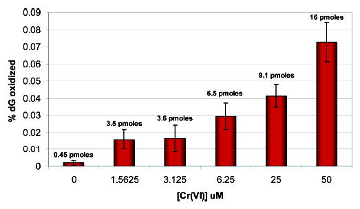
Dose-dependent formation of 8-oxoG in DNA with respect to Cr(VI) concentration.
Identification of Spiroiminodihydantoin (Sp) in Chromium-Oxidized Duplex DNA
Enzymatic digests of Cr(VI)/Asc-oxidized, duplex DNA oligonucleotides were carried out as described and analyzed by LC–ESI-MS for the formation of Sp. The retention time of Sp at a 0.4 mL/min flow rate using C18 reverse-phase chromatography was ×6.0 min with the other nucleosides eluting in the typical order of dC, dG, dT, and dA. A stable isotope-labeled standard, 18O-Sp, was synthesized as described previously (37) and identified by its characteristic mass spectral pattern as shown in Figure 7. Known concentrations of the 18O-Sp were spiked into the DNA digest of Cr(VI)/Asc-treated DNA as an internal standard to allow quantification. SIM was used to identify Sp formation in the DNA digests using the characteristic masses for the base, B + H2, or that of the nucleoside, M + H, and M + Na ions of Sp at m/z 184, 300, and 322, for 16O-Sp, and those at m/z 186, 302, and 324, for 18O-Sp. Figure 8 shows the LC/MS chromatogram ion current profiles at m/z 322 and 324 of a 2.5 μM DNA sample treated with 50 μM Cr(VI) and 500 μM ascorbate. The exact mass calculation of the M + Na peak for 16O-Sp using the 18O-Sp internal standard was 322.0764 compared to a calculated mass of 322.0770 to yield a 1.9 ppm mass accuracy. The identical retention times for 16O-Sp and 18O-Sp coupled with the exact mass calculations show the formation of spiroiminodihydantoin in the reaction of duplex DNA with Cr(VI) and ascorbate under these conditions.
Figure 7.

Characteristic mass spectra of the 18O-Sp isotopic standard. B = free base, and M = the free nucleoside.
Figure 8.

LC–ESI-MS (SIM) ion chromatogram profiles for a typical Cr(VI) oxidized DNA digest showing the m/z 324 of the spiked 18O-Sp internal standard (A) and the m/z 322 for 16O-Sp formed from the Cr(VI)/Asc reaction (B).
Quantification of Spiroiminodihydantoin (Sp) in Duplex DNA
The quantification of Sp in the Cr(VI)-oxidized, duplex DNA was performed by LC–ESI-MS using 18O-Sp as an internal standard. A calibration plot was obtained to judge the response of the mass spectrometer to known concentrations of 16O-Sp and 18O-Sp. Samples containing 0.1 nmoles of 18O-Sp were spiked with increasing amounts of 16O-Sp from 0.01 to 5.12 nmols. The ratio of the currents of these ions was plotted as a function of the concentration of 16O-Sp yielding a linear calibration plot as shown in Figure 9.
Figure 9.

LC–ESI-MS calibration curve for Sp.
Sp, formed from the oxidation reaction in DNA by Cr(VI)/ascorbate, was measured with known amounts of spiked 18O-Sp. The ions at m/z 322 and 324 (M + Na for 16O-Sp and 18O-Sp, respectively) were monitored. Duplex DNA (2.5 μM), oxidized by the greatest amount of Cr(VI)/Asc (50 μM/500 μM), converted 0.341 ± 0.013 nmoles or 1.55 ± 0.06% of all guanines to the further-oxidized lesion Sp. The lowest concentration of Cr(VI)/ Asc used in this study, 3.1 μM/31 μM, converted 0.069 ± 0.01 nmoles or 0.315 ± 0.05% of all guanines to the further-oxidized lesion Sp. A small amount of Sp and 8-oxoG was observed without Cr(VI) treatment. We have attributed this formation to the rigorous DNA digestion procedure necessary for Sp analysis or the presence of small amounts of contaminating, redox-active metals. Figure 10 shows a graphical representation of Sp formation with respect to 8-oxoG formation at the concentrations of Cr(VI) and ascorbate used in this study. The formation of Sp was observed to be approximately 20-fold greater than that of 8-oxoG.
Figure 10.

Formation of Sp and 8-oxoG from reaction of Cr(VI)/Asc with respect to the percentage of guanine oxidized in the DNA duplex oligonucleotide.
We have observed that Sp within DNA was refractory to normal enzymatic digestion reactions which necessitated the inclusion of a more rigorous digestion cocktail. Further, the enhanced polarity of Sp versus other nucleosides creates technical difficulties in the separation by HPLC from the hydrolyzed DNA matrix. These factors are likely to cause inherent errors in the quantification of Sp, and it is likely that the amount of Sp may be greater than that observed. However, these results clearly show that Sp can be formed directly from guanine by Cr(VI)/Asc oxidation in duplex DNA and, under these conditions, was significantly more prevalent than 8-oxoG.
Discussion
The reaction of Cr(VI) with a 10-fold excess of ascorbate resulted in guanine-specific oxidation in a duplex DNA oligonucleotide. The products of this oxidation reaction were determined using BER-specific lesion trapping, HPLC-ECD, and HPLC–ESI-MS. BER trapping with hOGG1 showed little or no formation of 8-oxoG, which correlated with the low levels of 8-oxoG subsequently identified using the more sensitive HPLC-ECD method. BER trapping was observed with the NEIL2 glycosylase, which showed a dose-dependence of trapping with Cr(VI)/Asc treatment. NEIL2 recognizes the Sp lesion in duplex DNA, and the formation of Sp was identified and quantified using HPLC–ESI-MS. The levels of Sp generated in the Cr(VI)/Asc system was approximately 20 times greater than that of 8-oxoG. These findings suggest that the Sp lesion may be a common lesion in DNA oxidation and could play a significant role in the initiation of cancer by carcinogenic Cr(VI) compounds. The presence of this lesion may explain many of the cellular effects observed for Cr(VI) exposure both in vitro and in vivo.
Reduction of Cr(VI) by Ascorbate
Previous EPR studies have characterized the Cr(VI)/Asc system used in this study in terms of the reactive intermediates produced during reduction (12). The reduction of Cr(VI) with a 10-fold molar excess of ascorbate has been observed to initially form a transient Cr(IV) species and the oxidized form of ascorbate, dehydroascorbate (DHA), followed by a second reduction reaction of Cr(IV) with Asc to give rise to the ascorbyl radical, Asc•−, and a kinetically inert Cr(III) species as shown in eqs 1 and 2 below:
| (1) |
| (2) |
On the basis of these results, we postulate that the species responsible for oxidation of guanine in DNA to form Sp and 8-oxoG is the Cr(IV) species formed in eq 1. Cr(IV) is a prime candidate for the DNA oxidant in this system since it is known to be highly oxidizing with standard reduction potentials for the chelate-stabilized Cr(IV)/Cr(III) couple estimated at 1.33–1.37 V (43). Cr(III) and Asc•− are generally less reactive. Under these conditions, no oxygen radicals have been observed (12), suggesting that the mechanism involves a direct, metal-mediated oxidation of DNA. The formation of Sp directly from guanine in DNA has previously only been observed for carbonate radical anions (44). To our knowledge, this study is the first example of Sp formation arising discretely from guanine in duplex DNA from reaction with a known carcinogenic metal.
The mechanism of Sp formation directly from guanine is a four-electron process with Sp being the terminal product that predominates at pH 7 or greater (45). 8-OxoG is the two electron intermediate in this oxidation process which is further oxidized to the 5-hydroxy-8-oxoguanine species, 5-OH-8-oxoG, and ultimately Sp. Figure 11 shows the putative reaction scheme for the formation of Sp from guanine with sequential one-electron oxidation by Cr(IV). A second terminal product of guanine oxidation, guanidinohydantoin (Gh), may also be formed during this oxidation reaction. However, these products are generally formed under more acidic conditions, while this study was carried out at neutral pH (45). We have not observed Gh under the current oxidation conditions, but we have yet to optimize the HPLC–ESI-MS parameters for identification of this product. It is also important to note that the oxidation process used here shows, for the first time, guanine-specific products caused by the direct oxidation of unmodified dsDNA by Cr(VI) without the introduction of exogenous H2O2.
Figure 11.

General reaction mechanism proposed for the formation of Sp from guanine.
Guanine-Specific Cleavage by Cr(VI)/Asc in Duplex DNA
A number of studies on the reaction of Cr(VI) with DNA have shown a bias toward reaction with guanine (46). These results have often been interpreted as the formation of chromium adducts with guanine through a phosphodiester backbone-liganded intermediate (47). However, no adduct of chromium with the guanine base has ever been discretely identified. We suggest that guanine-specific reactions in DNA give rise primarily to further-oxidized lesions such as Sp, although formation of these ternary adducts cannot be ruled out in our system.
Sp is known to be considerably more piperidine labile than 8-oxoG and has shown the ability to induce profound arrests in polymerase processing (24). The enhancement in cleavage that we have observed in guanines that are 5′ in a run of purines also supports this oxidative mechanism. We postulate that many of the effects of Cr(VI) treatment observed in cellular systems may be adequately explained by the formation of these further-oxidized lesions.
Formation of 8-OxoG in the Cr(VI)/Asc Oxidation Reaction
8-Oxo-2′ -deoxyguanine, 8-oxoG, is a well-characterized biomarker of oxidative damage in cellular and in vitro systems (48, 49). The formation of 8-oxoG in DNA has been observed with a vast array of oxidizing species, and its formation has been extensively followed using HPLC–MS, GC–MS, HPLC-ECD, and immuno-assays. 8-OxoG itself is mildly mutagenic and forms G:C → T:A transversion mutations approximately half the time during DNA replication in vitro (22, 30–33) but only about 5% of the time in vivo (31). The hOGG1 base excision repair pathway is the primary route for the excision and repair of 8-oxoG in human DNA, and genetic polymorphisms of this repair enzyme have been associated with increased risk of lung cancer (50). However, Ogg1 knockout mice have shown no change in the observable phenotype relative to wild-type mice, although a slight increase in mutation rate was observed in some tissues (51, 52). This lack of phenotype has been attributed to redundancy in base excision repair. The formation of 8-oxoG in DNA following an oxidant insult is undoubtedly an important consequence of DNA oxidation. However, the focus on 8-oxoG as the primary oxidized lesion of guanine may arise more from its ease of detection than from its biological significance.
8-OxoG has been shown to be sensitive to further oxidation, especially when compared to the parent guanine base (23, 24). The lowered reduction potential of this oxidized base with respect to guanine allows the lesion to act as a thermodynamic sink for electron donation in redox reactions and serve as a hot spot for further oxidation within a DNA sequence (25). The thermodynamics of 8-oxoG oxidation would thus suggest that a secondary oxidation of this product would predominate and result in the formation of a different subset of oxidized lesions, many of which may be more deleterious than the 8-oxoG lesion. A number of further-oxidized lesions of guanine have been identified arising from 8-oxoG oxidation in vitro which include that lesion studied here, spiroiminodihydantoin (Sp) and guanidinohydantoin (Gh), imidazalone, oxazolone, and oxaluric acid, to name a few (28, 29, 37, 42). We have previously shown that 8-oxoG lesions in DNA are prone toward specific further oxidation by high-valent chromium, Cr(V), complexes at the 8-oxoG site leading to the formation of Sp and Gh. Sp and Gh have proven to be much more mutagenic than 8-oxoG, causing G:C → T:A and G:C → C:G transversion mutations at a significantly greater frequency (30–33). As noted previously, the reaction of Cr(VI) with DNA in the presence of ascorbate can also form Cr/Asc/DNA ternary adducts (47). These ternary adducts have been shown to give rise to similar G:C → T:A transversion mutations as those observed for Sp and 8-oxoG (53).
Identification of 8-oxoG in Cr(VI)-treated systems in vitro had previously only been observed with addition of hydrogen peroxide to reaction mixtures of Cr(VI). In vivo, the formation of 8-oxoG has been reported as present in some cellular systems in low levels and absent in other studies. The results of this study may shed light on this contradiction, and we propose that as a labile intermediate of guanine oxidation, 8-oxoG, is either repaired or preferentially oxidized in DNA to further-oxidized lesions such as Sp. Thus, the 8-oxoG species would always be at low or even undetectable levels intracellulary.
Taken together, these data suggest that 8-oxoG may be an excellent and readily measurable biomarker of general oxidation, but the actual lesions responsible for the initiation of carcinogenesis may be the more mutagenic, further-oxidized lesions such as the Sp observed in these studies.
Sp Formation in the Cr(VI)/Asc Reaction With Unmodified Duplex DNA
The primary focus of this study was to identify and quantify the formation of the further-oxidized guanine lesions, spiroiminodihydantoin (Sp), arising from oxidation of guanine in unmodified duplex DNA. Clearly, the formation of these further-oxidized lesions from oxidative attack on guanine in DNA instead of in an 8-oxoG-modified DNA strand was necessary to establish their potential relevance in biological systems. To identify these lesions as arising in unmodified DNA from Cr(VI) oxidation, we exploited the specific affinity of trapping by the base excision repair enzyme NEIL2 and have further unambiguously identified their formation using HPLC–ESI-MS. The NEIL family of genes (Nei-like) have only recently been identified, and their affinities for oxidatively damaged bases are still being defined. We have recently shown that NEIL1 and NEIL2 both have high affinities for the further-oxidized guanine lesions Sp and Gh but little or no affinity for 8-oxoG in duplex DNA (36). When Sp formation was compared with 8-oxoG in this study, the prevalence of the glycosylase recognition was specific for further-oxidized lesions and not 8-oxoG, suggesting that Sp was formed at significantly greater levels than 8-oxoG.
The quantification of Sp by HPLC–ESI-MS is neither trivial nor ideal. Sp oligonucleotides have proven difficult to hydrolyze to their individual nucleosides. The generation of a tetrahedral sp3-hybridized carbon in Sp should significantly perturb DNA structure which slows enzymatic hydrolysis. Therefore, a cocktail of enzymes must be used to achieve digestion, while at the same time, the use of buffers must be minimized to give a clean mass spectrometric signal. An alternative method of chemical hydrolysis using HF in pyridine to hydrolyze the Sp base from oxidized DNA has been developed and successfully utilized by Ravanat and Cadet (54, 55). Finally, the retention time of Sp on a typical reverse-phase column is poor due to the polarity of Sp, leading to problematic identification/quantification of this species. It is likely, based on the rapidly increasing literature examples of Sp formation in a variety of different systems, that this may be a ubiquitous lesion at least as common as 8-oxoG. However, the poor enzymatic digestion and chromatographic behavior of this lesion may be a factor in it being overlooked in many of the systems. Therefore, while the quantification of Sp achieved here is not ideal, our approach has clearly shown the formation and existence of Sp by Cr(VI) oxidation in unmodified dsDNA and has shown that Sp is formed in quantities much greater than 8-oxoG in vitro.
Conclusion
The reaction of the human carcinogen Cr(VI) with the endogenous reductant ascorbate exhibits guanine-specific oxidation in normal duplex DNA. The primary lesion that was observed in this system was the further-oxidized lesion spiroiminodihydantoin, Sp. The formation of Sp, even at the lowest concentrations of Cr(VI) and ascorbate, was much greater than that of 8-oxoG (10–20-fold). This work suggest that the primary lesion responsible for initiation of carcinogenesis by Cr(VI) may be Sp, and the use of this species as a biomarker for Cr(VI) exposure may be more relevant than 8-oxoG. Further work to identify the formation of Sp in Cr(VI)-treated cellular systems is necessary to confirm the physiological significance of this lesion.
Acknowledgments
Funding for this study was provided by the National Institute of Environmental Health Sciences Grant (ES10437) to K.D.S. B.D.M. is partially supported by an NCRR, NIH Grant P20RR017670. The authors would like to thank Dr. Thomas Rosenquist for the generous gift of mNEIL2 and Dr. Fernando Cardozo-Pelaez, Dave Cox, and Celeste Bolin for assistance with 8-oxoG measurements.
Footnotes
Abbreviations: 8-oxoG, 7,8-dihydro-8-oxoguanine; Sp, spiroimin-odihydantoin; Gh, guanidinohydantoin; HPLC–ESI-MS, high-performance liquid chromatography electrospray ionization mass spectrometry; HPLC-ECD, high-performance liquid chromatography with electrochemical detection; Asc, ascorbate; hOGG1 human 8-oxoG-DNA glycosylase; mNEIL2, murine Nei-like DNA glycosylase 2; NaCNBH3, sodium cyanoborohydride; SIM, selected ion monitoring; Cr(V) –salen, N,N′-ethylenebis(salicylidene-animato)oxochromium(V); BER, base excision repair.
References
- 1.IARC Monograph on the Evaluation of Carcinogenic Risk to Humans. Chromium, Nickel and Welding (1990) Vol. 48, pp 49–508, IARC, Lyon, France. [PMC free article] [PubMed]
- 2.Hayes, R. B. (1982) Carcinogenic effects of chromium. In Biological and Environmental Aspects of Chromium (Langard, S., Ed.) pp 221–248, Elsevier Biomedical Press, New York.
- 3.Miller CA, Costa M. Characterization of DNA-protein complexes induced in intact cells by the carcinogen chromate. Mol Carcinog. 1988;1:125–133. doi: 10.1002/mc.2940010208. [DOI] [PubMed] [Google Scholar]
- 4.Cantoni O, Costa M. Analysis of the induction of alkali sensitive sites in the DNA by chromate and other agents that induce single strand breaks. Carcinogenesis. 1984;5:1207–1209. doi: 10.1093/carcin/5.9.1207. [DOI] [PubMed] [Google Scholar]
- 5.Cupo DY, Wetterhahn KE. Binding of chromium to chromatin and DNA from liver and kidney of rats treated with sodium dichromate and chromium(III) chloride in vivo. Cancer Res. 1985;145:1146–1151. [PubMed] [Google Scholar]
- 6.Hamilton JW, Wetterhahn KE. Chromium(VI)-induced DNA damage in chick embryo liver and blood cells in vivo. Carcinogenesis. 1986;7:2085–2088. doi: 10.1093/carcin/7.12.2085. [DOI] [PubMed] [Google Scholar]
- 7.Robison SH, Cantoni O, Costa M. Analysis of metal-induced DNA lesions and DNA-repair replication in mammalian cells. Mutat Res. 1984;131:173–182. doi: 10.1016/0167-8817(84)90058-0. [DOI] [PubMed] [Google Scholar]
- 8.Connett PH, Wetterhahn KE. Metabolism of the carcinogen chromate by cellular constituents. Struct Bonding. 1983;54:93–124. [Google Scholar]
- 9.Sugiyama M, Tsuzuki K, Ogura R. Effect of ascorbic acid on DNA damage, cytotoxicity, glutathione reductase and formation of paramagnetic chromium in Chinese hamster V-79 cells treated with sodium chromate(VI) J Biol Chem. 1991;266:3383–3386. [PubMed] [Google Scholar]
- 10.Goodgame DML, Joy AM. EPR study of the Cr(V) and radical species produced in the reduction of Cr(VI) by ascorbate. Inorg Chim Acta. 1987;135:115–118. [Google Scholar]
- 11.Shi X, Mao Y, Knapton AD, Ding M, Rojanasakul Y, Gannett PM, Dalal NS, Liu K. Reaction of Cr(VI) with ascorbate and hydrogen peroxide generates hydroxyl radicals and causes DNA damage: role of Cr(IV)-mediated Fenton-like reaction. Carcinogenesis. 1994;15:2475–2478. doi: 10.1093/carcin/15.11.2475. [DOI] [PubMed] [Google Scholar]
- 12.Stearns DM, Wetterhahn KE. Reaction of chromium(VI) with ascorbate produces chromium(V), chromium-(IV) and carbon-based radicals. Chem Res Toxicol. 1994;7:219–230. doi: 10.1021/tx00038a016. [DOI] [PubMed] [Google Scholar]
- 13.Wiegand HJ, Ottenwalder H, Bolt HM. The reduction of chromium(VI) to chromium(III) by glutathione: an intracellular redox pathway in the metabolism of the carcinogen chromate. Toxicology. 1984;33:341–348. doi: 10.1016/0300-483x(84)90050-7. [DOI] [PubMed] [Google Scholar]
- 14.Connett PH, Wetterhahn KE. In vitro reaction of the carcinogen chromate with cellular thiols and carboxylic acids. J Am Chem Soc. 1985;107:4282–4288. [Google Scholar]
- 15.Standeven AM, Wetterhahn KE. Ascorbate is the principal reductant of chromium(VI) in rat liver and kidney ultrafiltrates. Carcinogenesis. 1991;12:1733–1737. doi: 10.1093/carcin/12.9.1733. [DOI] [PubMed] [Google Scholar]
- 16.Pratviel G, Bernadou J, Meunier B. Carbon–hydrogen bonds of sugar units as targets for chemical nucleases and drugs. Angew Chem, Int Ed. 1995;34:746–769. [Google Scholar]
- 17.Burrows CJ, Muller JG. Oxidative nucleobase modifications leading to strand scission. Chem Rev. 1998;98:1109–1151. doi: 10.1021/cr960421s. [DOI] [PubMed] [Google Scholar]
- 18.Shi X, Sun X, Gannett PM, Dalal NS. Deferoxamine inhibition of Cr(V)-mediated radical generation and deoxyguanine hydroxylation: ESR and HPLC evidence. Arch Biochem Biophys. 1992;293:281–286. doi: 10.1016/0003-9861(92)90396-e. [DOI] [PubMed] [Google Scholar]
- 19.Misra M, Alcedo J, Wetterhahn KE. Two pathways for chromium(VI)-induced DNA damage in 14 day chick embryos: Cr-DNA binding in liver and 8-oxo-2′-deoxyguanosine in red blood cells. Carcinogenesis. 1994;15:2911–2917. doi: 10.1093/carcin/15.12.2911. [DOI] [PubMed] [Google Scholar]
- 20.Saito I, Takayama M, Sugiyama H, Nakatani K. Photoinduced DNA cleavage via electron transfer: demonstration that guanine residues located 5′ to guanine are the most electron-donating sites. J Am Chem Soc. 1995;117:6406–6407. [Google Scholar]
- 21.Yoshioka Y, Kawai H, Sato T, Yamaguchi K, Saito I. Ab initio molecular orbital study on the G-selectivity of GGG triplet in copper(I)-mediated one-electron oxidation. J Am Chem Soc. 2003;125:1968–1974. doi: 10.1021/ja028039m. [DOI] [PubMed] [Google Scholar]
- 22.Shibutani S, Takeshita M, Grollman AP. Insertion of specific bases during DNA synthesis past the oxidation damaged base 8-oxodG. Nature. 1991;349:431–434. doi: 10.1038/349431a0. [DOI] [PubMed] [Google Scholar]
- 23.Steenken S, Jovanovic SV, Bietti M, Bernhard K. The trap depth (in DNA) of 8-oxo-7,8-dihydro-2′-deoxyguanosine as derived from electron-transfer equilibria in aqueous solution. J Am Chem Soc. 2000;122:2372–2374. [Google Scholar]
- 24.Muller JG, Duarte V, Hickerson RP, Burrows CJ. Gel electrophoretic detection of 7,8-dihydro-8-oxoguanine and 7,8-dihydro-8-oxoadenine via oxidation by Ir(IV) Nucleic Acids Res. 1998;26:2247–2249. doi: 10.1093/nar/26.9.2247. [DOI] [PMC free article] [PubMed] [Google Scholar]
- 25.Delaney S, Barton JK. Long-range DNA charge transport. J Org Chem. 2003;68:6475–6483. doi: 10.1021/jo030095y. [DOI] [PubMed] [Google Scholar]
- 26.Sugden KD, Campo CK, Martin BD. Direct oxidation of guanine and 7,8-dihydro-8-oxo-guanine in DNA by a high valent chromium complex: a possible mechanism of chromate genotoxicity. Chem Res Toxicol. 2001;14:1315–1322. doi: 10.1021/tx010088+. [DOI] [PubMed] [Google Scholar]
- 27.Sugden KD, Martin BD. Guanine and 7,8-dihydro-8-oxo-guanine specific oxidation in DNA by chromium(V)″ Environ. Health Perspect. 2002;110:725–728. doi: 10.1289/ehp.02110s5725. [DOI] [PMC free article] [PubMed] [Google Scholar]
- 28.Luo W, Muller JG, Rachlin EM, Burrows CJ. Characterization of spiroiminodihydantoin as a product of one-electron oxidation of 8-oxo-7,8-dihydroguanosine. Org Lett. 2000;2:613–616. doi: 10.1021/ol9913643. [DOI] [PubMed] [Google Scholar]
- 29.Tretyakova NY, Niles JC, Burney S, Wishnok JS, Tannenbaum SR. Peroxynitrite-induced reactions of synthetic oligonucleotides containing 8-oxoguanine. Chem Res Toxicol. 1999;12:459–466. doi: 10.1021/tx980235c. [DOI] [PubMed] [Google Scholar]
- 30.Duarte V, Muller JG, Burrows CJ. Insertion of dGMP and dAMP during in vitro DNA synthesis opposite an oxidized form of 7,8-dihydro-8-oxoguanine. Nucleic Acids Res. 1999;27:496–502. doi: 10.1093/nar/27.2.496. [DOI] [PMC free article] [PubMed] [Google Scholar]
- 31.Henderson PT, Delaney JC, Muller JG, Neeley WL, Tannenbaum SR, Burrows CJ, Essigmann JM. The hydantoin lesions formed from oxidation of 7,8-dihydro-8-oxoguanine are potent sources of replication errors in vivo. Biochemistry. 2003;42:9257–9262. doi: 10.1021/bi0347252. [DOI] [PubMed] [Google Scholar]
- 32.Kornyushyna O, Berges AM, Muller JG, Burrows CJ. In vitro nucleotide misinsertion opposite the oxidized guanosine lesions spiroiminodihydantoin and guanidinohydantoin and DNA synthesis past the lesions using Escherichia coli DNA polymerase I (Klenow fragment) Biochemistry. 2002;41:15304–15314. doi: 10.1021/bi0264925. [DOI] [PubMed] [Google Scholar]
- 33.Henderson PT, Delaney JC, Tannenbaum SR, Essigmann JM. Oxidation of 7,8-dihydro-8-oxoguanine affords lesions that are potent sources of replication errors in vivo. Biochemistry. 2002;42:914–921. doi: 10.1021/bi0156355. [DOI] [PubMed] [Google Scholar]
- 34.Liu S, Medvedovic M, Dixon K. Mutational specificity in a shuttle vector replicating in chromium(VI)-treated mammalian cells. Environ Mol Mutagen. 1999;33:313–319. doi: 10.1002/(sici)1098-2280(1999)33:4<313::aid-em8>3.0.co;2-v. [DOI] [PubMed] [Google Scholar]
- 35.Feng Z, Hu W, Rom WN, Costa M, Tang MS. Chromium(VI) exposure enhances polycyclic aromatic hydrocarbon-DNA binding at the p53 gene in human lung cells. Carcinogenesis. 2003;24:771–778. doi: 10.1093/carcin/bgg012. [DOI] [PubMed] [Google Scholar]
- 36.Hailer MK, Slade PG, Rosenquist TA, Martin BD, Sugden KD. Recognition of the oxidized lesions spiroiminodihydantoin and guanidinohydantoin in DNA by mammalian base excision repair glycosylases NEIL1 and NEIL2. DNA Repair. 2005;4:41–50. doi: 10.1016/j.dnarep.2004.07.006. [DOI] [PubMed] [Google Scholar]
- 37.Luo W, Muller JG, Burrows CJ. The pH-dependent role of superoxide in riboflavin-catalyzed photooxidation of 8-oxo-7,8-dihydroguanosine. Organic Lett. 2001;3:2801–2804. doi: 10.1021/ol0161763. [DOI] [PubMed] [Google Scholar]
- 38.Bolin C, Stedeford T, Cardozo-Pelaez F. Single extraction protocol for the analysis of 8-hydroxy-2′-deoxyguanosine and the associated activity of 8-oxoguanine DNA glycosylase. J Neurosci Methods. 2004;136:69–76. doi: 10.1016/j.jneumeth.2003.12.025. [DOI] [PubMed] [Google Scholar]
- 39.Martin BD, Schoenhard JA, Sugden KD. Hypervalent chromium mimics reactive oxygen species as measured by the oxidant-sensitive dyes 2′,7′-dichlorofluorescein and dihydrorhodamine. Chem Res Toxicol. 1998;11:1402–1410. doi: 10.1021/tx9801559. [DOI] [PubMed] [Google Scholar]
- 40.Dubrovskaya V, Wetterhahn KE. Effects of Cr(VI) on the expression of the oxidative stress genes in human lung cells. Carcinogenesis. 1998;19:1401–1408. doi: 10.1093/carcin/19.8.1401. [DOI] [PubMed] [Google Scholar]
- 41.Krokan HE, Standal R, Slupphaug G. DNA glycosylases in the base excision repair of DNA. Curr Biol. 1997;7:397–407. doi: 10.1042/bj3250001. [DOI] [PMC free article] [PubMed] [Google Scholar]
- 42.Nash HM, Bruner SD, Scharer OD, Kawate T, Addona TA, Spooner E, Lane WS, Verdine GL. Cloning of a yeast 8-oxoguanine DNA glycosylase reveals the existence of a base-excision DNA-repair protein superfamily. Curr Biol. 1996;6:968–980. doi: 10.1016/s0960-9822(02)00641-3. [DOI] [PubMed] [Google Scholar]
- 43.Gould ES. Redox chemistry of chromium(IV) complexes. Coord Chem Rev 135/ 1994;136:651–684. [Google Scholar]
- 44.Joffe A, Geacintov NE, Shafirovich V. DNA lesions derived from site selective oxidation of guanine by carbonate radicals. Chem Res Toxicol. 2003;16:1528–1538. doi: 10.1021/tx034142t. [DOI] [PubMed] [Google Scholar]
- 45.Ye Y, Muller JG, Luo W, Mayne CL, Shallop AJ, Jones RA, Burrows CJ. Formation of 13C-, 15N-, and 18O-labeled guanidinohydantoin from guanosine oxidation with singlet oxygen. Implications for structure and mechanism. J Am Chem Soc. 2003;125:13926–13927. doi: 10.1021/ja0378660. [DOI] [PubMed] [Google Scholar]
- 46.Bridgewater LC, Manning FCR, Patierno SR. Base-specific arrest of in vitro DNA replication by carcinogenic chromium: relationship to DNA interstrand crosslinking. Carcinogenesis. 1994;15:2421–2427. doi: 10.1093/carcin/15.11.2421. [DOI] [PubMed] [Google Scholar]
- 47.Zhitkhovich A. Importance of chromium-DNA adducts in mutagenicity and toxicity of chromium(VI) Chem Res Toxicol. 2005;18:3–11. doi: 10.1021/tx049774+. [DOI] [PubMed] [Google Scholar]
- 48.Ames BN, Shigenaga M, Hagen TM. Oxidants, antioxidants and the degenerative disease of aging. Proc Natl Acad Sci USA. 1993;90:7915–7922. doi: 10.1073/pnas.90.17.7915. [DOI] [PMC free article] [PubMed] [Google Scholar]
- 49.Grollman AP, Moriya M. Mutagenesis by 8-oxo-guanine: an enemy within. Trends Genet. 1993;9:246–249. doi: 10.1016/0168-9525(93)90089-z. [DOI] [PubMed] [Google Scholar]
- 50.Park J, Chen L, Tockman MS, Elahi A, Lazarus P. The human 8-oxoguanine DNA N-glycosylase 1 (hOGG1) DNA repair enzyme and its association with lung cancer risk. Pharmacogenetics. 2004;14:103–109. doi: 10.1097/00008571-200402000-00004. [DOI] [PubMed] [Google Scholar]
- 51.Minowa O, Arai T, Hirano M, Monden Y, Nakai S, Fukuda M, Itoh M, Takano H, Hippou Y, Aburatani H, Masumura K, Nohmi T, Nishimura S, Noda T. Mmh/Ogg1 gene inactivation results in accumulation of 8-hydroxyguanine in mice. Proc Natl Acad Sci USA. 2000;97:4156–4161. doi: 10.1073/pnas.050404497. [DOI] [PMC free article] [PubMed] [Google Scholar]
- 52.Osterod M, Hollenbach S, Hengstler JG, Barnes DE, Lindahl T, Epe B. Age-related and tissue-specific accumulation of oxidative DNA base damage in 7,8-dihydro-8-oxoguanine-DNA glycosylase (Ogg1) deficient mice. Carcinogenesis. 2001;22:1459–1463. doi: 10.1093/carcin/22.9.1459. [DOI] [PubMed] [Google Scholar]
- 53.Quievryn G, Peterson E, Messer J, Zhitkhovich A. Genotoxicity and mutagenicity of chromium(VI)/ascorbate-generated DNA adducts in human and bacterial cells. Biochemistry. 2003;42:1062–1070. doi: 10.1021/bi0271547. [DOI] [PubMed] [Google Scholar]
- 54.Ravanat JL, Cadet J. Reaction of singlet oxygen with 2′-deoxyguanosine and DNA. Isolation and characterization of the main oxidation products. Chem Res Toxicol. 1995;8:379–388. doi: 10.1021/tx00045a009. [DOI] [PubMed] [Google Scholar]
- 55.Cadet J, Bellon S, Berger M, Bourdat AG, Douki T, Duarte V, Frelon S, Gasparutto D, Muller E, Ravanat JL, Sauvaigo S. Recent aspects of oxidative DNA damage: guanine lesions, measurements and substrate specificity of DNA repair glycosylases. Biol Chem. 2002;383:933–943. doi: 10.1515/BC.2002.100. [DOI] [PubMed] [Google Scholar]