



ABSTRACT
Objectives
The Schroth method is considered potentially valuable as an alternative intervention to control curve progression in mild to moderate adolescent idiopathic scoliosis. However, patients are often dependent on the subjective experience of the physiotherapist when practicing, as changes in spinal alignment and muscle activation are unknown. The aim of this study was to evaluate the effectiveness of the Schroth method for spinal alignment and muscle activation in adolescents with idiopathic scoliosis (AIS).
Methods
This prospective cross‐sectional study, conducted at the Scoliosis Engineering Center of Guangzhou Sport University (January 2020–October 2022), enrolled 36 AIS (4 males, 32 females; age 13.2 ± 1.8 years) presenting major thoracic curvature (Cobb angle: 10°–40°, Risser stage 0–4). Participants underwent a standardized 1‐week pretraining protocol before performing four Schroth exercises (“50 × Pezziball,” sail, muscle cylinder, and corrected standing). Real‐time spinal ultrasound angles (SUAs) and paraspinal muscle activation (via surface electromyography, sEMG) were quantified during habitual standing (baseline) and exercise conditions. Normality was verified using Kolmogorov–Smirnov tests. Within‐group comparisons employed paired t‐tests (effect sizes: Cohen's d; 95% CIs), while proximal thoracic (PT) compensation frequencies were assessed via chi‐squared tests.
Results
All four Schroth exercises significantly reduced the SUA of the primary thoracic curve compared to habitual standing (p < 0.05). The lumbar curve SUA decreased specifically after “50 × Pezziball” and muscle cylinder exercises (p < 0.05). PT curve compensation frequency rose from 16.7% (6/36) in habitual standing to 33.3% (12/36) and 30.6% (11/36) during “50 × Pezziball” and sail exercises, respectively (p < 0.05). sEMG demonstrated reduced paravertebral muscle (PSM) activation asymmetry at the thoracic apex (T8) during corrective standing, “50 × Pezziball,” and sail exercises (p < 0.05). However, “50 × Pezziball” and sail exercises triggered compensatory left‐dominant PSM imbalance at T2 (p < 0.05). At lumbar levels (L2–L5), performing 50 × Pezziball exercise exacerbated asymmetry (p < 0.05), whereas muscle cylinder exercises reversed the activation patterns of the PSM (p < 0.05).
Conclusion
While Schroth exercises effectively reduced thoracic curvature and improved T8 muscle symmetry, caution is warranted during “50 × Pezziball” and sail exercises due to increased PT compensation and lumbar/T2 muscle imbalances. Muscle cylinder exercises may mitigate adverse lumbar effects.
Keywords: adolescent idiopathic scoliosis, Schroth method, surface myoelectricity, three‐dimensional spinal ultrasound
The Schroth method as an alternative therapy for mild to moderate adolescent idiopathic scoliosis. This study focused on investigating changes in spinal alignment and paravertebral muscle (PSM) activation parameters during Schroth exercise. We found that Schroth exercise should be chosen with caution: potential effects of compensatory curves and paraspinal muscle activation imbalances.

1. Introduction
Adolescent idiopathic scoliosis (AIS), a three‐dimensional torsional deformity of unknown etiology, affects 1%–4% of adolescents globally [1]. While multifactorial pathogenesis is hypothesized, biomechanical studies emphasize the role of progressive asymmetric spinal loading and paraspinal muscle (PSM) dysfunction in curve progression [2, 3]. Specifically, convex‐side PSM hypotonia and concave‐side hypertonia create a self‐perpetuating cycle of vertebral rotation and lateral deviation [4]. Early intervention is essential, as curves measuring less than 30° at skeletal maturity exhibit a significantly lower risk of progression [5].
The Schroth method, a physiotherapeutic approach rooted in 3D postural correction, offers a noninvasive strategy for mild to moderate AIS [5, 6]. This method integrates three core principles: (1) Directional breathing mechanics to derotate the rib cage and spine by expanding the concave hemithorax during inhalation; (2) Asymmetric muscle activation through isometric contractions to strengthen hypotonic muscles on the convex side while elongating hypertonic muscles on the concave side; and (3) Proprioceptive re‐education using mirror feedback and tactile cues to reinforce corrected postural alignment [7]. By promoting symmetrical PSM engagement and spinal “auto‐correction,” the method theoretically interrupts the cycle of asymmetric loading and deformity progression. However, clinical outcomes are significantly contingent upon therapist proficiency and sustained patient compliance, underscoring the necessity for quantifiable neuromuscular biomarkers—such as surface electromyography (sEMG)‐derived activation ratios and ultrasonographic spinal alignment metrics—to standardize and personalize therapeutic protocols [7].
Emerging technologies offer solutions to these limitations. sEMG serves as a critical adjunct to therapeutic protocols for AIS, offering quantifiable neuromuscular control data to optimize muscle‐strengthening regimens and biofeedback interventions [8, 9]. However, therapeutic strategies must account for heterogeneous vertebral deformities beyond mere symmetrical PSM activation. Notably, concerns regarding cumulative ionizing radiation exposure from serial radiographs have raised oncogenic safety considerations in this pediatric population [10]. Three‐dimensional ultrasonography has emerged as a viable alternative for spinal curvature assessment, combining radiation‐free imaging, weight‐bearing positional analysis, cost‐effectiveness, and operator accessibility [11]. Recent advancements have established validated ultrasound‐derived markers (e.g., spinous process angle, lamina inclination) that demonstrate promising correlations with radiographic Cobb angle measurements (r = 0.82–0.91), positioning this modality as a superior option for follow‐up monitoring in scoliosis management [11].
Despite growing evidence supporting the biomechanical rationale of the Schroth method, critical knowledge gaps persist regarding its immediate structural and neuromuscular effects. This study addresses three unresolved scientific questions: (i) Real‐time coronal plane correction dynamics. Prior studies lack concurrent quantification of spinal curvature modulation and PSM activation during Schroth exercises. We integrate dynamic 3D ultrasonography and sEMG to resolve this gap, hypothesizing that directional breathing maneuvers reduce lateral curvature by ≥ 5° (spinal ultrasound angle, SUA) in real time. (ii) Neuromuscular symmetry restoration. The relationship between acute spinal rebalancing and PSM activation patterns remains undefined. We propose that Schroth exercises normalize convex‐concave sEMG amplitude disparities by ≥ 25% (vs. baseline), indicating balanced neuromuscular recruitment. (iii) Exercise‐specific compensatory trade‐offs. Compensatory mechanisms triggered by corrective exercises are poorly characterized. We evaluate proximal thoracic (PT) curve compensation and regional muscle imbalances to identify maneuvers that optimize efficacy while minimizing adverse effects.
Building upon clinical evidence [6], this study employs a dual‐modality approach: coronal plane ultrasonography to measure SUA changes during active self‐correction, and sEMG to analyze convex versus concave PSM activation ratios. This framework enables protocol optimization by linking structural corrections to neuromuscular adaptations.
2. Methods
2.1. Study Design
This cross‐sectional study was conducted in two parts. First, we explored the immediate effect of the Schroth method on the parameters of the coronal plane. Additionally, the effects of the aforementioned exercises on the sEMG characteristics of the paravertebral muscles were verified. Based on these findings, the immediate corrective effect of exercise was assessed by measuring muscle and skeletal variability.
Ethical approval was obtained from the Human Subject Committee of Guangzhou Sport University (2018LCL‐008) and the study protocol was registered with the Chinese Clinical Trial Registry (ChiCTR1800018310). Informed consent was obtained from each patient or legal guardian before recruitment. Data were collected according to the guidelines of the Declaration of Helsinki.
2.2. Sample Size Estimation
Sample size estimation was performed using G*Power (version 3.1, Germany). The effect size was calculated using the maximum Cohen value. The level of significance was set at 0.05, and the statistical power was set at 0.95 in a two‐tailed correlation test. The estimated required sample size was calculated to be 32 considering a possible dropout rate of 15%. Forty participants will be recruited.
2.3. Participants
Initially, 40 participants were recruited to account for potential attrition. Four participants withdrew due to scheduling conflicts (n = 2) or transient discomfort during exercises (n = 2), resulting in 36 completed cases for final analysis.
To investigate the characteristics of common types in this prospective observational cohort, we continuously selected 36 patients with major thoracic curvature, corresponding to Schroth's three‐curve classification [12]. The participants first visited the Scoliosis Engineering Center of Guangzhou Sport University from January 2020 to October 2022.
The inclusion criteria were as follows: newly diagnosed patients, those who had not received any treatment, those with a Cobb angle between 10° and 40°, and those with a Risser sign of grades 0–4. The exclusion criteria were as follows: patients with secondary scoliosis of known etiology, such as neuromuscular and congenital scoliosis, accompanied by skeletal dysplasia and connective tissue abnormalities, and those who underwent bracing or surgical correction.
2.4. Experimental Protocol
2.4.1. Assessments and Task Schedule
The selection of Schroth exercises followed established protocols [7, 12], incorporating the four core techniques (“50 × Pezziball,” prone, sail, and muscle cylinder) combined with respiratory training for comprehensive curve management across different spinal deformity patterns. Participants underwent a standardized 1‐week pretraining program to familiarize themselves with the exercises, focusing on maximal voluntary isometric contractions (MVICs) of the PSMs. Concurrently, standardized postural training for activities of daily living (ADLs) emphasizing upright and seated spinal alignment was integrated. Despite initial inclusion of all exercises, the prone position required exercises that were far away from the device and could not be scanned by the ultrasonic probe, necessitating exclusion of this exercise from scanning protocols to ensure technical feasibility.
2.5. Instrumentation and Measurement
2.5.1. Ultrasound Measurements
The Scolioscan system (model SCN802, Telefeld Medical Imaging Ltd., Hong Kong, China) was employed for standardized ultrasound imaging of the spinal column. This system, which has demonstrated reliable accuracy for assessing mild to moderate scoliosis through previous validation studies [13, 14], utilizes a 7.5‐MHz linear probe (75‐mm width) combined with real‐time 3D positional tracking sensors during freehand scanning. Participants maintained their habitual standing posture while performing Schroth exercises with arms extended and normal respiration, ensuring consistent spinal alignment throughout the procedure. All scans were conducted by a single experienced physician to maintain methodological consistency. Notably, while traditional ultrasonography typically focuses on T1‐L5 spinal segments, this protocol extended imaging cranially to C2 due to technical limitations preventing C1 visualization (Figure 1A). Post‐scanning processing generated a 2D coronal reconstruction of the spine, with the system automatically determining the midline through shadow analysis of SUAs and quantifying thoracic and lumbar curve SUA values (Figure 1B). This approach enabled evaluation of PT curve compensation dynamics during exercise interventions.
FIGURE 1.
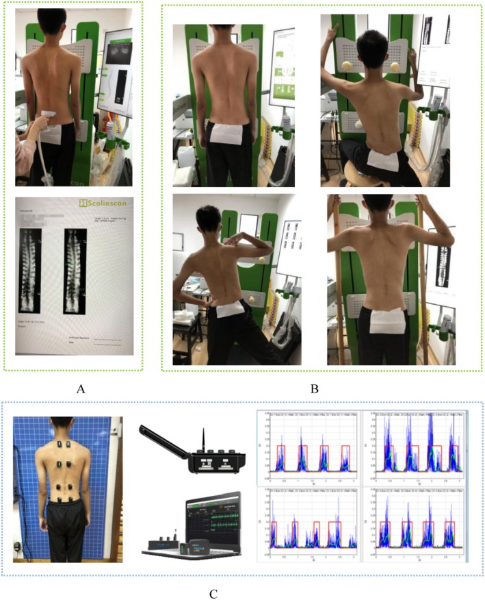
3D spinal ultrasound and sEMG are used to examine spinal morphology and paravertebral muscle activation when practiced with different movements. A is the SUA curve obtained using 3D spinal ultrasound. B denotes the position in which the participant is instructed to perform the standardized Schroth exercise. C is the surface electromyography of the paravertebral muscles of the participants.
2.5.2. sEMG Measurements
sEMG signals were recorded using active double parallel‐bar electrode sensors (Model Trigno, DelSys, Boston, MA, USA). Each Trigno IM wireless sensor collects signals independently from each other at a sampling frequency of 2000 Hz. The positive and negative electrodes were made of silicon rods and were placed 1 cm apart.
After the skin at the electrode sites was abraded with alcohol, the electrodes were positioned considering the direction and positioning of muscle fibers, as recommended in previous studies [15, 16]. The electrodes were placed bilaterally on the skin of the apical vertebrae of the thoracic and lumbar curves (erector spinae), T3 (trapezius transversus), and L5 (iliocostalis). Each electrode was placed 2.5 cm away from the spinous process.
2.5.3. sEMG Measurement Protocol
To evaluate the immediate effects of Schroth exercises, sEMG data were captured during four conditions:
Habitual standing posture: baseline measurement.
ADL (corrected standing posture): active derotational standing.
Three Schroth exercises: muscle cylinder, sail, and “50 × Pezziball” exercises.
For each condition, participants performed three trials with the following protocol:
Contraction phase: participants maintained MVIC during Schroth exercises for 6 s.
Rest phase: a 1‐min rest interval between trials to minimize fatigue.
Warm‐up: participants completed 1–2 practice trials before formal data collection to familiarize themselves with the exercises.
sEMG signals were analyzed using root mean square (RMS) values over the 6‐s MVIC window, normalized to the maximal amplitude recorded during the session.
2.5.4. sEMG Signal Processing
The total effective gain of the sEMG signal is 909 V/V ± 5% (bandwidth), and the baseline noise < 0.75 μV (RMS). A 16‐bit AD/board and EMG work acquisition software (DelSys Inc., Boston, MA, USA) was used to sample the surface EMG signal at 2000 Hz. All sensors are fixed to the skin through a double‐sided adhesive interface. A bandpass filter with a range of 10–500 Hz is used to eliminate undesired artifacts. sEMG signals were sampled at a rate of 2000 Hz, and the original EMG data were averaged by the RMS method to obtain the average amplitude of EMG signals. The RMS ratio of the subjects' EMG signal was calculated as follows:
2.6. Statistical Analysis
SPSS Statistics 25.0 for Windows (IBM, Armonk, NY, USA) was used for statistical analysis, as indicated by an average (95% CI). Mean values of SPA and sEMG ratio were tested for normality of distribution using the Kolmogorov–Smirnov test. If the values conform to the normal distribution, the difference between the values of different Schroth exercises and habitual standing positions within the group is compared using the independent sample t‐test. Count data measured the PT were expressed as the rate (%) using the chi‐square test. A p value of < 0.05 was considered statistically significant.
3. Results
3.1. Study Cohort Characteristics
The study enrolled 36 skeletally immature participants (4 male, 32 female) with AIS, aged 10–16 years (mean 13.2 ± 1.8 years). All participants met skeletal immaturity criteria (Risser stage 0–2) with female participants. Baseline spinous process ultrasound angle (SUA) measurements in habitual standing demonstrated a mean thoracic curvature of 29.4° ± 3.3° and lumbar curvature of 15.0° ± 3.3°.
3.2. Immediate Effects of Thoracic and Lumbar Curve
All four Schroth exercise modalities significantly reduced thoracic curve SUA compared with habitual standing (all p < 0.05; effect sizes: Cohen's d = 0.67–1.21, 95% CI 0.41–1.53). Lumbar curve SUA reduction achieved statistical significance following “50 × Pezziball” (p < 0.05; d = 0.53, 95% CI 0.12–0.94) and muscle cylinder exercises (p < 0.05; d = 0.49, 95% CI 0.08–0.90), but not with corrected standing (d = 0.18, 95% CI −0.20 to 0.56) or sail exercises (d = 0.22, 95% CI −0.16 to 0.60; Figure 2).
FIGURE 2.
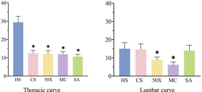
Immediate effects of Schroth exercises on spinal ultrasound angles (SUA) compared to habitual standing. Bars represent mean ± SD. Asterisks (*) indicate significant differences (p < 0.05, paired t‐tests). 50×, 50 × Pezziball; CS, corrected standing; HS, habitual standing; MC, muscle cylinder; SA, sail.
3.3. PT Curve Analysis
Significant differences in PT curve occurrences emerged between conditions: 6 cases (16.7%) during habitual standing versus 12 (33.3%) with “50 × Pezziball” (risk ratio [RR] = 2.00, 95% CI 1.12–3.57) and 11 (30.6%) with sail exercises (RR = 1.83, 95% CI 1.02–3.29; p < 0.05). No significant differences were observed between corrective standing (RR = 1.17, 95% CI 0.58–2.35), muscle cylinder exercises (RR = 1.33, 95% CI 0.71–2.51), and habitual standing (p > 0.05; Table 1).
TABLE 1.
Differences in ultrasound angles between corrective exercises and habitual standing positions (n = 36; %).
| Posture | PT compensation frequency (%) | χ 2 value (vs. HS) | p value |
|---|---|---|---|
| Habitual standing (HS) | 16.7 (6/36) | — | — |
| Corrected standing | 19.4 (7/36) | 0.12 | 0.729 |
| 50 × Pezziball | 33.3 (12/36) | 6.24 | 0.012* |
| Muscle cylinder | 22.2 (8/36) | 0.48 | 0.488 |
| Sail | 30.6 (11/36) | 4.92 | 0.027* |
Difference is statistically significant compared to habitual standing posture.
3.4. Electromyographic Findings
Surface electromyography revealed distinct PSM activation patterns: T2 marker analysis showed reduced RMS ratios during “50 × Pezziball” (d = −0.72, 95% CI −1.14 to −0.30) and sail exercises (d = −0.65, 95% CI −1.07 to −0.23) compared to habitual standing (p < 0.05), indicating left PSM predominance. Thoracic apex asymmetry (T8 RMS) decreased significantly during corrected standing (d = −0.41, 95% CI −0.83 to 0.01), “50 × Pezziball” (d = −0.89, 95% CI −1.32 to −0.46), and sail exercises (d = −0.55, 95% CI −0.97 to −0.13; p < 0.05). Lumbar compensation patterns diverged: “50 × Pezziball” increased L2/L5 RMS asymmetry (d = 0.61, 95% CI 0.18–1.04; p < 0.05), while muscle cylinder exercises demonstrated inverse asymmetry patterns (d = −0.38, 95% CI −0.80 to 0.04; Table 2).
TABLE 2.
sEMG ratios (convex/concave) at key spinal levels during exercises versus habitual standing.
| Spinal level | Posture | sEMG ratio (mean ± SD) | t‐value (vs. HS) | p value |
|---|---|---|---|---|
| T2 | Habitual standing | 1.41 ± 0.29 | — | — |
| 50 × Pezziball | 0.89 ± 0.21 | −3.72 | 0.001* | |
| Sail | 0.95 ± 0.24 | −3.14 | 0.003* | |
| T8 | Habitual standing | 1.58 ± 0.33 | — | — |
| Corrected standing | 1.17 ± 0.28 | −2.58 | 0.014* | |
| 50 × Pezziball | 0.89 ± 0.19 | −4.95 | < 0.001* | |
| Sail | 1.03 ± 0.25 | −2.89 | 0.007* | |
| L2 | Habitual standing | 1.24 ± 0.20 | — | — |
| 50 × Pezziball | 1.62 ± 0.35 | 3.88 | < 0.001* | |
| Muscle cylinder | 0.97 ± 0.18 | −2.34 | 0.026* | |
| L5 | 1.20 ± 0.16 | — | — | — |
| 50 × Pezziball | 1.44 ± 0.27 | 2.97 | 0.006* | |
| Muscle cylinder | 0.91 ± 0.14 | −3.01 | 0.005* |
Difference is statistically significant compared to habitual standing posture.
4. Discussion
4.1. Immediate Structural Corrections and Compensatory Mechanisms
This study investigated the acute biomechanical effects of Schroth‐based interventions on coronal plane alignment and paraspinal muscular (PSM) activation patterns in AIS. While our findings partially support the hypothesis that these exercises induce immediate biomechanical rebalancing and neuromuscular symmetry, they also reveal clinically significant complexities. The observed reductions in primary thoracic curve asymmetries during directional breathing maneuvers align with the proposed mechanism of rotational angular breathing counteracting apical vertebral rotation, validating the theoretical basis for spinal derotation. Similarly, improved convex‐concave PSM activation ratios in primary curves suggest targeted neuromuscular rebalancing—a critical prerequisite for interrupting asymmetric loading cycles.
However, inconsistent outcomes in secondary regions temper this optimism. The exercises such as the “50 × Pezziball” and sail maneuvers, though effective in imbalances in PT areas, underscore limitations in current protocol design. These divergent results (improved primary curve metrics vs. emerging secondary compensations) suggest that unidirectional corrective strategies may inadvertently redistribute biomechanical stresses rather than fully normalize spinal equilibrium. Furthermore, the variability in PSM activation patterns across participants underscores the need for individualized exercise prescriptions accounting for curve topology and neuromuscular heterogeneity. These findings collectively affirm the potential of Schroth exercises to modulate acute spinal mechanics but emphasize the necessity for advanced monitoring systems capable of detecting both intended corrections and unintended compensatory responses—a dual capability critical for optimizing therapeutic efficacy while mitigating iatrogenic risks.
According to the Schroth method, pelvic correction is assumed before the primary correction principle is performed to ensure optimal alignment of the pelvis and torso during major correction [7]. However, because x‐rays cause radiation damage, determining whether the theoretical correction effect is achieved during each training session is impossible. Therapists rely on their clinical experience to observe whether a patient is targeting a specific posture during practice.
The four most commonly used exercises in the Schroth method are the “50 × Pezziball,” prone, sail, and muscle cylinder exercises. All these exercises can be performed for pelvic and postural corrections in most curves. First, when the participants practiced the Schroth exercises, 3D ultrasound revealed reduced SUA levels. This does not mean that all exercises would make the PSM sEMG symmetrical. Critically, asymmetry responses varied by exercise type, exemplified by the elevated T2/lumbar sEMG asymmetry observed during high‐repetition Pezziball protocols (50×) compared to habitual posture.
4.2. Asymmetric Neuromuscular Activation: Cause or Consequence?
Emerging evidence suggests asymmetrical PSM activation plays a critical role in scoliosis pathogenesis. Studies demonstrate increased sEMG activity on the convex side of spinal curves, potentially driving rotational deformities during early‐stage scoliosis [17]. Concurrently, the concave side exhibits progressive neuromuscular alterations, including muscle atrophy (26% reduction in cross‐sectional area vs. convex side), type I fiber deficiency (38% lower proportion), and histopathological changes such as fatty infiltration (15%–22% increase)—likely secondary adaptations to chronic biomechanical stress [17]. Two competing hypotheses explain convex‐side EMG hyperactivity: (1) compensatory strengthening to counteract curve progression [18] versus (2) fatigue‐induced overactivation from sustained postural demands [19]. Current consensus favors a fiber‐type redistribution model, with convex regions dominated by fatigue‐resistant type I fibers (72% prevalence vs. 54% in concave regions) that generate persistent asymmetric tension—a putative driver of curve aggravation [11, 17]. Conversely, concave‐side type I fiber depletion (↓18% vs. controls) may reflect neuromuscular dysfunction secondary to mechanical loading abnormalities [20]. Notably, while EMG quantifies activation patterns, it does not directly measure muscle force generation capacity [19]. Our findings extend this pathophysiological framework to Rigo‐type A scoliosis. Patients demonstrated 41% greater paraspinal activation at apical vertebrae during habitual standing, potentially reflecting compensatory mechanisms against vertebral axis deviation. Exercise‐induced asymmetries appeared task‐dependent, with “50 × Pezziball” and sail exercises producing left shoulder elevation (28°–35° increase) that mechanically mimics Chêneau brace correction [21]. This exercise increased PT curve incidence by 2.1‐fold (95% CI 1.4–3.2) while exacerbating T2‐level muscle imbalance (RMS ratio Δ = 0.38, p = 0.01), suggesting trade‐offs between primary curve correction and secondary compensatory effects.
4.3. Exercise‐Specific Efficacy: Balancing Structure and Function
The observed right‐sided PSM predominance during muscle cylinder exercises aligns with established lumbar stabilization mechanisms [7]. This activation pattern likely reflects targeted engagement of the quadratus lumborum to counterbalance gravitational forces while modulating lumbar curvature. The reversal of lumbar RMS ratios at inferior spinal levels further supports the exercise's capacity to induce directional neuromuscular adaptation, though the precise neurophysiological pathways warrant further investigation.
While Schroth interventions demonstrated coronal plane alignment improvements, heterogeneous muscle activation patterns persisted across spinal regions. These findings partially validate the hypothesis of enhanced neuromuscular symmetry, suggesting the method's structural corrections may precede functional muscular rebalancing. The observed rostrocaudal activation disparities imply region‐specific neuromuscular demands during therapeutic movements.
Other exercises associated with the Schroth method used in this study included the correction of posture while standing in ADLs, the observed decrease in the SUA of the main curve, and the improved asymmetry of PSM activation, suggesting that these exercises are gentle and acceptable in daily life. Note that the asymmetric activation of PSMs on both sides of the RMS may be explained by 3D changes in spine morphology in patients with scoliosis. Therefore, attention should be paid to the variables of the spine. During the practice, we used radiation‐free 3D ultrasound to verify the aforementioned exercises. Ultrasound measurements showed that the Schroth method had an immediate corrective effect on the main curve correction in these patients; however, the compensatory upper thoracic curve should be considered during the main thoracic curve exercise. Therefore, the development of a tool to visualize the fidelity of spinal and paravertebral muscle changes in motion could improve the predictability of clinical correction.
4.4. Toward Precision Rehabilitation: Integrating Biofeedback Technologies
The real‐time coupling of 3D ultrasound and sEMG metrics demonstrated in this study holds immediate translational value for clinical practice. First, the observed heterogeneity in PSM activation patterns across exercises (e.g., asymmetric T2 activation during “50 × Pezziball”) suggests that standardized Schroth protocols may require personalization based on individual curve dynamics. Therapists could prioritize exercises that simultaneously reduce SUA and improve sEMG symmetry (e.g., sail exercises) while avoiding maneuvers that exacerbate compensatory curves (e.g., PT curve compensation during thoracic corrections). Second, the ability to quantify immediate corrections via ultrasound allows clinicians to implement a “test–retest” framework during sessions: if an exercise fails to reduce SUA by ≥ 5° or normalize convex‐concave sEMG ratios (e.g., > 1.5:1), alternative strategies (e.g., brace‐assisted corrections) could be trialed in real time. Finally, integrating sEMG biofeedback into Schroth training—where patients visually monitor muscle activation symmetry—may enhance motor learning and reduce reliance on therapist experience. This is particularly critical for adolescents, who often struggle with proprioceptive awareness of corrected postures. However, our finding that certain exercises improved structural alignment without normalizing PSM activation (e.g., muscle cylinder exercises) underscores a key clinical paradox: short‐term curve corrections may not equate to neuromuscular rebalancing. Thus, therapists should pair ultrasound‐guided structural assessments with sEMG biofeedback to ensure both biomechanical and neuromuscular adaptations. For instance, exercises achieving SUA reduction but sEMG asymmetry could be supplemented with targeted convex‐side strengthening (e.g., isometric holds) to address underlying asymmetry of PSM.
4.5. Prospect of Clinical Application
The integration of 3D ultrasonography and sEMG biofeedback into Schroth‐based rehabilitation opens transformative avenues for clinical management of AIS. Beyond immediate postural corrections, these technologies could enable dynamic, patient‐specific exercise prescriptions. For instance, portable ultrasound devices paired with wearable sEMG sensors may allow home‐based rehabilitation, where adolescents perform Schroth exercises under real‐time guidance via mobile apps. Such systems could track compliance and automatically adjust exercise difficulty based on SUA reduction thresholds (e.g., maintaining > 30% symmetry improvement over 10 sessions). This democratizes access to precision rehabilitation, particularly in resource‐limited settings lacking specialized therapists.
4.6. Limitations and Strengths
This study innovatively integrates 3D ultrasonography and sEMG to provide real‐time, radiation‐free monitoring of structural and neuromuscular responses to Schroth exercises—a significant methodological advancement over traditional radiographic approaches. The dual‐modality design allows clinicians to disentangle biomechanical corrections from compensatory muscle recruitment patterns, addressing a critical gap in AIS rehabilitation research. Furthermore, the inclusion of standardized exercises across curve severities (SUA: 15°–33°) enhances the generalizability of immediate correction effects.
However, several limitations warrant consideration. First, the exclusive focus on three‐curve AIS patterns limits insights into single/double‐curve subtypes, which may exhibit distinct neuromuscular compensation mechanisms. Second, the small sample size (n = 36) restricts subgroup analyses to identify optimal exercise prescriptions for specific Risser stages or menarchal statuses. Third, the cross‐sectional design precludes conclusions about long‐term therapeutic sustainability; future longitudinal studies should track whether transient SUA reductions translate to Cobb angle stabilization. Finally, while sEMG captures activation asymmetry, it cannot differentiate between adaptive strengthening (e.g., convex‐side hypertrophy) and pathological overuse—a distinction critical for tailoring muscle‐specific interventions.
Despite these constraints, this study establishes a foundational framework for technology‐enhanced, personalized Schroth rehabilitation. The reproducible metrics (SUA, sEMG ratios) and immediate biofeedback protocols offer scalable tools for multicenter trials aimed at optimizing exercise efficacy across diverse AIS populations.
5. Conclusion
In this trial of the Schroth method in participants with three curve, the SUA of the main thoracic curve was reduced. Caution should be exercised when performing the “50 × Pezziball” and sail exercises with asymmetric activation of the paravertebral muscles and compensation of the PT curvature.
Author Contributions
Xiaohui Zhang: conceptualization, methodology, investigation, funding acquisition, writing – original draft. Jinmiao Lv: investigation, formal analysis. Xianglan Li: investigation, validation. Baiyan Lin: investigation, formal analysis. Yinzi Huang: formal analysis, software. Yan Lin: data curation, visualization. Min Hu: funding acquisition, conceptualization. Bagen Liao: writing – review and editing, conceptualization, methodology.
Disclosure
Patients and/or the public were not involved in the design, conduct, reporting, or dissemination plans of this research.
Ethics Statement
Ethical approval was obtained from the Human Subject Committee of Guangzhou Sport University (2018LCL‐008).
Consent
Consent obtained directly from patient(s).
Conflicts of Interest
The authors declare no conflicts of interest.
Acknowledgments
The authors thank the staff and patients of Scoliosis Clinic at Guangzhou Sport University for their involvement in this study.
Funding: This work was supported by Key Research Platform and Project of Guangdong Provincial Department of Education (Grant No. 2024ZDZX2064), Higher Education Teaching Reform Project of Guangdong Provincial Department of Education (Strategy and practice exploration of innovative training mode for sports rehabilitation professionals under the guidance of supply‐side reform), The Basic and Applied Basic Research Project of Guangzhou City (Grant No. 2023A04J0554), Guangzhou Sports Science and Technology Collaborative Innovation Center (Grant No. 2023B04J0466), and Open Fund of the Guangdong Provincial Key Laboratory of Physical Activity and Health Promotion (Grant No. 2021B1212040014).
Data Availability Statement
Data are available on reasonable request.
References
- 1. Fong D. Y. T., Lee C. F., Cheung K. M. C., et al., “A Meta‐Analysis of the Clinical Effectiveness of School Scoliosis Screening,” Spine (Phila Pa 1976) 35, no. 10 (2010): 1061–1071. [DOI] [PubMed] [Google Scholar]
- 2. Smit T. H., “On Growth and Scoliosis,” European Spine Journal 33 (2024): 2439–2450. [DOI] [PubMed] [Google Scholar]
- 3. Stokes I. A. F., “Analysis and Simulation of Progressive Adolescent Scoliosis by Biomechanical Growth Modulation,” European Spine Journal 16, no. 10 (2007): 1621–1628. [DOI] [PMC free article] [PubMed] [Google Scholar]
- 4. Weinstein S. L. and Ponseti I. V., “Curve Progression in Idiopathic Scoliosis,” Journal of Bone and Joint Surgery. American Volume 65 (1983): 447–455. [PubMed] [Google Scholar]
- 5. Schreiber S., Parent E. C., Khodayari Moez E., et al., “Schroth Physiotherapeutic Scoliosis‐Specific Exercises Added to the Standard of Care Lead to Better Cobb Angle Outcomes in Adolescents With Idiopathic Scoliosis – An Assessor and Statistician Blinded Randomized Controlled Trial,” PLoS One 11, no. 12 (2016): e0168746. [DOI] [PMC free article] [PubMed] [Google Scholar]
- 6. Park J.‐H., Jeon H.‐S., and Park H.‐W., “Effects of the Schroth Exercise on Idiopathic Scoliosis: A Meta‐Analysis,” European Journal of Physical and Rehabilitation Medicine 54, no. 3 (2018): 440–449. [DOI] [PubMed] [Google Scholar]
- 7. Berdishevsky H., Lebel V. A., Bettany‐Saltikov J., et al., “Physiotherapy Scoliosis‐Specific Exercises – A Comprehensive Review of Seven Major Schools,” Scoliosis and Spinal Disorders 11 (2016): 20. [DOI] [PMC free article] [PubMed] [Google Scholar]
- 8. De Oliveira A. S., Gianini P. E. S., Camarini P. M. F., and Bevilaqua‐Grossi D., “Electromyographic Analysis of Paravertebral Muscles in Patients With Idiopathic Scoliosis,” Spine 36, no. 5 (2011): E334–E339. [DOI] [PubMed] [Google Scholar]
- 9. Kwok G., Yip J., Cheung M.‐C., and Yick K.‐L., “Evaluation of Myoelectric Activity of Paraspinal Muscles in Adolescents With Idiopathic Scoliosis During Habitual Standing and Sitting,” BioMed Research International 2015 (2015): 958450. [DOI] [PMC free article] [PubMed] [Google Scholar]
- 10. Simony A., Hansen E. J., Christensen S. B., Carreon L. Y., and Andersen M. O., “Incidence of Cancer in Adolescent Idiopathic Scoliosis Patients Treated 25 Years Previously,” European Spine Journal 25, no. 10 (2016): 3366–3370. [DOI] [PubMed] [Google Scholar]
- 11. Lai K. K., Lee T. T., Lee M. K., Hui J. C., and Zheng Y. P., “Validation of Scolioscan Air‐Portable Radiation‐Free Three‐Dimensional Ultrasound Imaging Assessment System for Scoliosis,” Sensors (Basel, Switzerland) 21, no. 8 (2021): 2858. [DOI] [PMC free article] [PubMed] [Google Scholar]
- 12. Weiss H.‐R., “The Method of Katharina Schroth: History, Principles and Current Development,” Scoliosis 6 (2011): 17. [DOI] [PMC free article] [PubMed] [Google Scholar]
- 13. Wong Y.‐s., Lai K K.‐l., Zheng Y.‐p., et al., “Is Radiation‐Free Ultrasound Accurate for Quantitative Assessment of Spinal Deformity in Idiopathic Scoliosis (IS): A Detailed Analysis With EOS Radiography on 952 Patients,” Ultrasound in Medicine and Biology 45, no. 11 (2019): 2866–2877. [DOI] [PubMed] [Google Scholar]
- 14. Lee T. Y., Yang D., Lai K. K., et al., “Three‐Dimensional Ultrasonography Could Be a Potential Non‐Ionizing Tool to Evaluate Vertebral Rotation of Subjects With Adolescent Idiopathic Scoliosis,” JOR Spine 6, no. 3 (2023): e1259. [DOI] [PMC free article] [PubMed] [Google Scholar]
- 15. Cheung J., Veldhuizen A. G., Halberts J. P. K., Sluiter W. J., and Van Horn J. R., “Geometric and Electromyographic Assessments in the Evaluation of Curve Progression in Idiopathic Scoliosis,” Spine (Phila Pa 1976) 31, no. 3 (2006): 322–329. [DOI] [PubMed] [Google Scholar]
- 16. Cheung J., Halbertsma J. P. K., Veldhuizen A. G., et al., “A Preliminary Study on Electromyographic Analysis of the Paraspinal Musculature in Idiopathic Scoliosis,” European Spine Journal 14, no. 2 (2005): 130–137. [DOI] [PMC free article] [PubMed] [Google Scholar]
- 17. Chan W. W. Y., Fu S.‐N., Chong T.‐F., et al., “Associations Between Paraspinal Muscle Characteristics and Spinal Curvature in Conservatively Treated Adolescent Idiopathic Scoliosis: A Systematic Review,” Spine Journal 24, no. 4 (2024): 692–720. [DOI] [PubMed] [Google Scholar]
- 18. Riddle H. F. and Roaf R., “Muscle Imbalance in the Causation of Scoliosis,” Lancet 268, no. 6877 (1955): 1245–1247. [DOI] [PubMed] [Google Scholar]
- 19. Wilczyński J., “Relationship Between Muscle Tone of the Erector Spinae and the Concave and Convex Sides of Spinal Curvature in Low‐Grade Scoliosis Among Children,” Children (Basel, Switzerland) 8, no. 12 (2021): 1168. [DOI] [PMC free article] [PubMed] [Google Scholar]
- 20. Mannion A. F., Meier M., Grob D., and Müntener D., “Paraspinal Muscle Fibre Type Alterations Associated with Scoliosis: An Old Problem Revisited with New Evidence,” European Spine Journal 7, no. 4 (1998): 289–293. [DOI] [PMC free article] [PubMed] [Google Scholar]
- 21. Kotwicki T. and Cheneau J., “Biomechanical Action of a Corrective Brace on Thoracic Idiopathic Scoliosis: Cheneau 2000 Orthosis,” Disability and Rehabilitation. Assistive Technology 3, no. 3 (2008): 146–153. [DOI] [PubMed] [Google Scholar]
Associated Data
This section collects any data citations, data availability statements, or supplementary materials included in this article.
Data Availability Statement
Data are available on reasonable request.