


Figure 3.
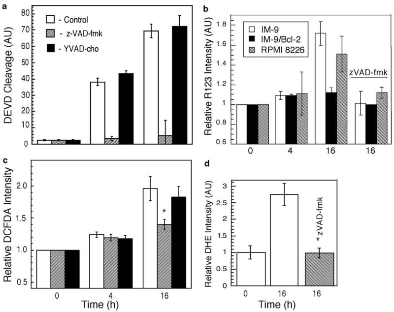
zVAD-fmk blocks DEVD cleavage activity and late ROS increase. (a) IM-9 cells were lysed at the indicated times following irradiation and assayed for caspase-3 activity toward the peptide substrate Ac-DEVD-p-nitroanilide, which was normalized to that of control cells. (b) The effect of IR on Δψm was examined in IM-9 and RPMI-8226 cells, irradiated in the absence or presence of zVAD-fmk. Δψm was measured using R123 as described in the ‘Materials and methods’ section. Data were normalized against untreated cells and represent the mean values of three separate experiments. Cells were stained with 1 μM CM-H2DCFDA (c) or 5 mM DHE (d) and cellular fluorescence levels were analyzed by flow cytometry, similar to Figure 2. The data shown represent the mean values of two duplicate experiments. The values shown are for control, zVAD, and YVAD-treated cells as represented by empty, shaded, and filled symbols, respectively. *P<0.05, as determined by t-test