



ABSTRACT
Beiging of adipocytes is characteristic of a higher number of mitochondria, the central hub of metabolism in the cell. However, studies show that beiging can improve metabolic health or cause metabolic disorders. Here we discuss a liver-fat crosstalk for iron flux associated with healthy beiging of adipocytes. Deletion of the transcription factor FoxO1 in adipocytes (adO1KO mice) induces a higher iron flux from the liver to white adipose tissue, concurrent with augmented mitochondrial biogenesis that increases iron demands. In addition, adO1KO mice adopt an alternate mechanism to sustain mitophagy, which enhances mitochondrial quality control, thereby improving mitochondrial respiratory capacity and metabolic health. However, the liver-fat crosstalk is not detectable in adipose Atg7 knockout (ad7KO) mice, which undergo beiging of adipocytes but have metabolic dysregulation. Autophagic clearance of mitochondria is blocked in ad7KO mice, which accumulates dysfunctional mitochondria and elevates mitochondrial content but lowers mitochondrial respiratory capacity. Mitochondrial biogenesis is comparable in the control and ad7KO mice, and the iron influx into adipocytes and iron efflux from the liver remain unchanged. Therefore, activation of the liver-fat crosstalk is critical for mitochondrial quality control that underlies healthy beiging of adipocytes.
KEYWORDS: Adipose beiging, FoxO1, Atg7, liver-fat crosstalk, iron flux
Fat or adipose tissue plays an important role in metabolic homeostasis. Brown adipocytes are packed with mitochondria that are rich in uncoupling protein 1 (UCP1) that facilitates energy metabolism through thermogenesis, and with small multilocular lipid droplets [1]. White adipocytes, however, have lower mitochondrial content but large unilocular lipid droplets [2]. Activation of brown adipose tissue or beiging of white adipose tissue increases energy expenditure and improves metabolic health [3–5]. Yet, beiging of adipocytes in autophagy-deficient mice is associated with metabolic disorder, elevated inflammation and oxidative stress, and even increased death rate at young age [6–8]. Our recent study reveals a liver-fat crosstalk for iron flux in healthy beiging where mitochondrial homeostasis is well maintained, but the crosstalk is absent from unhealthy beiging of adipocytes that lose mitochondrial quality control [9]. The liver-fat crosstalk may serve as an important indicator of healthy beiging of adipocytes.
As the central hub of metabolism, mitochondria control adipocyte differentiation and function [2]. Adipocyte differentiation is associated with increased mitochondrial number and proteins involved in fatty acid metabolism [10]. In addition, beiging of adipocytes leads to elevated mitochondrial content and respiratory activity [3–5]. In line with the essential role of iron in mitochondrial enzymes [11], deprivation of iron causes mitochondrial dysfunction and impairs adipocyte differentiation [12–14]. In response to stimuli of adaptive thermogenesis, beiging of adipose tissue increases iron demands and uptake in adipocyte [5,15]. By contrast, hormonal blockage of iron mobility or use of iron-deficient diets dampens adipose beiging capacity and thermogenesis in mice [16,17]. These studies underline the importance of iron status for adipose beiging and function.
Iron homeostasis is controlled by the processes of absorption, utilization, and storage [18,19]. Currently it remains largely undefined how iron flux is coordinated across tissues during beiging of adipose tissue. To address this question, we generated a mouse model that undergoes adipose beiging by conditionally deleting the transcription factor FoxO1 in adipocytes (adO1KO) [5]. AdO1KO adipocytes show a significant elevation in cellular iron (non-heme) and a healthy beiging phenotype, including the upregulation of adipose beiging markers (e.g., UCP1 and Cidea), increase in mitochondrial proteins and respiration, and enhancements in metabolism and energy expenditure [5]. Consistently, iron uptake proteins (e.g., TfR1 and DMT1) are elevated in adipose tissue from adO1KO mice [5]. However, the iron-storage protein ferritin is downregulated in adipose tissue from adO1KO mice versus control mice, because augmented mitochondrial biogenesis increases mitochondrial iron demand that is known to induce cytosolic “iron depletion” [5,9,20,21]. To meet the higher iron requirement for augmented mitochondrial biogenesis during adipocyte beiging [9], adO1KO mice adopt a higher iron influx into adipocytes (via TfR1 and DMT1) and increase iron efflux out of the liver (via Fpn) [5]. As such, hepatic iron content and ferritin level are downregulated while iron contents in adipose tissue and the serum are overall higher in adO1KO mice than the controls [5]. Of note, iron content and iron-regulatory proteins in the spleen and small intestine are comparable in the control and adO1KO mice. Thus, a liver-fat crosstalk is critical to meet the higher iron demands imposed by augmented mitochondrial biogenesis during healthy beiging of adipocytes in adO1KO mice.
Mitochondrial homeostasis is also regulated by mitophagy, a housekeeping process that removes dysfunctional mitochondria via autophagy [22,23]. To maintain autophagic clearance of dysfunctional mitochondria, adO1KO mice adopt an alternate mechanism by upregulating Atg7 and Fundc1 but downregulating Bnip3 [9]. Augmented mitochondrial biogenesis plus the sustained mitophagy constitutes the mechanism of quality control, resulting in healthy beiging characterized by a higher mitochondrial content and respiratory capacity, as well as metabolic improvement in adO1KO mice [5,9].
Interestingly, loss of adipocyte Atg7 (ad7KO) also led to accumulation of mitochondria and elevated UCP1 in the adipose tissues, a typical phenotype of adipocyte beiging [9]. However, the ad7KO mice did not show metabolic improvement; instead, the mice exhibited metabolic disorders along with compromised mitochondrial function, indicative of an unhealthy browning of adipose tissue [9]. The ad7KO mice showed higher mitochondrial COXIV protein level in the adipose tissue (Figure 1, A-B), consistent with the observation of mitochondrial acumulation under electron microscopy [9]. However, there was no significant change in mitochondrial biogenesis mediated by Pgc1α or Pgc1β (Figure 1, A-B). Additionally, adipose mitophagy receptors were upregulated in ad7KO mice, by 4.1-fold (p<0.001) for Bnip3 and 2.7-fold (p<0.01) for Fundc1, although Pink1 was elevated insignificantly (1.5-fold, p>0.05), in support of the notion that ablation of autophagy results in accumulation of mitophagy receptor proteins (Figure 1, C-D) [24]. To this end, mitochondrial dynamics proteins, Drp1 and OPA1, which are required for Bnip3 and Fundc1 mediated autophagic clearance of mitochondria [25,26]. were elevated by 3.8-fold (p < 0.001) and 2.2-fold (p < 0.05), respectively, in adipose tissues from ad7KO mice (Figure 1, E-F). The upregulation of dynamics proteins may arise from mitigated turnover owing to autophagy ablation [27]. Inhibitory phosphorylation of Drp1 at Ser637 (pDrp1-S637) was increased (3.2-fold, p < 0.01), but it showed no significant difference after being normalized against total Drp1, suggesting that the elevated pDrp1-S637 was due to the accumulation of total Drp1 (Figure 1, E-F).
Figure 1.
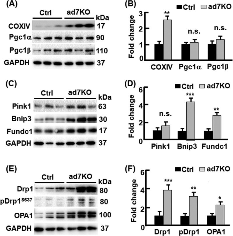
Mitochondrial regulation in ad7KO mice. (A-B) Western blotting (panel A) and densitometric (panel B) analyses of mitochondrial biogenesis markers (Pgc1α and Pgc1β) and mitochondrial content marker (COXIV) in visceral adipose tissues from control and ad7KO male mice. n=6. (C-D) Western blotting (panel C) and densitometric (panel D) analyses of mitophagy markers (Pink1, Bnip3, and Fundc1) in visceral adipose tissues from control and ad7KO male mice. n=6. (E-F) Western blotting (panel E) and densitometric (panel F) analyses of mitochondrial dynamics proteins (Drp1, pDrp1-S637, and OPA1) in visceral adipose tissues from control and ad7KO male mice. n=6. *p < 0.05, **p < 0.01; ***p<0.001.
In line with deficiency of autophagy and mitophagy, the ad7KO mice showed a substantial accumulation of dysfunctional mitochondria (Figure 1, and reference[9]). As the essential element of mitochondrial enzymes and complexes [11], iron was elevated in the adipose tissue from ad7KO mice versus control mice (Figure 2, A), which was similar to adO1KO mice that exhibit higher mitochondrial content and healthy browning of adipose tissue [5]. In contrast to adO1KO mice [5], however, ad7KO mice showed no significant changes in liver and serum iron contents versus control mice (Figure 2, B-C). Further analysis of iron-regulatory proteins demonstrated that iron influx into adipose tissue and iron efflux from the liver remained unchanged (Figure 2, D-G). Consistently, the proteins responsible for iron uptake (TfR1 and DMT1), export (FPN), and storage (ferritin, FTH) were comparable in ad7KO and control mice (Figure 2, D-G). These data suggest that blocking autophagy (or mitophagy) in ad7KO mice leads to unhealthy beiging of adipose tissue, where there is no change in mitochondrial biogenesis and iron demand, nor does it require activation of the liver-fat crosstalk for iron supply. Because mitochondrial iron is less accessible than cytosolic iron [20,21], the overall higher cellular iron level due to mitochondrial accumulation does not change ferritin (an iron status marker) expression in the adipose tissue from ad7KO mice (Figure 2, D-E).
Figure 2.
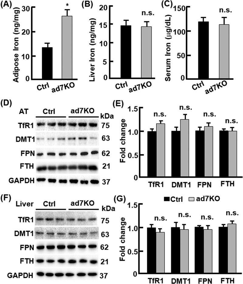
Iron metabolism in ad7KO mice. (A-C) the contents of non-heme iron in visceral adipose tissue (panel A), the liver (panel B), and serum (panel C) in control and ad7KO male mice. n= 6-9. (D-E) Western blotting (panel D) and densitometric (panel E) analyses of iron regulating proteins in visceral adipose tissues from control and ad7KO male mice. n=6. (F-G) Western blotting (panel F) and densitometric (panel G) analyses of iron regulating proteins in liver tissues from control and ad7KO male mice. n=6. *p < 0.05; n.s., not significant.
Our study discovers a liver-fat crosstalk for iron flux that distinguishes healthy vs. unhealthy beiging of adipose tissue (Figure 3). Deletion of adipose FoxO1 leads to healthy beiging of adipose tissue, where mitophagy is sustained via alternate mechanism and mitochondrial biogenesis is augmented [9]. Activation of the liver-fat crosstalk coordinates a higher iron flux from the liver into adipose tissue to meet an increased iron demand for augmented mitochondrial biogenesis (Figure 3). As a result, mitochondrial function and metabolic health are thus enhanced in adO1KO mice [5,9]. In the ad7KO mice, however, beiging of adipose tissue is unhealthy and the liver-fat crosstalk is not activated (Figure 3). Deletion of adipose Atg7 causes mitophagy defects while mitochondrial biogenesis remains unchanged (Figure 1, and reference [9]). This leads to poor quality control of mitochondria and, in line with mice lacking Atg3 that accumulate dysfunctional mitochondria in adipose tissue and develop metabolic disorders [6], the Ad7KO mice exhibited higher mitochondrial content but impaired metabolism (Figure 1, and reference [9]). As mitochondrial biogenesis is not activated by ad7KO and iron requirement is not increased in adipose tissue, the liver-fat crosstalk becomes unnecessary in ad7KO mice (Figures 1-3).
Figure 3.
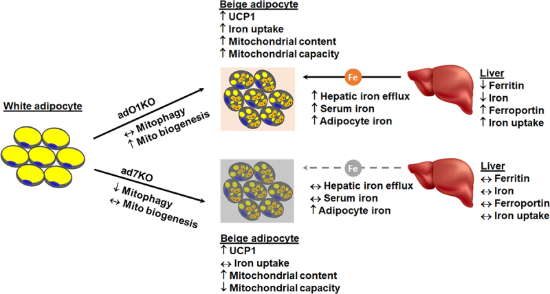
Schematic view of the liver-fat crosstalk during beiging of adipocytes. In adO1KO mice, active crosstalk between the liver and adipose tissue supplies iron required for augmented mitochondrial biogenesis during beiging of adipocytes. Increased mitochondrial biogenesis plus sustained mitophagy upregulates mitochondrial content and respiratory capacity, improving systemic metabolism. In ad7KO mice, however, a higher mitochondrial content results from impairment in mitophagy and accumulation of poor-quality-controlled mitochondria, thereby reducing mitochondrial capacity. As mitochondrial biogenesis is unchanged, the liver-fat crosstalk is silenced. ↔, unchanged; ↑, upregulated; ↓, downregulated. The gray dashed line indicates the absence of activation of a liver-fat crosstalk for iron supply.
Outstanding questions remain as to: (1) whether the liver-fat crosstalk for iron flux serves as a shared mechanism for adipose beiging induced by different stimuli such as fasting, exercise, and cold exposure [28–30]. (2) What molecules and signaling pathways are involved in the regulation of the liver-fat crosstalk. One of the top candidates can be hepcidin, the hormone that mitigates iron efflux [18]. Administration of hepcidin was shown to impair adipocyte beiging [16]. However, a recent study suggests that hepcidin is required for adipose beiging as knockout of hepcidin suppresses the beiging phenotype and thermogenesis [31]. (3) In addition to hepatocytes, macrophages play an important role in iron storage and recycling [19]. Whether and how macrophages interact with adipocytes during adipose beiging process warrant future investigation. (4) Fasting induces adipose browning via autophagy induction, which is associated with an increase in Ucp1, COXIV, mitochondrial biogenesis via Pgc1a, and mitochondrial activities [32]. The autophagy-mediated adipose browning was in line with the phenotype in adO1KO mice, where an alternate mechanism is adopted to mediate autophagy/mitophagy and mitochondrial quality control during adipose browning [9]. Whether and how autophagy stimulation by fasting affects iron flux in the liver and fat tissue are worthy of future investigation. Tackling these questions will advance our understanding of iron metabolism in adipose tissue, and it may inform new strategies to achieve healthy beiging and metabolic improvement.
Materials and methods
Mice
Atg7,fl°x/fl°x adipoqCreER,T2 and adipoqCreERT2:: Atg7 fl°x/fl°x mice were described previously.[9,33,34] Adult AdipoqCreERT2:: Atg7 fl°x/fl°x mice (13-15 week old) were treated with tamoxifen (50 mg/kg body weight) by intraperitoneal injection once a day for 5 consecutive days to obtain ad7KO mice; age-matched Atg7 fl°x/fl°x were treated with tamoxifen likewise and used as the control mice.[9] All the mice were housed in plastic cages on a 12-h (7:00 am – 7:00 pm) light–dark photocycle, with free access to water and regular chow diet. For tissue collection, the mice were euthanized by inhalation of CO2. The procedures of mouse handling and treatments met the NIG guidelines and were approved by the Institutional Animal Care and Use Committees at the University of Florida (UF).
Iron assay
Total non-heme iron was analyzed with Iron Assay Kits (Cat # MAK025) from Sigma according to the manufacturer’s instructions.[5]
Immunoblotting
The procedure was performed as described.[5,9] Specifically, tissue and cell lysates were prepared with PLC lysis buffer (30 mM Hepes, pH 7.5, 150 mM NaCl, 10% glycerol, 1% Triton X-100, 1.5 mM MgCl2, 1 mM EGTA, 10 mM NaPPi, 100 mM NaF, 1 mM Na3VO4) supplemented with protease inhibitor cocktail (Roche), and 1 mM PMSF. Total protein concentrations of the lysates were determined using a DC protein assay kit (Bio-Rad). Antibody (catalog number) information: GAPDH (600004-1-lg), Pgc1α (66369-1-lg), Pgc1β (22378-1-AP), COXIV (11242-1-AP), Fundc1 (28519-1-AP), Pink1 (23274-1-AP), Drp1 (12957-1-AP), and OPA1 (27733-1-AP) antibodies were purchased from Proteintech (Rosemont, IL, USA); antibodies against DMT1 (bs-3577R-TR) and TfR1 (bs-0988R) from Bioss Antibodies; Ferroportin antibody (NBP1-21503) from Novus Biologicals; FTH (SAB2108662-100UL) and p-Drp1-S637 (SAB5701801) antibodies from Sigma-Millipore (Billerica, MA, USA); and Bnip3 antibody (Ab10433) from Abcam (Boston, MA, USA).
Statistical analysis
Measurements were duplicated or triplicated, with 6–9 mice included in each group. Data were presented as mean ± SD. Unless the use of female mice were specified, the animal studies were conducted on males. Differences between groups and treatments were validated by one-way analysis of variance or a two-sided t-test. A value of p < 0.05 was considered statistically significant.
Funding Statement
This work was supported in part by the American Heart Association [18TPA34230082 to Z.C.], and the U.S. Departement of Agricuture (USDA) National Institute of Food and Agriculture [1020373 to Z.C].
Disclosure statement
No conflicts of interest are declared by the author(s).
Reference
- 1.Harms M, Seale P.. Brown and beige fat: development, function and therapeutic potential. Nature medicine 2013; 19:1252–10. [DOI] [PubMed] [Google Scholar]
- 2.Zhu QZ, An YA, Scherer PE.. Mitochondrial regulation and white adipose tissue homeostasis. Trends in cell biology 2022; 32:351–64. [DOI] [PMC free article] [PubMed] [Google Scholar]
- 3.Cero C, Lea HJ, Zhu KY, Shamsi F, Tseng YH, Cypess AM.. beta3-Adrenergic receptors regulate human brown/beige adipocyte lipolysis and thermogenesis. Jci Insight 2021; 6. [DOI] [PMC free article] [PubMed] [Google Scholar]
- 4.Sohn JH, Ji Y, Cho CY, et al. Spatial regulation of reactive oxygen species via g6pd in brown adipocytes supports thermogenic function. Diabetes 2021; 70:2756–70. [DOI] [PubMed] [Google Scholar]
- 5.Shi L, Tao Z, Zheng L, et al. FoxO1 regulates adipose transdifferentiation and iron influx by mediating Tgfbeta1 signaling pathway. Redox biology 2023; 63:102727. [DOI] [PMC free article] [PubMed] [Google Scholar]
- 6.Cai J, Pires KM, Ferhat M, Chaurasia B, Buffolo MA, Smalling R, et al. autophagy ablation in adipocytes induces insulin resistance and reveals roles for lipid peroxide and nrf2 signaling in adipose-liver crosstalk. Cell reports 2018; 25:1708–17 e5. [DOI] [PMC free article] [PubMed] [Google Scholar]
- 7.Singh R, Xiang Y, Wang Y, Baikati K, Cuervo AM, Luu YK, et al. Autophagy regulates adipose mass and differentiation in mice. The Journal of clinical investigation 2009; 119:3329–39. [DOI] [PMC free article] [PubMed] [Google Scholar]
- 8.Mizunoe Y, Sudo Y, Okita N, Hiraoka H, Mikami K, Narahara T, et al. Involvement of lysosomal dysfunction in autophagosome accumulation and early pathologies in adipose tissue of obese mice. Autophagy 2017; 13:642–53. [DOI] [PMC free article] [PubMed] [Google Scholar]
- 9.Shi L, Yang J, Tao Z, Zheng L, Bui TF, Alonso RL, et al. Loss of FoxO1 activates an alternate mechanism of mitochondrial quality control for healthy adipose browning. Clin Sci 2024; 138:371–85. [DOI] [PMC free article] [PubMed] [Google Scholar]
- 10.Wilson-Fritch L, Burkart A, Bell G, Mendelson K, Leszyk J, Nicoloro S, et al. Mitochondrial biogenesis and remodeling during adipogenesis and in response to the insulin sensitizer rosiglitazone. Molecular and cellular biology 2003; 23:1085–94. [DOI] [PMC free article] [PubMed] [Google Scholar]
- 11.Ward DM, Cloonan SM.. mitochondrial iron in human health and disease. Annual Review of Physiology, Vol 81 2019; 81:453–82. [DOI] [PMC free article] [PubMed] [Google Scholar]
- 12.Moreno-Navarrete JM, Ortega F, Moreno M, Ricart W, Fernández-Real JM.. Fine-tuned iron availability is essential to achieve optimal adipocyte differentiation and mitochondrial biogenesis. Diabetologia 2014; 57:1957–67. [DOI] [PubMed] [Google Scholar]
- 13.Suzuki T, Komatsu T, Shibata H, Tanioka A, Vargas D, Kawabata-Iwakawa R, et al. Crucial role of iron in epigenetic rewriting during adipocyte differentiation mediated by JMJD1A and TET2 activity. Nucleic Acids Research 2023; 51:6120–42. [DOI] [PMC free article] [PubMed] [Google Scholar]
- 14.Walter PB, Knutson MD, Paler-Martinez A, Lee S, Xu Y, Viteri FE, et al. Iron deficiency and iron excess damage mitochondria and mitochondrial DNA in rats. Proceedings of the National Academy of Sciences of the United States of America 2002; 99:2264–9. [DOI] [PMC free article] [PubMed] [Google Scholar]
- 15.Yook JS, You M, Kim Y, Zhou M, Liu Z, Kim YC, et al. The thermogenic characteristics of adipocytes are dependent on the regulation of iron homeostasis. The Journal of Biological Chemistry 2021; 296:100452. [DOI] [PMC free article] [PubMed] [Google Scholar]
- 16.Yook JS, You M, Kim J, Toney AM, Fan R, Puniya BL, et al. Essential role of systemic iron mobilization and redistribution for adaptive thermogenesis through HIF2-alpha/hepcidin axis. Proceedings of the National Academy of Sciences of the United States of America 2021; 118. [DOI] [PMC free article] [PubMed] [Google Scholar]
- 17.Yook JS, Thomas SS, Toney AM, You M, Kim YC, Liu Z, et al. Dietary Iron Deficiency Modulates Adipocyte Iron Homeostasis, Adaptive Thermogenesis, and Obesity in C57BL/6 Mice. The Journal of nutrition 2021; 151:2967–75. [DOI] [PMC free article] [PubMed] [Google Scholar]
- 18.Nemeth E, Ganz T.. Hepcidin and Iron in Health and Disease. Annual review of medicine 2023; 74:261–77. [DOI] [PMC free article] [PubMed] [Google Scholar]
- 19.Roemhild K, von Maltzahn F, Weiskirchen R, Knuchel R, von Stillfried S, Lammers T.. Iron metabolism: pathophysiology and pharmacology. Trends Pharmacol Sci 2021; 42:640–56. [DOI] [PMC free article] [PubMed] [Google Scholar]
- 20.Nie G, Sheftel AD, Kim SF, Ponka P.. Overexpression of mitochondrial ferritin causes cytosolic iron depletion and changes cellular iron homeostasis. Blood 2005; 105:2161–7. [DOI] [PubMed] [Google Scholar]
- 21.Chitambar CR. Cellular iron metabolism: mitochondria in the spotlight. Blood 2005; 105:1844–5. [DOI] [PubMed] [Google Scholar]
- 22.Onishi M, Yamano K, Sato M, Matsuda N, Okamoto K.. Molecular mechanisms and physiological functions of mitophagy. The EMBO journal 2021; 40:e104705. [DOI] [PMC free article] [PubMed] [Google Scholar]
- 23.Cheng Z. FoxO transcription factors in mitochondrial homeostasis. The Biochemical journal 2022; 479:525–36. [DOI] [PMC free article] [PubMed] [Google Scholar]
- 24.Park CW, Hong SM, Kim ES, Kwon JH, Kim KT, Nam HG, et al. BNIP3 is degraded by ULK1-dependent autophagy via MTORC1 and AMPK. Autophagy 2013; 9:345–60. [DOI] [PMC free article] [PubMed] [Google Scholar]
- 25.Chen M, Chen ZH, Wang YY, Tan Z, Zhu CZ, Li YJ, et al. Mitophagy receptor FUNDC1 regulates mitochondrial dynamics and mitophagy. Autophagy 2016; 12:689–702. [DOI] [PMC free article] [PubMed] [Google Scholar]
- 26.Lee Y, Lee HY, Hanna RA, Gustafsson AB.. Mitochondrial autophagy by Bnip3 involves Drp1-mediated mitochondrial fission and recruitment of Parkin in cardiac myocytes. American journal of physiology Heart and circulatory physiology 2011; 301:H1924–31. [DOI] [PMC free article] [PubMed] [Google Scholar]
- 27.Purnell PR, Fox HS.. Autophagy-mediated turnover of dynamin-related protein 1. BMC Neurosci 2013; 14:86. [DOI] [PMC free article] [PubMed] [Google Scholar]
- 28.Smorlesi A, Frontini A, Giordano A, Cinti S.. The adipose organ: white-brown adipocyte plasticity and metabolic inflammation. Obesity reviews: an official journal of the International Association for the Study of Obesity 2012; 13 Suppl 2:83–96. [DOI] [PubMed] [Google Scholar]
- 29.Li G, Xie C, Lu S, Nichols RG, Tian Y, Li L, et al. Intermittent Fasting Promotes White Adipose Browning and Decreases Obesity by Shaping the Gut Microbiota. Cell metabolism 2017; 26:672–85 e4. [DOI] [PMC free article] [PubMed] [Google Scholar]
- 30.Ding H, Zheng S, Garcia-Ruiz D, Hou D, Wei Z, Liao Z, et al. Fasting induces a subcutaneous-to-visceral fat switch mediated by microRNA-149-3p and suppression of PRDM16. Nature communications 2016; 7:11533. [DOI] [PMC free article] [PubMed] [Google Scholar]
- 31.Deschemin JC, Ransy C, Bouillaud F, Chung S, Galy B, Peyssonnaux C, et al. Hepcidin deficiency in mice impairs white adipose tissue browning possibly due to a defect in de novo adipogenesis. Scientific reports 2023; 13. [DOI] [PMC free article] [PubMed] [Google Scholar]
- 32.Martinez-Lopez N, Tarabra E, Toledo M, Garcia-Macia M, Sahu S, Coletto L, et al. System-wide Benefits of Intermeal Fasting by Autophagy. Cell metabolism 2017; 26:856–71 e5. [DOI] [PMC free article] [PubMed] [Google Scholar]
- 33.Komatsu M, Waguri S, Ueno T, Iwata J, Murata S, Tanida I, et al. Impairment of starvation-induced and constitutive autophagy in Atg7-deficient mice. Journal of Cell Biology 2005; 169:425–34. [DOI] [PMC free article] [PubMed] [Google Scholar]
- 34.Sassmann A, Offermanns S, Wettschureck N.. Tamoxifen-inducible Cre-mediated recombination in adipocytes. Genesis 2010; 48:618–25. [DOI] [PubMed] [Google Scholar]