



Abstract
The burden of early onset neonatal sepsis due to group B Streptococcus (GBS) has declined substantially over the past 20 years as a result of widespread use of intrapartum antibiotic prophylaxis. However, GBS remains the leading cause of neonatal sepsis in the United States. And unlike other diseases prevented through measures that may decrease or even eliminate the pathogen from the population, GBS colonization among pregnant women — and therefore the risk of transmission to newborns — has remained steady over time. It is important, therefore, that obstetricians and neonatologists remain vigilant about preventing and diagnosing GBS infections.
Keywords: Streptococcus agalactiae, Streptococcus group B, Infant, Newborn, Sepsis, Meningitis
GBS DISEASE IN INFANTS
GBS emerged in the 1970s as the leading cause of neonatal sepsis and meningitis in the United States.1–3 Two distinct clinical syndromes among infants became apparent: early onset disease that presented within the first week of life, and late-onset disease that manifested between 1 week and 3 months.3 Before the initiation of prevention strategies, the burden of early onset disease was much greater than that of late-onset disease, with approximately 80% of GBS disease in infants occurring within the first week of life.4 The annual incidence of early onset disease in the United States in the early 1990s (before the widespread implementation of preventive measures) was 1.7 cases per 1000 live births. The incidence of early-onset disease, however, has declined dramatically over the past 20 years, and in recent years it has remained less than 0.4 cases per 1000 live births.5 It is noteworthy that the incidence of late-onset disease (0.4 cases per 1000 live births) has remained relatively stable over the past 20 years6 (Fig. 1).
Fig. 1.
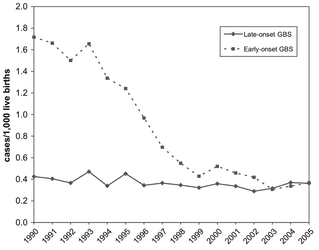
Trends in early- and late-onset neonatal group B streptococcal infections in active bacterial core surveillance areas, United States, 1990–2005. (Adapted from Jordan HT, Farley MM, Craig A, et al. Revisiting the need for vaccine prevention of late-onset neonatal group B streptococcal disease. Pediatr Infect Dis J 2008;27:1060; with permission.)
Early onset GBS infections are vertically transmitted.7 The organism may ascend from the vagina to the amniotic fluid during the intrapartum period. Once in the amniotic fluid, the bacteria may replicate and colonize the infant’s skin or mucus membranes, or may be aspirated into the lungs of the infant, leading to an invasive infection. Exposure to the bacteria also may occur during passage through the birth canal. Less commonly, GBS can cross intact membranes, leading to intrauterine infection in the fetus, which may result in stillbirth or advanced infection at the time of delivery.8,9
In contrast to early onset disease, late-onset GBS infection is not always acquired from the mother. Maternal colonization, does play an important role; one study found approximately 50% of infants with late-onset disease had been colonized at birth, and their mothers had been colonized with an identical serotype.10 However, the source of infection among infants born to noncolonized women is unclear. Nosocomial transmission has been demonstrated.11,12 Horizontal transmission from mother to infant after the perinatal period may occur, and case reports also have identified breast milk as a source of infection for late-onset disease.13–16
Early onset disease often presents within the first hours of life, and most cases occur within the first 24 to 48 hours. Most infants exhibit signs of respiratory distress. Cardiovascular instability and apnea also are observed frequently. The most common clinical syndrome of early onset disease is bacteremia, with or without pneumonia. Less commonly, early onset GBS infection may manifest as meningitis.4,17,18 Early onset disease is characterized by rapid clinical deterioration.
Late-onset disease can present at any time between 1 week and 3 months of life. The median age of onset in a large review of cases in the United States was 36 days.6 The clinical presentation of late-onset GBS infections may be similar to that of early-onset disease. Meningitis is relatively more common in infants with late-onset disease compared with early onset disease, comprising more than 25% of late-onset cases.6,18 Sepsis, however, is the most commonly observed clinical syndrome for both early and late-onset disease. Late-onset infections also may present as septic arthritis or osteomyelitis, although such manifestations are relatively rare. The course of late-onset disease is generally less fulminant than that of early onset disease.
In the 1970s, mortality rates associated with early onset disease were reported to be 50% or higher, whereas late onset-disease had a case fatality rate of approximately 20%.19 Advances in neonatal medical care led to improved outcomes for both clinical syndromes, with mortality rates around 4% to 6% for early and late-onset GBS infections in recent years.6,18,20 Mortality rates among preterm infants are up to eight times higher than those of full-term infants; the risk of mortality increases with decreasing gestational age.6,18,20,21 Despite improved survival rates, long-term sequelae resulting from GBS infections, particularly meningitis, represent a major morbidity. One long-term follow-up study of survivors of GBS meningitis found that approximately 50% of the children had some deficit, and 29% exhibited severe neurologic sequelae.22
RISK FACTORS FOR GBS DISEASE
Early Onset GBS Disease
The primary risk factor for early onset disease is maternal vaginal colonization with GBS. Colonization with GBS during pregnancy can be transient, intermittent, or persistent.23–25 Colonization is asymptomatic, and the gastrointestinal tract likely serves as a reservoir for vaginal colonization.24,26,27 In the United States, approximately 10% to 30% of pregnant woman are colonized with GBS.28 Rates of GBS colonization are higher among black women,27,29–32 a factor which likely contributes to the disproportionate burden of GBS disease observed among black infants.5 HIV infection does not appear to influence likelihood of GBS colonization among pregnant women.33 The risk for early onset disease increases with heavier colonization in the mother.34,35 GBS bacteriuria is considered a marker of heavy maternal colonization and has been associated with an increased risk of neonatal disease.36–38
In the absence of any intervention, approximately 1% to 2% of infants born to colonized mothers develop early onset GBS infections.17,39,40 Additional factors beyond maternal GBS colonization, however, play a role in risk for early onset disease. Maternal chorioamnionitis, intrapartum maternal fever (a marker of chorioamnionitis) and prolonged rupture of membranes are all strongly associated with early onset GBS disease.10,41–43 These obstetric risk factors may reflect increased and prolonged infant exposure to GBS before delivery. Some studies have identified certain obstetric procedures, such as the use of intrauterine fetal monitoring devices41,44 or greater than or equal to five vaginal examinations during labor,41,45 to be associated with early onset disease.
Preterm infants, in addition to suffering worse outcomes from GBS disease, are at 3- to 30-fold greater risk of developing early onset infections compared with full-term infants, with the highest risk at lower gestational ages (Fig. 2).44,46,47 The risk among preterm infants likely is related to decreased transfer of maternal antibodies.48 Although preterm infants have a much greater risk of early onset GBS disease, approximately 75% of cases occur among term infants.28,49 This is in contrast to other pathogens causing neonatal sepsis (such as Escherichia coli) that predominantly affect preterm infants.
Fig. 2.
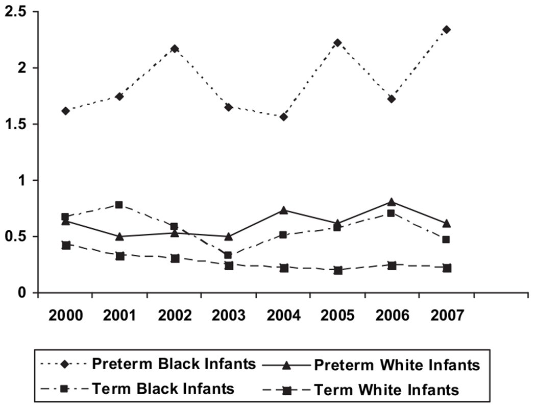
Early-onset group B streptococcal incidence rates in active bacterial core surveillance areas stratified by race and term, 2000—2007. (Adapted from CDC. Trends in perinatal group B streptococcal disease—United States, 2000–2006. MMWR Morb Mortal Wkly Rep 2009;58:109–12.)
Early onset GBS disease disproportionately affects black infants, even after stratifying by term to adjust for the higher rates of preterm delivery among blacks (see Fig. 2).5,46 Higher rates of maternal colonization may be a contributing factor; yet the racial disparity has persisted despite widespread use of intrapartum antibiotics. The implementation of universal screening and intrapartum antibiotic prophylaxis (IAP) has been relatively high among women of all races, and differences in implementation do not account for the observed racial disparities in infant GBS disease.49
Young maternal age is associated with an increased risk of early onset disease,43,46 likely as a result of lower levels of anti-GBS antibodies in younger age groups of women.29 Low maternal antibody levels have been shown to convey an increased risk of early onset disease in the infant.50–52 Previous delivery of an infant with GBS disease—which may also be associated with low maternal antibody levels—is a risk factor for early onset GBS disease in subsequent pregnancies.53–55
Late-onset GBS Disease
The risk factors for late-onset disease are not as well characterized as those for early onset disease. Maternal colonization, while not as strongly associated as it is with early onset disease, is a risk factor for late-onset GBS sepsis.56 As with early onset disease, prematurity has been found to be an important risk factor for late-onset GBS disease.56 Black race is also a risk factor for late-onset disease.46 The incidence of late-onset GBS disease is three times higher among black infants in the United States compared with white infants, and the increased risk persists after stratifying by gestational age.6 As with early onset disease, the cause of this racial disparity is unclear.
GBS PREVENTION IN THE UNITED STATES
Clinical trials in the 1980s demonstrated that IAP could reduce the risk of early onset GBS infections in infants.57,58 The efficacy of IAP for preventing early onset disease is nearly 90%.59,60 Use of IAP increased throughout the 1990s, with widespread uptake following the publication of consensus guidelines for the prevention of perinatal GBS disease developed by the Centers for Disease Control and Prevention (CDC), the American College of Obstetricians and Gynecologists, and the American Academy of Pediatrics (AAP) in 1996.40 The guidelines advocated one of two methods to identify candidates for IAP:
Culture-based screening for pregnant women between 35 and 37 weeks gestation to identify women colonized with GBS
Risk-based screening for women with intrapartum risk factors (including gestational age less than 37 weeks, temperature of at least 100.4°, or rupture of membranes for at least 18 hours).
IAP also was recommended for women with GBS bacteriuria at any point during the pregnancy, and women with a history of a previous infant with invasive GBS disease. The 1996 guidelines were implemented widely across the Untied States, with IAP administered in approximately 27% of deliveries in 1998 and 1999.59 Early onset GBS disease declined by approximately 70% during the 1990s.28
In 2002, the GBS prevention guidelines were revised. The fundamental change in the updated guidelines was a recommendation for universal culture-based screening, since a population-based study had demonstrated the superiority of this approach over risk factor-based screening.59 The risk-based approach was recommended only in cases where the maternal GBS status was unknown or unavailable at the time of delivery. The 2002 guidelines also incorporated recommendations for the prevention of GBS disease in the context of preterm labor, and for revised second-line antibiotic agents for penicillin-allergic women. The 2002 guidelines stated that IAP was not indicated in the context of planned cesarean deliveries before the onset of labor or rupture of membranes.28
A large population-based cohort study conducted in 2003 and 2004 demonstrated that the acceptance of universal screening as recommended in the 2002 guidelines was widespread. Van Dyke and colleagues49 reported that the proportion of pregnant women screened before delivery increased from 48% in 1998 and1999 to 85% in 2003 and 2004. The proportion of women with an indication for IAP who actually received intrapartum antibiotics increased from 74% to 85% during the same time period (Fig. 3).
Fig. 3.
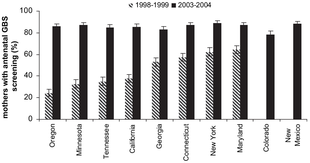
Proportion of pregnant women with antenatal screening for GBS in active bacterial core surveillance areas across the United States. (From Van Dyke MK, Phares CR, Lynfield R, et al. Evaluation of universal antenatal screening for Group B Streptococcus. NEJM 2009;360:2630; with permission.)
The study by Van Dyke and colleagues49 also identified areas for improvement in the implementation of the GBS prevention guidelines. Although the 2002 guidelines recommended that women with threatened preterm labor should be screened on admission if GBS culture was not performed in the previous 4 weeks, among women with threatened preterm delivery and unknown GBS status, only 18% of those who progressed to delivery and 31% of those who did not progress to delivery underwent GBS screening on admission. Furthermore, despite preterm delivery being an indication for IAP in women with unknown GBS colonization status, only 63% of women who delivered preterm with unknown colonization received IAP. Suboptimal implementation also was noted in the choice of antibiotic agent for penicillin-allergic women. Only 14% of penicillin-allergic women at low risk for anaphylaxis received the recommended agent cefazolin. Clindamycin was the most commonly used agent among penicillin-allergic mothers, given to 70% of women at low risk for anaphylaxis and 84% of those at high risk. However, the effectiveness of clindamycin as an IAP agent is unknown; there are very limited data on the pharmacokinetics of clindamycin in pregnancy, and GBS is becoming increasingly resistant to clindamycin.18,61
Despite these limitations, the incidence of early onset disease was reduced further following the issuance of the 2002 guidelines. In recent years, the incidence has remained fairly stable at 0.34 to 0.37 cases per 1000 live births.5,18 Late-onset disease unfortunately is not impacted by IAP, and incidence rates have remained fairly stable of the past 20 years (see Fig. 1).
Neonatal Management Challenges in the era of GBS Prevention
As the use of IAP for prevention of GBS disease became more widespread in the 1990s, there was initially substantial apprehension about whether exposure to IAP would complicate detection of sepsis in the newborn or impact the management approach of pediatricians. The 1996 and 2002 guidelines for GBS prevention included a sample algorithm for the management of infants exposed to intrapartum antibiotics that recommended a full diagnostic evaluation and empiric treatment for infants considered to be at very high risk of sepsis, a more limited evaluation in those with a lesser risk, and observation only for infants whose mother received at least 4 hours of an appropriate antibiotics. These recommendations aimed to identify infants at high risk for early onset GBS disease, while avoiding unnecessary diagnostic evaluations in neonates. Nonetheless, the management of infants in the era of universal screening and IAP remains challenging. Studies in recent years have provided additional data that permit a refining of the neonatal management recommendations in the GBS prevention guidelines.
Presentation of Early Onset GBS Disease After Exposure to IAP
Uncertainty about how IAP might affect the clinical presentation of early onset GBS sepsis led to concerns that prevention efforts could impair the ability of pediatricians to diagnose GBS disease. Exposure to antibiotics potentially could mask the signs of sepsis in a newborn, or delay the onset until after the infant had been discharged home, resulting in true cases of early onset disease going undetected until late in the clinical course. Several studies conducted after 1996, however, have found no significant difference in the clinical presentation of early onset GBS sepsis between infants exposed to IAP and those not exposed. Although some cases of early onset disease are detected in infants with no signs of sepsis who are screened because of risk factors for GBS sepsis,21,62–64 most infants with GBS sepsis present with signs of infection within the first 24 to 48 hours of life, a pattern unaffected by exposure to IAP.21,62–65
Management of Infants Exposed to IAP
During the early phase of GBS prevention efforts, there was also concern that pediatricians might react to the uncertainty of the clinical management of infants exposed to IAP by conducting excessive evaluations and treating infants unnecessarily with antibiotics.66,67 A population-based study in Connecticut in 1996 found that infants of women who received IAP were no more likely to receive an antibiotic and did not have a significant increase in length of stay compared with infants born to women who did not receive IAP.68 In contrast, a study conducted in 19 hospitals in Utah from 1998 to 2002 found that infants born to mothers who received any intravenous antibiotic were more likely to receive intravenous antibiotics for more than 72 hours and had significantly longer lengths of stay and hospital costs, compared with infants whose mothers did not receive any intravenous antibiotics. This study was limited by use of administrative data only, which did not permit the authors to determine whether maternal antibiotics were administered intrapartum and thus were administered for GBS prevention or some other reason.69 Further studies on the impact of the GBS prevention guidelines on the management of at-risk infants are needed.
The 2002 guidelines recommend that infants of mothers who receive intrapartum antibiotics for suspected chorioamnionitis undergo a full diagnostic evaluation and receive empiric antibiotic therapy for possible GBS disease. Chorioamnionitis is an important risk factor for early onset GBS disease, and may indicate active infection in the fetus before delivery. Intrapartum fever—often a symptom of chorioamnionitis—has been identified as risk factor for failure of IAP to prevent GBS disease in the newborn.60,70 Given the association between epidural labor analgesia and fever, however, there is concern that chorioamnionitis may be overdiagnosed, resulting in unnecessary diagnostic evaluations of neonates.71 Unpublished data from CDC surveillance sites across the United States in 2003 and 2004 demonstrated that while 67% of parturients received epidural labor analgesia, only 4% of those had fever, and the proportion of women diagnosed with chorioamnionitis (3%) was the same among the women with and without epidural labor analgesia. Thus, clinical chorioamnionitis is relatively uncommon—even among women with epidural labor analgesia—but warrants particular concern for clinicians caring for newborns.
Diagnostic Evaluations Among Infants Exposed to IAP
The 1996 and 2002 GBS prevention guidelines recommended a limited evaluation (complete blood cell [CBC] count with differential and blood culture) for infants exposed to IAP who are either less than 35 weeks gestational age or whose mother received less than 4 hours of IAP before delivery. The rationale behind the gestational age limit was that in the setting of IAP, it may be difficult to properly ascertain signs of sepsis in very preterm infants, and that CBC count and blood culture may aid in making the diagnosis. The recommendation regarding duration of IAP stems from evidence that IAP given for less than 4 hours is less effective at preventing colonization and early onset GBS disease compared with antibiotics given for longer durations60,72(S. Schrag, PhD, unpublished data, 2009). Therefore, IAP given for less than 4 hours might be insufficient to decrease the risk of disease, but may impact the clinical presentation in a manner that would make it more difficult to diagnose sepsis.
Limited data available on the performance of CBC count as a screening test, however, suggest that while the test has a relatively high negative predictive value, the positive predictive value is quite limited, particularly among infants with no sings of sepsis.73–75 A large study of neonatal sepsis evaluations among infants weighing at least 2000 g at birth conducted in 1995 and 1996 demonstrated that the presence of clinical signs of sepsis is a highly sensitive indicator of sepsis of all etiologies.63 All infants with early onset GBS sepsis had clinical signs of infection within 8–12 hours of birth, regardless of whether their mother had received intrapartum antibiotics. The findings suggest that for many infants, careful clinicial observation performs well as a screen for early onset GBS sepsis.
False-Negative Cases of Early Onset GBS Disease
Studies reporting on patterns of early onset GBS disease in the era of widespread use of IAP have found approximately 60% to 80% of cases occur among infants born to women with a negative antenatal GBS screen.21,49,64 False-negative cases are bound to occur; GBS culture at 35 to 37 weeks has a 95% to 97% negative predictive value for absence of colonization at the time of delivery. Therefore, a small proportion of women will not be colonized at the time of their screening and acquire GBS colonization between the time of the culture and the onset of labor.76 Indeed, if the implementation of the guidelines were optimized (all pregnant women screened, and all women with indications for IAP receiving IAP), then those cases that continue to occur would highly likely be born to mothers who had screened negative, reflecting the inherent limitations of the prevention strategy. Clinicians should understand that a negative maternal GBS screen indicates that an infant is at low–but not zero—risk of GBS disease. GBS sepsis should be suspected in any infant with signs of sepsis, regardless of maternal screening results.
POTENTIAL UNINTENDED CONSEQUENCES OF GBS PREVENTION
Non-GBS Sepsis
Although GBS prevention efforts have very effectively decreased early onset GBS infections in the United States, there has been concern about the overall impact of widespread IAP on early onset neonatal sepsis. One question raised is whether the decrease in GBS infections is a true reduction in disease, or whether antibiotics are simply resulting in negative cultures while GBS continues to cause clinical sepsis. Nationally representative hospital discharge data, however, demonstrated a steady decrease in clinical sepsis rates from 1990 to 2002, with a marked decline in clinical sepsis among term infants during the 2 years following the issuance of the 1996 GBS prevention guidelines.77
Another concern is whether the decrease in GBS disease has been accompanied by an increase in other potentially more lethal neonatal pathogens. The predominant etiologies of neonatal sepsis have tended to change over time, and the leading causes of neonatal sepsis before the emergence of GBS in the 1970s were gram-negative pathogens, including E coli.78,79 As the incidence of early onset GBS disease has declined over the past 15 years, the relative contribution of E coli and other non-GBS pathogens has increased. Yet, in terms of disease incidence, most studies have found overall stable80–87 or decreasing88,89 rates of sepsis caused by non-GBS pathogens during time periods in which the use of IAP has increased. An increased rate of E coli infections has been noted among preterm and low birth weight or very low birth weight infants,90–94 although it is unclear whether GBS prevention efforts have contributed to the observed rise in E coli infections among preterm infants. Of note, a large, multicenter study of sepsis in preterm infants that reported an increase in E coli incidence between 1991 to 1993 and 1998 to 2000, found no significant change between 1998 to 2000 and 2002 to 2003.95 The results of this study and one other also suggest that early onset sepsis caused by coagulase-negative staphylococci may be on the rise among preterm or very low birth weight infants.87,95 Continued monitoring of trends in non-GBS pathogens among preterm and low birth weight infants is needed. Available evidence to date does not suggest any rise in non-GBS sepsis among term infants.
Another worry about widespread use of IAP is that it may select for ampicillin-resistant non GBS-pathogens, resulting in sepsis that is more difficult to treat. Some studies have found an increasing proportion of ampicillin-resistant isolates among preterm or very low birth weight infants with E coli sepsis over time.85,93,96 A large multicenter study, however, found no significant change in the proportion of ampicillin-resistant E coli causing sepsis among very low birth weight infants between 1998 to 2000 and 2002 to 200395 In addition, ampicillin-resistant E coli is increasing in settings beyond the newborn nursery.97 Therefore it is unclear to what extent IAP may be contributing to changes in resistance patterns.
Other studies have reported an association between IAP exposure and infection with ampicillin-resistant E coli and other gram-negative pathogens among infants of all gestational ages45,90,98–100 and among preterm infants93,94 These studies, however, have used infants infected with nonresistant pathogens as a control group; such a design does not account for the ampicillin-susceptible infections that are prevented by IAP and therefore tends to overestimate an association between antibiotic exposure and antibiotic resistance.101 A large, case–control study that enrolled early onset E coli cases and uninfected infants born at the same hospitals found no association between IAP exposure and infection with ampicillin-resistant E coli.102
GBS Antibiotic Resistance
Another issue often raised regarding IAP is whether the extensive use of antibiotics in GBS-colonized women may exert selective pressure on the organism resulting in antibiotic resistance. Fortunately, GBS remains susceptible to penicillin and ampicillin, as well as most cephalosporins. In recent years there have been reports of isolates with increasing minimum inhibitory concentrations (MICs) to beta-lactam antibiotics, including 14 noninvasive isolates from adults in Japan,103 and 22 invasive isolates recovered from patients of varying ages in the United States.104 The isolates were not clustered geographically or temporally (collected from 1995 to 2005 in Japan and 1999 to 2005 in the United States). Alterations in a penicillin-binding protein, which may explain the elevated MICs, were found in all of the isolates from Japan and four of those from the United States. The measured MICs are just at the susceptibility threshold, and their clinical significance is as yet unclear. Although clinicians can confidently continue to use beta-lactam antibiotics for the prevention and treatment of GBS infections, continued surveillance of susceptibility patterns is warranted.
GBS resistance to macrolides, on the other hand, is clearly on the rise. Various studies in the United States have found the proportion of colonizing and invasive GBS isolates resistant to clindamycin to be in the range of 10% to 20%,61,105–108 and rates of resistance as high as 41% have been reported.109 Most studies have also reported increases in erythromycin resistance over time. The decreasing susceptibility of GBS to clindamycin and erythromycin underscores the importance of careful choice of IAP agent for penicillin-allergic GBS-colonized women at high risk for anaphylaxis.
2010 GBS GUIDELINES REVISION
The GBS prevention guidelines are currently under revision, and updated recommendations are expected by the end of 2010. A working group involving representatives from CDC, the Committee on Infectious Disease and Committee on the Fetus and Newborn of the AAP, the American College of Obstetricians and Gynecologists, the American College of Nurse Midwives, microbiologists, pharmacologists and other key stakeholders has been reviewing available data on the implementation and impact of the 2002 guidelines, as well as advances in laboratory methods for GBS detection. The revised guidelines will aim to strengthen and clarify recommendations where available data suggest that implementation has been suboptimal, such as preterm deliveries and the management of penicillin-allergic women. Recommendations regarding newer laboratory procedures, such as pigmented growth media and molecular methods for GBS detection will be included.
Efforts are underway to clarify and expand the scope of the algorithm and recommendations for the clinical care of infants as it pertains to GBS prevention, as reflected in the proposed revised algorithm in Fig. 4. Recognizing that an increasing proportion of cases of early onset GBS disease occur among infants born to women with negative GBS screens, the proposed revised algorithm will apply to all infants, regardless of maternal GBS status or exposure to IAP. Any infant with signs of sepsis should undergo a full diagnostic evaluation (including CBC count with differential and platelets, a blood culture, a chest radiograph if respiratory symptoms are present, and a lumbar puncture if the patient is stable enough to tolerate the procedure) and receive antibiotic treatment. Similarly, any infant whose mother had chorioamnionitis should have a diagnostic evaluation and receive antibiotic therapy. In the case of a well-appearing infant whose mother had chorioamnionitis, the evaluation need not include a chest radiograph or a lumbar puncture. Chorioamnionitis is diagnosed clinically and may be overdiagnosed in the setting of fevers resulting from epidural labor analgesia. Thus, clinicians caring for infants born to women with suspected chorioamnionitis may wish to confer with obstetric providers regarding the likelihood of true chorioamnionitis in the mother to avoid unnecessary evaluations and antibiotics in the neonate.
Fig. 4.
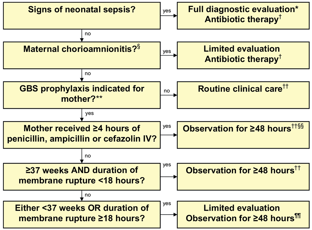
Algorithm for recommended management of newborns at risk for group B streptococcal disease.
* Includes CBC with differential, platelets, blood culture, chest radiograph (if respiratory abnormalities are present), and LP (if patient stable enough to tolerate procedure and sepsis is suspected).
† Antibiotic therapy should be directed towards the most common causes of neonatal sepsis including GBS and other organisms (including gram negative pathgoens), and should take into account local antibiotic resistance patterns.
§ Consultation with obstetric providers is important to determine the level of clinical suspicion for chorioamnionitis. Chorioamnionitis is diagnosed clinically and some of the signs are non-specific.
¶ Includes blood culture (at birth), and CBC with differential and platelets. Some experts recommend a CBC with differential and platelets at 6-12 hours of age.
** GBS prophylaxis indicated if one or more of the following: (1) mother GBS positive at 35-37 weeks’ gestation, (2) GBS status unknown with one or more intrapartum risk factors including <37 weeks’ gestation, ROM ≥ 18 hours or T ≥100.4°F (38.0°C), (3) GBS bacteriuria during current pregnancy, (4) history of a previous infant with GBS disease.
†† If signs of sepsis develop, a full diagnostic evaluation should be done and antibiotic therapy initiated.
§§ If ≥37 weeks’ gestation, observation may occur at home after 24 hours if there is a knowledgeable observer and ready access to medical care.
¶¶ Some experts recommend a CBC with differential and platelets at 6-12 hours of age.
For well-appearing infants with no exposure to maternal chorioamnionitis, recommended management will depend on whether IAP was indicated for the mother. If IAP was not indicated and an infant is well appearing, then routine care is recommended. If IAP was indicated, then neonatal management will be guided by the adequacy of IAP and the presence of certain risk factors for early onset disease. If the mother received penicillin, ampicillin, or cefazolin for at least 4 hours, then the infant should be observed for at least 48 hours. If the IAP was less than 4 hours in duration, or another agent such as clindamycin or vancomycin (for which there are very limited data on effectiveness for preventing vertical GBS transmission), then the IAP is considered to be inadequate. However, in the absence of other risk factors, including gestational age less than 37 weeks or rupture of membranes greater than 18 hours, infants exposed to inadequate IAP may be managed through careful clinical observation for at least 48 hours; this is in contrast to the 2002 recommendation that all such infants have a screening CBC and blood culture. If the IAP was inadequate and the delivery was either preterm or complicated by prolonged rupture of membranes, then the infant should have a limited diagnostic evaluation; antibiotics are not warranted unless the laboratory or clinical findings are suggestive of sepsis. Adherence to the revised neonatal recommendations would be expected to result in fewer unnecessary diagnostic evaluations in well-appearing newborns. The recommendations and algorithm presented here are proposed changes to the 2002 guidelines and may undergo further revision before dissemination.
FUTURE OF GBS PREVENTION: VACCINES
Maternal immunization against GBS is an appealing approach to preventing perinatal GBS disease. A vaccine that effectively prevents or diminishes colonization in parturient women and results in transplacental transfer of antibody to infants could obviate the need for antenatal GBS screening and use of IAP. Additionally, a GBS vaccine might help prevent stillbirth caused by GBS, late-onset GBS disease, and invasive GBS infections in mothers. An effective vaccine also may help diminish the persistent racial disparities in neonatal GBS disease.
The protective effect of maternal antibodies against neonatal GBS disease has been demonstrated in several studies.48,110,111 Several candidate vaccines targeting GBS surface proteins have been developed, and phase 1 and 2 human studies of capsular polysaccharide-protein conjugate vaccines have found the vaccines to be safe and immunogenic.112–114 A recent double-blind randomized trial of a conjugate GBS (serotype 3) vaccine among nonpregnant women of reproductive age resulted in a significant delay in acquisition of the serotype contained in the vaccine among vaccine recipients compared with those who did not receive the vaccine.115 Although GBS vaccines have shown promise, there remain hurdles to overcome. Clinical trials among pregnant women are complicated by ethical and medicolegal concerns. Furthermore, the primary outcome of interest—early onset GBS sepsis—has become a relatively rare endpoint as a result of antenatal screening and IAP. Trials using immunologic responses or maternal colonization may be more realistic approaches to obtaining vaccine licensure.114
SUMMARY
Much progress has been made toward decreasing the burden of early onset GBS disease in the United States. The incidence has declined by more than 80% since the preprevention era. Maternal GBS colonization, however, remains as common as during the preprevention era, and the risk for GBS infection in an infant born to a colonized mother who failed to receive IAP or received inadequate IAP is as high as it was in the early 1970s. Prevention efforts must be ongoing to maintain and further reduce the incidence of early onset disease. Further research is needed to identify effective means to reduce racial disparities for GBS disease and to decrease the burden of late-onset GBS disease, including vaccine trials.
The currently available GBS prevention strategies are highly effective at preventing early onset disease, but imperfect. Even with ideal implementation of universal screening and IAP, some cases will continue to occur among infants born to women who became colonized after their antenatal GBS screen, or who had a precipitous delivery that did not allow sufficient time for effective IAP. Clinicians must understand the risk factors for early onset GBS disease and the limitations of the prevention strategies to rapidly detect and treat cases that continue to occur in the era of GBS prevention through IAP. Revised GBS prevention guidelines will provide updated recommendations regarding the care of newborns in relation to GBS prevention.
Acknowledgments
The findings and conclusions in this paper are those of the authors and do not necessarily represent the views of the Centers for Disease Control and Prevention.
REFERENCES
- 1.Baker CJ, Barrett FF, Gordon RC, et al. Suppurative meningitis due to streptococci of Lancefield group B: a study of 33 infants. J Pediatr 1973;82(4):724–9. [DOI] [PubMed] [Google Scholar]
- 2.Barton LL, Feigin RD, Lins R. Group B beta hemolytic streptococcal meningitis in infants. J Pediatr 1973;82(4):719–23. [DOI] [PubMed] [Google Scholar]
- 3.Franciosi RA, Knostman JD, Zimmerman RA. Group B streptococcal neonatal and infant infections. J Pediatr 1973;82(4):707–18. [DOI] [PubMed] [Google Scholar]
- 4.Schuchat A. Epidemiology of group B streptococcal disease in the United States: shifting paradigms. Clin Microbiol Rev 1998;11(3):497–513. [DOI] [PMC free article] [PubMed] [Google Scholar]
- 5.CDC. Trends in perinatal group B streptococcal disease—United States, 2000–2006. MMWR Morb Mortal Wkly Rep 2009;58(5):109–12. [PubMed] [Google Scholar]
- 6.Jordan HT, Farley MM, Craig A, et al. Revisiting the need for vaccine prevention of late-onset neonatal group B streptococcal disease: a multistate, population-based analysis. Pediatr Infect Dis J 2008;27(12):1057–64. [DOI] [PubMed] [Google Scholar]
- 7.Baker CJ, Barrett FF. Transmission of group B streptococci among parturient women and their neonates. J Pediatr 1973;83(6):919–25. [DOI] [PubMed] [Google Scholar]
- 8.Desa DJ, Trevenen CL. Intrauterine infections with group B beta-haemolytic streptococci. Br J Obstet Gynaecol 1984;91(3):237–9. [DOI] [PubMed] [Google Scholar]
- 9.Katz V, Bowes WA Jr. Perinatal group B streptococcal infections across intact amniotic membranes. J Reprod Med 1988;33(5):445–9. [PubMed] [Google Scholar]
- 10.Dillon HC Jr, Khare S, Gray BM. Group B streptococcal carriage and disease: a 6-year prospective study. J Pediatr 1987;110(1):31–6. [DOI] [PubMed] [Google Scholar]
- 11.Easmon CS, Hastings MJ, Clare AJ, et al. Nosocomial transmission of group B streptococci. Br Med J 1981;283(6289):459–61. [DOI] [PMC free article] [PubMed] [Google Scholar]
- 12.Anthony BF, Okada DM, Hobel CJ. Epidemiology of the group B streptococcus: maternal and nosocomial sources for infant acquisitions. J Pediatr 1979;95(3):431–6. [DOI] [PubMed] [Google Scholar]
- 13.Olver WJ, Bond DW, Boswell TC, et al. Neonatal group B streptococcal disease associated with infected breast milk. Arch Dis Child 2000;83(1):F48–9. [DOI] [PMC free article] [PubMed] [Google Scholar]
- 14.Dinger J, Muller D, Pargac N, et al. Breast milk transmission of group B streptococcal infection. Pediatr Infect Dis J 2002;21(6):567–8. [DOI] [PubMed] [Google Scholar]
- 15.Godambe S, Shah PS, Shah V. Breast milk as a source of late onset neonatal sepsis. Pediatr Infect Dis J 2005;24(4):381–2. [DOI] [PubMed] [Google Scholar]
- 16.Wang LY, Chen CT, Liu WH, et al. Recurrent neonatal group B streptococcal disease associated with infected breast milk. Clin Pediatr (Phila) 2007;46(6):547–9. [DOI] [PubMed] [Google Scholar]
- 17.NIH. Summary of the workshop on perinatal infections due to group B Streptococcus. J Infect Dis 1977;136(1):137–52. [DOI] [PubMed] [Google Scholar]
- 18.Phares CR, Lynfield R, Farley MM, et al. Epidemiology of invasive group B streptococcal disease in the United States, 1999–2005. JAMA 2008;299(17):2056–65. [DOI] [PubMed] [Google Scholar]
- 19.Baker CJ, Barrett FF. Group B streptococcal infections in infants. The importance of the various serotypes. JAMA 1974;230(8):1158–60. [PubMed] [Google Scholar]
- 20.Schrag SJ, Zywicki S, Farley MM, et al. Group B streptococcal disease in the era of intrapartum antibiotic prophylaxis. N Engl J Med 2000;342(1):15–20. [DOI] [PubMed] [Google Scholar]
- 21.Puopolo KM, Madoff LC, Eichenwald EC. Early-onset group B streptococcal disease in the era of maternal screening. Pediatrics 2005;115(5):1240–6. [DOI] [PubMed] [Google Scholar]
- 22.Edwards MS, Rench MA, Haffar AA, et al. Long-term sequelae of group B streptococcal meningitis in infants. J Pediatr 1985;106(5):717–22. [DOI] [PubMed] [Google Scholar]
- 23.Lewin EB, Amstey MS. Natural history of group B streptococcus colonization and its therapy during pregnancy. Am J Obstet Gynecol 1981;139(5):512–5. [DOI] [PubMed] [Google Scholar]
- 24.Hoogkamp-Korstanje JA, Gerards LJ, Cats BP. Maternal carriage and neonatal acquisition of group B streptococci. J Infect Dis 1982;145(6):800–3. [DOI] [PubMed] [Google Scholar]
- 25.Hansen SM, Uldbjerg N, Kilian M, et al. Dynamics of Streptococcus agalactiae colonization in women during and after pregnancy and in their infants. J Clin Microbiol 2004;42(1):83–9. [DOI] [PMC free article] [PubMed] [Google Scholar]
- 26.Badri MS, Zawaneh S, Cruz AC, et al. Rectal colonization with group B Streptococcus: relation to vaginal colonization of pregnant women. J Infect Dis 1977;135(2):308–12. [DOI] [PubMed] [Google Scholar]
- 27.Meyn LA, Krohn MA, Hillier SL. Rectal colonization by group B Streptococcus as a predictor of vaginal colonization. Am J Obstet Gynecol 2009;201(1):76, e1–e7. [DOI] [PMC free article] [PubMed] [Google Scholar]
- 28.Schrag S, Gorwitz R, Fultz-Butts K, et al. Prevention of perinatal group B streptococcal disease. Revised guidelines from CDC. MMWR Recomm Rep 2002;51(RR-11):1–22. [PubMed] [Google Scholar]
- 29.Campbell JR, Hillier SL, Krohn MA, et al. Group B streptococcal colonization and serotype-specific immunity in pregnant women at delivery. Obstet Gynecol 2000;96(4):498–503. [DOI] [PubMed] [Google Scholar]
- 30.Newton ER, Butler MC, Shain RN. Sexual behavior and vaginal colonization by group B Streptococcus among minority women. Obstet Gynecol 1996;88:577–82. [DOI] [PubMed] [Google Scholar]
- 31.Stapleton RD, Kahn JM, Evans LE, et al. Risk factors for group B streptococcal genitourinary tract colonization in pregnant women. Obstet Gynecol 2005;106(6):1246–52. [DOI] [PubMed] [Google Scholar]
- 32.Katz VL, Moos MK, Cefalo RC, et al. Group B streptococci: results of a protocol of antepartum screening and intrapartum treatment. Am J Obstet Gynecol 1994;170(2):521–6. [DOI] [PubMed] [Google Scholar]
- 33.El Beitune P, Duarte G, Maffei CM, et al. Group B Streptococcus carriers among HIV-1 infected pregnant women: prevalence and risk factors. Eur J Obstet Gynecol Reprod Biol 2006;128(1–2):54–8. [DOI] [PubMed] [Google Scholar]
- 34.Yancey MK, Duff P, Kubilis P, et al. Risk factors for neonatal sepsis. Obstet Gynecol 1996;87(2):188–94. [DOI] [PubMed] [Google Scholar]
- 35.Regan JA, Klebanoff MA, Nugent RP, et al. Colonization with group B streptococci in pregnancy and adverse outcome. VIP Study Group. Am J Obstet Gynecol 1996;174(4):1354–60. [DOI] [PubMed] [Google Scholar]
- 36.Liston TE, Harris RE, Foshee S, et al. Relationship of neonatal pneumonia to maternal urinary and neonatal isolates of group B streptococci. South Med J 1979;72(11):1410–2. [DOI] [PubMed] [Google Scholar]
- 37.Wood EG, Dillon HC Jr. A prospective study of group B streptococcal bacteriuria in pregnancy. Am J Obstet Gynecol 1981;140(5):515–20. [DOI] [PubMed] [Google Scholar]
- 38.Persson K, Christensen KK, Christensen P, et al. Asymptomatic bacteriuria during pregnancy with special reference to group B streptococci. Scand J Infect Dis 1985;17(2):195–9. [DOI] [PubMed] [Google Scholar]
- 39.Boyer KM, Gotoff SP. Strategies for chemoprophylaxis of GBS early onset infections. Antibiot Chemother 1985;35:267–80. [DOI] [PubMed] [Google Scholar]
- 40.CDC. Prevention of perinatal group B streptococcal disease: a public health perspective. Centers for Disease Control and Prevention. MMWR Recomm Rep 1996;45(RR-7):1–24. [PubMed] [Google Scholar]
- 41.Adams WG, Kinney JS, Schuchat A, et al. Outbreak of early onset group B streptococcal sepsis. Pediatr Infect Dis J 1993;12(7):565–70. [DOI] [PubMed] [Google Scholar]
- 42.Boyer KM, Gadzala CA, Burd LI, et al. Selective intrapartum chemoprophylaxis of neonatal group B streptococcal early onset disease. I. Epidemiologic rationale. J Infect Dis 1983;148(5):795–801. [DOI] [PubMed] [Google Scholar]
- 43.Schuchat A, Deaver-Robinson K, Plikaytis BD, et al. Multistate case–control study of maternal risk factors for neonatal group B streptococcal disease. The Active Surveillance Study Group. Pediatr Infect Dis J 1994;13(7):623–9. [DOI] [PubMed] [Google Scholar]
- 44.Adair CE, Kowalsky L, Quon H, et al. Risk factors for early-onset group B streptococcal disease in neonates: a population-based case–control study. CMAJ 2003;169(3):198–203. [PMC free article] [PubMed] [Google Scholar]
- 45.Schuchat A, Zywicki SS, Dinsmoor MJ, et al. Risk factors and opportunities for prevention of early-onset neonatal sepsis: a multicenter case–control study. Pediatrics 2000;105:21–6. [DOI] [PubMed] [Google Scholar]
- 46.Schuchat A, Oxtoby M, Cochi S, et al. Population-based risk factors for neonatal group B streptococcal disease: results of a cohort study in metropolitan Atlanta. J Infect Dis 1990;162(3):672–7. [DOI] [PubMed] [Google Scholar]
- 47.Oddie S, Embleton ND. Risk factors for early onset neonatal group B streptococcal sepsis: case–control study. BMJ 2002;325(7359):308. [DOI] [PMC free article] [PubMed] [Google Scholar]
- 48.Baker CJ, Kasper DL. Correlation of maternal antibody deficiency with susceptibility to neonatal group B streptococcal infection. N Engl J Med 1976;294(14):753–6. [DOI] [PubMed] [Google Scholar]
- 49.Van Dyke MK, Phares CR, Lynfield R, et al. Evaluation of universal antenatal screening for group B Streptococcus. N Engl J Med 2009;360(25):2626–36. [DOI] [PubMed] [Google Scholar]
- 50.Baker CJ, Edwards MS, Kasper DL. Role of antibody to native type III polysaccharide of group B Streptococcus in infant infection. Pediatrics 1981;68(4):544–9. [PubMed] [Google Scholar]
- 51.Vogel LC, Boyer KM, Gadzala CA, et al. Prevalence of type-specific group B streptococcal antibody in pregnant women. J Pediatr 1980;96(6):1047–51. [DOI] [PubMed] [Google Scholar]
- 52.Christensen KK, Christensen P, Dahlander K, et al. Quantitation of serum antibodies to surface antigens of group B streptococci types Ia, Ib, and III: low antibody levels in mothers of neonatally infected infants. Scand J Infect Dis 1980;12(2):105–10. [DOI] [PubMed] [Google Scholar]
- 53.Christensen KK, Dahlander K, Linden V, et al. Obstetrical care in future pregnancies after fetal loss in group B streptococcal septicemia. A prevention program based on bacteriological and immunological follow-up. Eur J Obstet Gynecol Reprod Biol 1981;12(3):143–50. [DOI] [PubMed] [Google Scholar]
- 54.Faxelius G, Bremme K, Kvist-Christensen K, et al. Neonatal septicemia due to group B streptococci–perinatal risk factors and outcome of subsequent pregnancies. J Perinat Med 1988;16(5-6):423–30. [DOI] [PubMed] [Google Scholar]
- 55.Carstensen H, Christensen KK, Grennert L, et al. Early-onset neonatal group B streptococcal septicaemia in siblings. J Infect 1988;17(3):201–4. [DOI] [PubMed] [Google Scholar]
- 56.Lin FY, Weisman LE, Troendle J, et al. Prematurity is the major risk factor for late-onset group B Streptococcus disease. J Infect Dis 2003;188(2):267–71. [DOI] [PubMed] [Google Scholar]
- 57.Boyer KM, Gotoff SP. Prevention of early-onset neonatal group B streptococcal disease with selective intrapartum chemoprophylaxis. New Engl J Med 1986;314(26):1665–9. [DOI] [PubMed] [Google Scholar]
- 58.Garland SM, Fliegner JR. Group B Streptococcus (GBS) and neonatal infections: the case for intrapartum chemoprophylaxis. Aust N Z J Obstet Gynaecol 1991;31(2):119–22. [DOI] [PubMed] [Google Scholar]
- 59.Schrag SJ, Zell ER, Lynfield R, et al. A population-based comparison of strategies to prevent early onset group B streptococcal disease in neonates. New Engl J Med 2002;347(4):233–9. [DOI] [PubMed] [Google Scholar]
- 60.Lin FY, Brenner RA, Johnson YR, et al. The effectiveness of risk-based intrapartum chemoprophylaxis for the prevention of early onset neonatal group B streptococcal disease. Am J Obstet Gynecol 2001;184(6):1204–10. [DOI] [PubMed] [Google Scholar]
- 61.Castor ML, Whitney CG, Como-Sabetti K, et al. Antibiotic resistance patterns in invasive group B streptococcal isolates. Infect Dis Obstet Gynecol 2008;2008:727505. [DOI] [PMC free article] [PubMed] [Google Scholar]
- 62.Bromberger P, Lawrence JM, Braun D, et al. The influence of intrapartum antibiotics on the clinical spectrum of early onset group B streptococcal infection in term infants. Pediatrics 2000;106:244–50. [DOI] [PubMed] [Google Scholar]
- 63.Escobar GJ, Li DK, Armstrong MA, et al. Neonatal sepsis workups in infants ≥ 2000 grams at birth: a population-based study. Pediatrics 2000;106:256–63. [DOI] [PubMed] [Google Scholar]
- 64.Pulver LS, Hopfenbeck MM, Young PC, et al. Continued early onset group B streptococcal infections in the era of intrapartum prophylaxis. J Perinatol 2009;29(1):20–5. [DOI] [PubMed] [Google Scholar]
- 65.Pinto NM, Soskolne EI, Pearlman MD, et al. Neonatal early onset group B streptococcal disease in the era of intrapartum chemoprophylaxis: residual problems. J Perinatol 2003;23(4):265–71. [DOI] [PubMed] [Google Scholar]
- 66.Mercer BM, Ramsey RD, Sibai BM. Prenatal screening for group B Streptococcus. II. Impact of antepartum screening and prophylaxis on neonatal care. Am J Obstet Gynecol 1995;173:842–6. [DOI] [PubMed] [Google Scholar]
- 67.Peralta-Carcelen M, Fargason CA Jr, Cliver SP, et al. Impact of maternal group B streptococcal screening on pediatric management in full-term newborns. Arch Pediatr Adolesc Med 1996;150(8):802–8. [DOI] [PubMed] [Google Scholar]
- 68.Balter S, Zell ER, O’Brien KL, et al. Impact of intrapartum antibiotics on the care and evaluation of the neonate. Pediatr Infect Dis J 2003;22(10):853–7. [DOI] [PubMed] [Google Scholar]
- 69.Glasgow TS, Speakman M, Firth S, et al. Clinical and economic outcomes for term infants associated with increasing administration of antibiotics to their mothers. Paediatr perinat epidemiol 2007;21(4):338–46. [DOI] [PubMed] [Google Scholar]
- 70.Ascher DP, Becker JA, Yoder BA, et al. Failure of intrapartum antibiotics to prevent culture-proved neonatal group B streptococcal sepsis. J Perinatol 1993;13(3):212–6. [PubMed] [Google Scholar]
- 71.Lieberman E, Lang JM, Frigoletto F Jr, et al. Epidural analgesia, intrapartum fever, and neonatal sepsis evaluation. Pediatrics 1997;99(3):415–9. [DOI] [PubMed] [Google Scholar]
- 72.de Cueto M, Sanchez MJ, Sampedro A, et al. Timing of intrapartum ampicillin and prevention of vertical transmission of group B streptococcus. Obstet gynecol 1998;91(1):112–4. [DOI] [PubMed] [Google Scholar]
- 73.Gerdes JS, Polin RA. Sepsis screen in neonates with evaluation of plasma fibronectin. Pediatr Infect Dis J 1987;6(5):443–6. [DOI] [PubMed] [Google Scholar]
- 74.Greenberg DN, Yoder BA. Changes in the differential white blood cell count in screening for group B streptococcal sepsis. Pediatr Infect DisJ 1990;9(12):886–9. [DOI] [PubMed] [Google Scholar]
- 75.Ottolini MC, Lundgren K, Mirkinson LJ, et al. Utility of complete blood count and blood culture screening to diagnose neonatal sepsis in the asymptomatic at risk newborn. Pediatr Infect Dis J 2003;22(5):430–4. [DOI] [PubMed] [Google Scholar]
- 76.Yancey MK, Schuchat A, Brown LK, et al. The accuracy of late antenatal screening cultures in predicting genital group B streptococcal colonization at delivery. Obstet gynecol 1996;88(5):811–5. [DOI] [PubMed] [Google Scholar]
- 77.Lukacs S, Schuchat A, Schoendorf K. National estimates of newborn sepsis: United States, 1990–2002. Presented at the annual meeting of Society for Pediatric and Perinatal Epidemiologic Research. June 14–15, 2004, Salt Lake City (Utah). [Google Scholar]
- 78.McCracken GH Jr. Group B streptococci: the new challenge in neonatal infections. J Pediatr 1973;82(4):703–6. [DOI] [PubMed] [Google Scholar]
- 79.Freedman RM, Ingram DL, Gross I, et al. A half century of neonatal sepsis at Yale: 1928 to 1978. Am J Dis Child 1981;135(2):140–4. [DOI] [PubMed] [Google Scholar]
- 80.Main EK, Slagle T. Prevention of early onset invasive neonatal group B streptococcal disease in a private hospital setting: the superiority of culture-based protocols. Am J Obstet Gynecol 2000;182(6):1344–54. [DOI] [PubMed] [Google Scholar]
- 81.Baltimore RS, Huie SM, Meek JI, et al. Early onset neonatal sepsis in the era of group B streptococcal prevention. Pediatrics 2001;108(5):1094–8. [DOI] [PubMed] [Google Scholar]
- 82.Cordero L, Sananes M, Ayers LW. Bloodstream infections in a neonatal intensive care unit: 12 years’ experience with an antibiotic control program. Infect Control Hosp Epidemiol 1999;20(4):242–6. [DOI] [PubMed] [Google Scholar]
- 83.Angstetra D, Ferguson J, Giles WB. Institution of universal screening for group B Streptococcus (GBS) from a risk management protocol results in reduction of early-onset GBS disease in a tertiary obstetric unit. Aust N Z J Obstet Gynaecol 2007;47(5):378–82. [DOI] [PubMed] [Google Scholar]
- 84.Sutkin G, Krohn MA, Heine RP, et al. Antibiotic prophylaxis and nongroup B streptococcal neonatal sepsis. Obstet Gynecol 2005;105(3):581–6. [DOI] [PubMed] [Google Scholar]
- 85.Alarcon A, Pena P, Salas S, et al. Neonatal early onset Escherichia coli sepsis: trends in incidence and antimicrobial resistance in the era of intrapartum antimicrobial prophylaxis. Pediatr Infect Dis J 2004;23(4):295–9. [DOI] [PubMed] [Google Scholar]
- 86.Rentz AC, Samore MH, Stoddard GJ, et al. Risk factors associated with ampicillin-resistant infection in newborns in the era of group B streptococcal prophylaxis. Arch Pediatr Adolesc Med 2004;158(6):556–60. [DOI] [PubMed] [Google Scholar]
- 87.Edwards RK, Jamie WE, Sterner D, et al. Intrapartum antibiotic prophylaxis and early onset neonatal sepsis patterns. Infect Dis Obstet Gynecol 2003;11(4):221–6. [DOI] [PMC free article] [PubMed] [Google Scholar]
- 88.Isaacs D, Royle JA. Intrapartum antibiotics and early onset neonatal sepsis caused by group B Streptococcus and by other organisms in Australia. Australasian Study Group for Neonatal Infections. Pediatr Infect Dis J 1999;18(6):524–8. [DOI] [PubMed] [Google Scholar]
- 89.Daley AJ, Isaacs D. Ten-year study on the effect of intrapartum antibiotic prophylaxis on early onset group B streptococcal and Escherichia coli neonatal sepsis in Australasia. Pediatr Infect Dis J 2004;23(7):630–4. [DOI] [PubMed] [Google Scholar]
- 90.Joseph TA, Pyati SP, Jacobs N. Neonatal early onset Escherichia coli disease. The effect of intrapartum ampicillin. Arch Pediatr Adolesc Med 1998;152(1):35–40. [DOI] [PubMed] [Google Scholar]
- 91.Towers CV, Carr MH, Padilla G, et al. Potential consequences of widespread antepartal use of ampicillin. Am J Obstet Gynecol 1998;179(4):879–83. [DOI] [PubMed] [Google Scholar]
- 92.Levine EM, Ghai V, Barton JJ, et al. Intrapartum antibiotic prophylaxis increases the incidence of gram-negative neonatal sepsis. Infect Dis Obstet Gynecol 1999;7(4):210–3. [DOI] [PMC free article] [PubMed] [Google Scholar]
- 93.Bizzarro MJ, Dembry LM, Baltimore RS, et al. Changing patterns in neonatal Escherichia coli sepsis and ampicillin resistance in the era of intrapartum antibiotic prophylaxis. Pediatrics 2008;121(4):689–96. [DOI] [PubMed] [Google Scholar]
- 94.Stoll BJ, Hansen N, Fanaroff AA, et al. Changes in pathogens causing early onset sepsis in very low birth weight infants. N Engl J Med 2002;347(4):240–7. [DOI] [PubMed] [Google Scholar]
- 95.Stoll BJ, Hansen NI, Higgins RD, et al. Very low birth weight preterm infants with early onset neonatal sepsis: the predominance of gram-negative infections continues in the National Institute of Child Health and Human Development Neonatal Research Network, 2002–2003. Pediatr Infect Dis J 2005;24(7):635–9. [DOI] [PubMed] [Google Scholar]
- 96.Hyde TB, Hilger TM, Reingold A, et al. Trends in incidence and antimicrobial resistance of early onset sepsis: population-based surveillance in San Francisco and Atlanta. Pediatrics 2002;110(4):690–5. [DOI] [PubMed] [Google Scholar]
- 97.Al-Hasan MN, Lahr BD, Eckel-Passow JE, et al. Antimicrobial resistance trends of Escherichia coli bloodstream isolates: a population-based study, 1998-2007. J Antimicrob Chemother 2009;64(1):169–74. [DOI] [PMC free article] [PubMed] [Google Scholar]
- 98.Mercer BM, Carr TL, Beazley DD, et al. Antibiotic use in pregnancy and drug-resistant infant sepsis. Am J Obstet Gynecol 1999;181(4):816–21. [DOI] [PubMed] [Google Scholar]
- 99.Terrone DA, Rinehart BK, Einstein MH, et al. Neonatal sepsis and death caused by resistant Escherichia coli: possible consequences of extended maternal ampicillin administration. Am J Obstet Gynecol 1999;180:1345–8. [DOI] [PubMed] [Google Scholar]
- 100.Towers CV, Briggs GG. Antepartum use of antibiotics and early onset neonatal sepsis: the next 4 years. Am J Obstet Gynecol 2002;187(2):495–500. [DOI] [PubMed] [Google Scholar]
- 101.Moore MR, Schrag SJ, Schuchat A. Effects of intrapartum antimicrobial prophylaxis for prevention of group B streptococcal disease on the incidence and ecology of early onset neonatal sepsis. Lancet Infect Dis 2003;3(4):201–13. [DOI] [PubMed] [Google Scholar]
- 102.Schrag SJ, Hadler JL, Arnold KE, et al. Risk factors for invasive, early onset Escherichia coli infections in the era of widespread intrapartum antibiotic use. Pediatrics 2006;118(2):570–6. [DOI] [PubMed] [Google Scholar]
- 103.Kimura K, Suzuki S, Wachino J, et al. First molecular characterization of group B streptococci with reduced penicillin susceptibility. Antimicrob Agents Chemother 2008;52(8):2890–7. [DOI] [PMC free article] [PubMed] [Google Scholar]
- 104.Dahesh S, Hensler ME, Van Sorge NM, et al. Point mutation in the group B streptococcal pbp2x gene conferring decreased susceptibility to beta-lactam antibiotics. Antimicrob Agents Chemother 2008;52(8):2915–8. [DOI] [PMC free article] [PubMed] [Google Scholar]
- 105.Manning SD, Foxman B, Pierson CL, et al. Correlates of antibiotic-resistant group B streptococcus isolated from pregnant women. Obstet Gynecol 2003;101(1):74–9. [DOI] [PubMed] [Google Scholar]
- 106.Varman M, Romero JR, Cornish NE, et al. Characterization and mechanisms of resistance of group B streptococcal isolates obtained at a community hospital. Eur J Clin Microbiol Infect Dis 2005;24(6):431–3. [DOI] [PubMed] [Google Scholar]
- 107.Chohan L, Hollier LM, Bishop K, et al. Patterns of antibiotic resistance among group B streptococcus isolates: 2001–2004. Infect Dis Obstet Gynecol 2006;2006:57492. [DOI] [PMC free article] [PubMed] [Google Scholar]
- 108.Panda B, Iruretagoyena I, Stiller R, et al. Antibiotic resistance and penicillin tolerance in ano-vaginal group B streptococci. J Matern Fetal Neonatal Med 2009;22(2):111–4. [DOI] [PubMed] [Google Scholar]
- 109.Borchardt SM, DeBusscher JH, Tallman PA, et al. Frequency of antimicrobial resistance among invasive and colonizing group B streptococcal isolates. BMC Infect Dis. 2006;6:57. [DOI] [PMC free article] [PubMed] [Google Scholar]
- 110.Lin FY, Philips JB 3rd, Azimi PH, et al. Level of maternal antibody required to protect neonates against early-onset disease caused by group B Streptococcus type Ia: a multicenter, seroepidemiology study. J Infect Dis 2001;184(8):1022–8. [DOI] [PubMed] [Google Scholar]
- 111.Lin FY, Weisman LE, Azimi PH, et al. Level of maternal IgG anti-group B Streptococcus type III antibody correlated with protection of neonates against early-onset disease caused by this pathogen. J Infect Dis 2004;190(5):928–34. [DOI] [PubMed] [Google Scholar]
- 112.Baker CJ, Edwards MS. Group B streptococcal conjugate vaccines. Arch Dis Child 2003;88(5):375–8. [DOI] [PMC free article] [PubMed] [Google Scholar]
- 113.Heath PT, Feldman RG. Vaccination against group B Streptococcus. Expert Rev Vaccines 2005;4(2):207–18. [DOI] [PubMed] [Google Scholar]
- 114.Edwards MS. Group B streptococcal conjugate vaccine: a timely concept for which the time has come. Hum Vaccin 2008;4(6):444–8. [DOI] [PubMed] [Google Scholar]
- 115.Hillier S, Ferris D, Fine D, et al. Women receiving group B Streptococcus serotype III tetanus toxoid (GBS III-TT) vaccine have reduced vaginal and rectal acquisition of GBS type III. Presented at Infectious Diseases Society of America Annual Meeting. Philadelphia, October 29 to November 1, 2009. [Google Scholar]