


Abstract
目的
探讨人脂肪间充质干细胞(ADSC)来源外泌体对脓毒症小鼠肺血管内皮细胞(PMVEC)损伤的影响及其机制。
方法
采用实验研究方法。取空军军医大学第一附属医院收治的3例行腹部手术切除患者(均为女性, 年龄10~25岁)遗弃的新鲜脂肪组织进行原代人ADSC的分离培养, 于第5天采用倒置相差显微镜观察细胞形态。取第3代ADSC, 采用流式细胞术检测ADSC中CD29、CD34、CD44、CD45、CD73和CD90的表达情况。选取第3~5代ADSC, 采用差速超高速离心法获取其上清液中的外泌体, 采用透射电镜和纳米颗粒跟踪分析法及蛋白质印迹法分别检测外泌体形状、粒径大小及CD9、CD63、肿瘤易感基因101(TSG101)和β肌动蛋白的蛋白表达。取24只成年雄性BALB/c小鼠, 按照随机数字表法(分组方法下同)分成正常对照组、单纯盲肠结扎穿孔(CLP)组和CLP+ADSC外泌体组, 每组8只, 分别进行相应处理。术后24 h, 采用酶联免疫吸附测定法检测小鼠血清中肿瘤坏死因子α(TNF-α)和白细胞介素1β(IL-1β)水平, 采用苏木精-伊红染色和髓过氧化物酶染色检测小鼠肺组织形态, 采用免疫荧光法检测小鼠肺组织细胞中8-羟基脱氧鸟苷(8-OHdG)的表达。取1个月龄雌雄不拘C57小鼠, 采用组织块法获取原代PMVEC。取第3代PMVEC, 采用免疫荧光法和流式细胞术检测PMVEC中CD31的表达。取第3代PMVEC, 与ADSC来源外泌体共培养12 h, 采用PKH26试剂盒检测PMVEC吞噬外泌体的情况。取第3代PMVEC, 将细胞分为空白对照组、单纯巨噬细胞上清液组和巨噬细胞上清液+ADSC外泌体组, 每组3孔, 分别进行相应处理。24 h后, 采用流式细胞术检测细胞中活性氧含量, 采用免疫荧光法检测细胞中8-OHdG表达, 采用Transwell实验测定细胞单分子层通透性。以上样本数均为3。对数据行单因素方差分析和LSD-t检验。
结果
分离的原代ADSC培养至第5天, 呈梭形密集生长, 呈典型的漩涡样排列。第3代ADSC中CD29、CD44、CD73和CD90阳性细胞百分比均 > 90%, CD34和CD45阳性细胞百分比均 < 5%。第3~5代ADSC来源外泌体呈典型的双凹盘状结构, 外泌体平均粒径为103 nm, 外泌体中CD9、CD63和TSG101的蛋白表达均呈阳性, β肌动蛋白的蛋白表达呈阴性。术后24 h, 与正常对照组比较, 单纯CLP组小鼠血清中TNF-α和IL-1β的含量均明显增加(t值分别为28.76、29.69, P < 0.01);与单纯CLP组比较, CLP+ADSC外泌体组小鼠血清中TNF-α和IL-1β的含量均明显降低(t值分别为9.90、4.76, P < 0.05或P < 0.01)。术后24 h, 正常对照组小鼠肺组织结构清晰完整、无炎症细胞浸润, 单纯CLP组小鼠的肺组织较正常对照组水肿明显、炎症细胞浸润现象明显, CLP+ADSC外泌体组小鼠肺组织较单纯CLP组水肿症状明显减轻、炎症细胞浸润明显减少。术后24 h, 3组小鼠肺组织中CD31呈阳性表达;单纯CLP组小鼠肺组织中8-OHdG阳性细胞荧光强度较正常对照组显著增大, CLP+ADSC外泌体组小鼠肺组织中8-OHdG阳性细胞荧光强度较单纯CLP组明显下降。第3代PMVEC的免疫荧光结果显示CD31呈阳性表达, 流式细胞术鉴定结果显示CD31阳性细胞占比为99.5%。共培养12 h, ADSC来源外泌体成功被PMVEC吞噬, 进入胞质。第3代PMVEC培养24 h后, 与空白对照组比较, 单纯巨噬细胞上清液组PMVEC的活性氧荧光强度明显增加(t=15.73, P < 0.01);与单纯巨噬细胞上清液组比较, 巨噬细胞上清液+ADSC外泌体组PMVEC的活性氧荧光强度明显降低(t=4.72, P < 0.01)。第3代PMVEC培养24 h, 与空白对照组相比, 单纯巨噬细胞上清液组PMVEC的8-OHdG阳性荧光强度明显增强, 巨噬细胞上清液+ADSC外泌体组PMVEC的8-OHdG阳性荧光强度介于空白对照组和单纯巨噬细胞上清液组之间。第3代PMVEC培养24 h, 与空白对照组比较, 单纯巨噬细胞上清液组的PMVEC单分子层细胞通透性明显增加(t=6.34, P < 0.01);与单纯巨噬细胞上清液组比较, 巨噬细胞上清+ADSC外泌体组的PMVEC单分子层细胞通透性明显降低(t=2.93, P < 0.05)。
结论
ADSC来源外泌体可改善小鼠肺组织氧化性损伤, 降低由巨噬细胞上清液诱导的PMVEC的活性氧含量、8-OHdG表达以及细胞通透性。
Keywords: 脓毒症, 急性肺损伤, 活性氧, 肺血管内皮细胞, 8-羟基脱氧鸟苷
Abstract
Objective
To investigate the effects of exosomes from human adipose-derived mesenchymal stem cells (ADSCs) on pulmonary vascular endothelial cells (PMVECs) injury in septic mice and its mechanism.
Methods
The experimental research method was adopted. The primary ADSCs were isolated and cultured from the discarded fresh adipose tissue of 3 patients (female, 10-25 years old), who were admitted to the First Affiliated Hospital of Air Force Medical University undergoing abdominal surgery, and the cell morphology was observed by inverted phase contrast microscope on the 5th day. The expressions of CD29, CD34, CD44, CD45, CD73, and CD90 of ADSCs in the third passage were detected by flow cytometry. The third to the fifth passage of ADSCs were collected, and their exosomes from the cell supernatant were obtained by differential ultracentrifugation, and the shape, particle size, and the protein expressions of CD9, CD63, tumor susceptibility gene 101 (TSG101), and β-actin of exosomes were detected, respectively, by transmission electron microscopy, nano-particle tracking analysis and Western blotting. Twenty-four adult male BALB/c mice were adopted and were divided into normal control group, caecal ligation perforation (CLP) alone group, and CLP+ADSC-exosome group with each group of 8 according to random number table (the same grouping method below) and were treated accordingly. At 24 h after operation, tumor necrosis factor (TNF-α) and interleukin 1β (IL-1β) levels of mice serum were detected by enzyme-linked immunosorbent assay, and lung tissue morphology of mice was detected by hematoxylin-eosin and myeloperoxidase staining, and the expression of 8-hydroxy-deoxyguanosine (8-OHdG) of mouse lung cells was detected by immunofluorescence method. Primary PMVECs were obtained from 1-month-old C57 mice regardless gender by tissue block method. The expression of CD31 of PMVECs was detected by immunofluorescence and flow cytometry. The third passage of PMVECs was co-cultured with ADSCs derived exosomes for 12 h, and the phagocytosis of exosomes by PMVECs was detected by PKH26 kit. The third passage of PMVECs were adopted and were divided into blank control group, macrophage supernatant alone group, and macrophage supernatant+ADSC-exosome group, with 3 wells in each group, which were treated accordingly. After 24 h, the content of reactive oxygen species in cells was detected by flow cytometry, the expression of 8-OHdG in cells was detected by immunofluorescence, and Transwell assay was used to determine the permeability of cell monolayer. The number of samples in above were all 3. Data were statistically analyzed with one-way analysis of variance and least significant difference t test.
Results
The primary ADSCs were isolated and cultured to day 5, growing densely in a spindle shape with a typical swirl-like. The percentages of CD29, CD44, CD73 and CD90 positive cells of ADSCs in the third passage were all > 90%, and the percentages of CD34 and CD45 positive cells were < 5%. Exosomes derived from ADSCs of the third to fifth passages showed a typical double-cavity disc-like structure with an average particle size of 103 nm, and the protein expressions of CD9, CD63 and TSG101 of exosomes were positive, while the protein expression of β-actin of exosomes was negative. At 24 h after operation, compared with those in normal control group, both the levels of TNF-α and IL-1β of mice serum in CLP alone group were significantly increased (with t values of 28.76 and 29.69, respectively, P < 0.01); compared with those in CLP alone group, both the content of TNF-α and IL-1β of mice serum in CLP+ADSC-exosome group was significantly decreased (with t values of 9.90 and 4.76, respectively, P < 0.05 or P < 0.01). At 24 h after surgery, the pulmonary tissue structure of mice in normal control group was clear and complete without inflammatory cell infiltration; compared with those in normal control group, the pulmonary tissue edema and inflammatory cell infiltration of mice in CLP alone group were more obvious; compared with those in CLP alone group, the pulmonary tissue edema and inflammatory cell infiltration of mice in CLP+ADSC-exosome group were significantly reduced. At 24 h after operation, endothelial cells in lung tissues of mice in 3 groups showed positive expression of CD31; compared with that in normal control group, the fluorescence intensity of 8-OHdG positive cells of the lung tissues of mice in CLP alone group was significantly increased, and compared with that in CLP alone group, the fluorescence intensity of 8-OHdG positive cells in the lung tissues of mice in CLP+ADSC-exosome group was significantly decreased. The PMVECs in the 3rd passage showed CD31 positive expression by immunofluorescence, and the result of flow cytometry showed that CD31 positive cells accounted for 99.5%. At 12 h after co-culture, ADSC-derived exosomes were successfully phagocytose by PMVECs and entered its cytoplasm. At 12 h after culture of the third passage of PMVECs, compared with that in blank control group, the fluorescence intensity of reactive oxygen species of PMVECs in macrophage supernatant alone group was significantly increased (t=15.73, P < 0.01); compared with that in macrophage supernatant alone group, the fluorescence intensity of reactive oxygen species of PMVECs in macrophage supernatant+ADSC-exosome group was significantly decreased (t=4.72, P < 0.01). At 12 h after culture of the third passage of PMVECs, and the 8-OHdG positive fluorescence intensity of PMVECs in macrophage supernatant alone group was significantly increased; and compared with that in blank control group, the 8-OHdG positive fluorescence intensity of PMVECs in macrophage+ADSC-exosome supernatant group was between blank control group and macrophage supernatant alone group. At 12 h after culture of the third passage PMVECs, compared with that in blank control group, the permeability of PMVECs monolayer in macrophage supernatant alone group was significantly increased (t=6.34, P < 0.01); compared with that in macrophage supernatant alone group, the permeability of PMVECs monolayer cells in macrophage supernatant+ADSC-exosome group was significantly decreased (t=2.93, P < 0.05).
Conclusions
Exosomes derived from ADSCs can ameliorate oxidative damage in mouse lung tissue, decrease the level of reactive oxygen species, 8-OHdG expression, and permeability of PMVECs induced by macrophage supernatant.
Keywords: Sepsis, Acute lung injury, Reactive oxygen species, Pulmonary vascular endothelial cells, 8-hydroxydeoxyguanosine
脓毒症是ICU的常见病, 其发病率和病死率高, 病理机制复杂, 常引发MODS、甚至死亡[1-4]。急性肺损伤(ALI)是脓毒症常见的严重并发症, 常发展为ARDS[5-6]。肺血管内皮细胞(PMVEC)是构成肺血管通透性的主要屏障, 对维持肺血管壁的完整性具有特殊意义[7]。ALI发生时, 机体迅速产生各种炎症介质及大量活性氧[8-9], 可引起广泛的肺血管内皮损伤, 进而导致肺血管通透性增高以及肺水肿等, 最终加重肺损伤。研究证实, 血管内皮细胞不仅是炎症反应中的被动靶细胞, 也是效应细胞, 在微循环障碍、脓毒症休克及多器官功能损伤中起着重要作用[10-15]。因此, 有效抑制促炎介质释放、减少氧化应激反应是干预脓毒症的关键[16], 也是减轻内皮细胞损伤的关键。干细胞及外泌体已被报道在小鼠损伤后的组织修复和再生中发挥作用[17-20]。ADSC来源外泌体作为一种重要的细胞通信手段, 在体内、体外都能发挥不依赖于ADSC的抗炎和保护作用[21-25]。本研究通过对小鼠进行盲肠结扎穿孔(CLP)构建脓毒症动物模型, 诱导小鼠发生急性肺损伤, 然后采用ADSC来源外泌体进行干预以观察其对PMVEC氧化性损伤及细胞通透性的影响, 并探讨其可能机制。
1. 材料与方法
本实验研究遵循空军军医大学动物实验伦理委员会和国家有关实验动物管理和使用的规定。
1.1. 动物及主要试剂与仪器来源
24只健康清洁级成年雄性BALB/c小鼠(体重20~25 g)与5只健康清洁级1个月龄雌雄不拘C57小鼠(体重10~15 g), 均由空军军医大学实验动物中心提供, 许可证号:SYXK(军)2012-0022。
内皮细胞专用培养基(内含体积分数5%胎牛血清、体积分数1%内皮细胞生长因子等为独立包装配套试剂, 可根据实验条件进行全部或部分添加)购于美国ScienCell公司, 兔源CD31多克隆抗体、Alexa Fluor 488标记的山羊抗兔IgG抗体、花青素标记的驴抗小鼠IgG抗体、兔源髓过氧化物酶(MPO)多克隆抗体、HE染液购自武汉赛维尔生物科技有限公司, Alexa Fluor 488标记的驴抗小鼠IgG多克隆抗体、小鼠源8-羟基脱氧鸟苷(8-OHdG)单克隆抗体购自美国Abcam公司, 藻红蛋白标记的小鼠抗人CD29、CD34、CD44、CD45、CD73和CD90单克隆抗体购自美国Becton Dickinson公司, LPS、Ⅰ型胶原酶、PKH26试剂盒、生物素标记的牛血清白蛋白(BSA)购自美国Sigma公司, 兔抗人CD9、CD63、肿瘤易感基因101(TSG101)蛋白和β肌动蛋白单克隆抗体购自武汉三鹰生物技术有限公司, 辣根过氧化物酶标记的山羊抗兔IgG抗体、兔源CD31多克隆抗体、异硫氰酸荧光素(FITC)标记的山羊抗兔IgG抗体、FITC直标抗小鼠同型对照IgG抗体购自武汉博士德生物工程有限公司, 小鼠IL-1β和TNF-α ELISA试剂盒购自美国BD公司, 成人ADSC专用培养基购自广州赛业公司, DMEM/F12培养基、DMEM培养基购自美国HyClone公司, 胎牛血清购自美国Gibco公司, RAW264.7小鼠单核巨噬细胞白血病细胞系购自美国典型培养物保藏中心, Transwell小室购自美国Corning公司, 活性氧检测试剂盒、二辛丁酸蛋白浓度测定试剂盒、4′, 6-二脒基-2-苯基吲哚(DAPI)购自北京碧云天生物技术研究所, 兔SP检测试剂盒(SP-9001, 链霉卵白素-生物素法检测系统)购于北京中杉金桥生物技术有限公司。
Steri-Cycle 371型二氧化碳培养箱购自美国Thermo Fisher Scientific公司, OptimaTMXPN型超高速离心机购自美国Beckman公司, ChemiDOC型凝胶成像系统购自美国Bio-Rad公司, FACS Aria Ⅲ型流式细胞仪购自美国BD公司, EVOS FL Auto 2型激光扫描共聚焦显微镜购自美国Invitrogen公司, Infinite M200 Pro型全波长多功能酶标仪购自瑞士TECAN公司, FSX100型全自动生物图像导航仪购自日本Olympus公司, 倒置相差显微镜购自德国ZEISS公司, ZetaVIEW S/N 17-310型纳米颗粒跟踪分析仪(NTA)购自美国PARTICLE METRIX公司。
1.2. ADSC及其外泌体实验
该部分实验的具体步骤, 同文献[26]。
1.2.1. ADSC的分离培养及鉴定
经患者知情同意, 取2020年6—9月3例空军军医大学第一附属医院烧伤与皮肤外科腹部手术患者(均为女性, 10~25岁)切除后遗弃的新鲜脂肪组织, 经分离培养后得到原代细胞, 采用ADSC专用培养基重新悬浮细胞, 进行常规培养, 第5天于20倍倒置相差显微镜下观察细胞形态。取第3代细胞, 培养24 h后分别加入藻红蛋白标记的鼠抗人CD29、CD34、CD44、CD45、CD73和CD90单克隆抗体(稀释比均为1∶1 000), 使用流式细胞仪检测其表达情况。本实验重复3次。
1.2.2. ADSC来源外泌体的制备与鉴定
1.2.2.1. 外泌体制备
取第3~5代ADSC, 采用差速超高速离心法获取其上清液中的外泌体。将沉淀溶解于无菌PBS中, 取少量前述溶液, 采用二辛丁酸蛋白浓度测定试剂盒检测其浓度, 剩余部分冻存于-80 ℃冰箱备用。
1.2.2.2. 外泌体鉴定
取外泌体, 通过冷链运输至上海华盈生物有限公司, 在40 000倍的透射电镜和NTA下分别检测外泌体形状及大小。另取ADSC及其外泌体, 采用免疫印迹法检测CD9、CD63、TSG101和β肌动蛋白的表达, 其中一抗为兔抗人CD9、CD63、TSG101和β肌动蛋白单克隆抗体(稀释比均为1∶1 000), 二抗为辣根过氧化物酶标记的山羊抗兔IgG二抗(稀释比为1∶3 000)。于凝胶成像分析系统进行相关蛋白条带灰度扫描分析。本实验重复3次。
1.3. 动物实验
1.3.1. 脓毒症动物模型的建立与分组及标本采集
取24只BALB/c小鼠, 适应性饲养1周后按照随机数字表法(分组方法下同)分成正常对照组、单纯CLP组和CLP+ADSC外泌体组, 每组8只。单纯CLP组和CLP+ADSC外泌体组小鼠均行CLP手术诱导脓毒症[20, 27], 其中CLP+ADSC外泌体组小鼠在诱导脓毒症后, 即刻进行尾静脉注射100 mL质量浓度为1 μg/mL的ADSC来源外泌体, 单纯CLP组小鼠尾静脉注射等量无菌PBS;正常对照组小鼠仅尾静脉注射等量无菌PBS。术后24 h, 摘除其眼球取血备用;然后用颈椎脱臼法处死小鼠, 取其肺组织采用40 g/L多聚甲醛固定, 常规石蜡包埋, 制成厚度为5 μm切片备用。
1.3.2. ELISA法检测血清中TNF-α和IL-1β水平
取1.3.1中的血标本, 静置30 min, 于4 ℃条件下以1 600×g离心15 min, 小心吸取上层血清, 按照ELISA说明书步骤检测小鼠血清中的TNF-α和IL-1β水平。
1.3.3. HE及MPO免疫组织化学染色检测肺组织形态
取1.3.1中石蜡切片, 进行常规HE染色, 采用生物图像导航仪在20倍光学显微镜下观察肺组织形态并拍照。另取石蜡切片, PBS冲洗3次, 采用体积分数3%过氧化氢封闭, PBS洗涤3次。采用50 g/L的BSA溶液室温封闭30 min, 切片上滴加兔源MPO多克隆一抗(稀释比为1∶100), 在湿盒中于4 ℃孵育过夜。PBS洗涤3次, 按照SP-9001试剂盒说明书进行免疫组织化学染色。采用生物图像导航仪在20倍显微镜下观察肺组织形态并拍照。
1.3.4. 免疫荧光法检测肺组织细胞中8-OHdG和CD31表达
取1.3.1中石蜡切片, 脱蜡后采用柠檬酸进行修复, PBS洗涤3次。30~50 g/L的BSA溶液常温下封闭30 min, 加入小鼠源8-OHdG单克隆及兔源CD31多克隆一抗(稀释比均为1∶400), 于湿盒内4 ℃孵育过夜。PBS洗涤3次, 每次5 min。加入Alexa Fluor 488标记的山羊抗兔IgG二抗、花青素标记的驴抗小鼠IgG二抗(稀释比均为1∶200), 室温避光孵育1 h, PBS洗涤3次。加入含防荧光淬灭剂的DAPI染液避光室温孵育5 min, 封片。将切片置于40倍激光扫描共聚焦显微镜下观察肺组织氧化性损伤情况并采集图像。
1.4. 细胞实验
1.4.1. PMVEC培养及鉴定
取C57小鼠5只, 参照文献[28]麻醉后进行处死, 浸泡于体积分数75%乙醇中, 取小鼠的肺组织, 于超净台中采用组织块法获取PMVEC, 采用内皮细胞专用培养基在37 ℃、含体积分数5%二氧化碳培养箱中进行常规细胞培养。
采用免疫荧光法进行鉴定。取培养的第3代PMVEC, 接种于12孔板中, 待细胞生长至80%融合后, 弃去培养液, 40 g/L多聚甲醛固定10 min, 50 g/L BSA溶液封闭30 min, 加入兔源CD31多克隆一抗(稀释比1∶100), 在湿盒内于4 ℃孵育过夜。PBS洗涤3次, 加入FITC标记的山羊抗兔IgG多克隆二抗(稀释比1∶200)避光室温孵育1 h。PBS洗涤3次后, 加入含防荧光淬灭剂的DAPI染液, 室温避光孵育5 min。激光扫描共聚焦显微镜40倍镜下观察CD31表达并采集图像。
采用流式细胞术进行检测。取原代培养的PMVEC, 置于2个流式管中, 每管约2×10⁶个细胞, 一管加入FITC标记的CD31抗体, 另一管加入FITC直标抗小鼠同型对照IgG。于4 ℃避光孵育30 min, PBS洗涤3次, 200×g离心5 min。弃上清液后, 加入0.5 mL PBS重新悬浮细胞, 上流式细胞仪检测CD31表达。
1.4.2. PMVEC吞噬ADSC来源外泌体实验
取第3代PMVEC, 调整细胞浓度为5×10⁴个/mL, 以每孔100 μL接种于96孔板中, 培养至细胞生长达60%融合。取100 μL从1.4.1中提取的ADSC来源外泌体, 加入4 μL PKH26红色荧光细胞连接试剂, 室温避光孵育5 min后, 于4 ℃下以100 000×g离心1 h, 获取沉淀, 并用100 μL PBS重新悬浮。取前述20 μL PKH26标记的ADSC来源外泌体悬液, 加入到前述生长达60%融合的PMVEC培养液中, 避光孵育12 h, 用PBS清洗3次, 40 g/L多聚甲醛固定15 min, 加入1 μL质量浓度0.5 μg/mL的DAPI染细胞核。200倍激光扫描共聚焦显微镜下观察PMVEC对PKH26标记的ADSC来源外泌体的吞噬情况。样本数为3。
1.4.3. PMVEC分组及处理
取巨噬细胞RAW264.7, 采用含体积分数10%胎牛血清的DMEM培养基进行常规细胞培养, 待细胞生长至80%融合时, 加入终质量浓度为1 μg/mL的LPS, 继续培养12 h, 取其上清液(以下称巨噬细胞上清液)备用。取第3代PMVEC, 按照1×10⁵个/mL密度接种于12孔板中, 待细胞生长至80%融合时, 更换无血清内皮细胞专用培养基进行细胞培养12 h。将细胞分为空白对照组、单纯巨噬细胞上清液组和巨噬细胞上清液+ADSC外泌体组, 每组3孔。空白对照组细胞中仅加入内皮细胞专用培养基;单纯巨噬细胞上清液组细胞中加入巨噬细胞上清液;巨噬细胞上清液+ADSC外泌体组先加入终质量浓度30 μg/mL ADSC来源外泌体预孵育2 h, 弃上清液后加入巨噬细胞上清液和终质量浓度为30 μg/mL ADSC来源外泌体。
1.4.4. PMVEC中活性氧含量
采用流式细胞术进行检测。按1.4.3实验分组及处理细胞24 h, 弃上清液, 加入胰蛋白酶消化、收集细胞, PBS洗涤2次, 根据细胞内活性氧检测试剂盒说明书加入活性氧荧光探针(稀释比为1∶1 000), 37 ℃避光孵育30 min, PBS洗涤3次, 离心后弃上清液, 加PBS重新悬浮细胞, 流式细胞仪检测荧光强度, 表示活性氧含量。样本数为3。
1.4.5. PMVEC中8-OHdG表达
采用免疫荧光法进行检测。取第3代PMVEC, 将对数生长期细胞以8×10⁴个/mL接种于12孔板中, 用含体积分数5% 胎牛血清的内皮细胞专用培养基进行常规细胞培养, 待细胞生长至80%融合时, 更换无血清的内皮细胞专用培养基继续培养12 h。取PMVEC, 同1.4.3进行分组及处理, 培养24 h后, 以40 g/L多聚甲醛固定液在室温下固定10~15 min, PBS洗涤3次, 每次5 min。采用30 g/L BSA溶液, 于37 ℃条件下封闭30 min。加入50 μL小鼠源8-OHdG单克隆一抗(稀释比为1∶100), 4 ℃孵育过夜。PBS洗涤3次, 加入Alexa Fluor 488标记的驴抗小鼠IgG二抗(稀释比为1∶500), 37 ℃孵育1 h。PBS洗涤3次, 加入含防荧光淬灭剂的DAPI染液避光室温孵育5 min, 封片, 于40倍倒置相差荧光显微镜观察、拍照。
1.4.6. PMVEC单分子层通透性
采用Transwell实验测定。取第3代PMVEC, 按照1×10⁵个/mL密度接种于预先用10 g/L的明胶浸润的Transwell小室的上室(膜直径为6.5 mm, 孔径为3 μm)。待Transwell小室上室的PMVEC生长至80%融合时, 同1.4.3进行分组及处理。培养24 h, 在每组Transwell小室的上室中加入100 μL的生物素标记的BSA, 继续培养30 min, 分别取Transwell小室中上室和下室各100 μL培养基, 采用ELISA法测定生物素标记的BSA浓度, 以此反映PMVEC单层细胞通透性的大小。样本数为3。
1.5. 统计学处理
采用GraphPad Prism 8.0统计软件进行数据分析。所有计量资料数据均符合正态分布, 以x ± s表示。组间总体比较采用单因素方差分析, 两两比较采用LSD-t检验。P < 0.05为差异有统计学意义。
2. 结果
2.1. ADSC及其外泌体鉴定
分离的原代细胞, 于次日贴壁, 呈圆点状。继续培养至第5天, 细胞成梭形密集生长, 呈典型的漩涡样排列。第3代细胞中的CD29、CD44、CD73和CD90阳性细胞百分比均 > 90%, CD34和CD45阳性细胞百分比均 < 5%, 符合ADSC特性。
第3~5代ADSC来源外泌体的透射电镜结果显示, 外泌体呈典型的双凹盘状结构, 直径约100 nm;NTA结果显示, 外泌体粒径为30~185 nm, 平均粒径为103 nm;外泌体中CD9、CD63和TSG101的表达均呈阳性, β肌动蛋白表达呈阴性, 而β肌动蛋白在ADSC中有表达。
2.2. 人ADSC来源外泌体对脓毒症小鼠的影响
2.2.1. 炎症反应
术后24 h, 正常对照组、单纯CLP组和CLP+ADSC外泌体组小鼠血清中TNF-α和IL-1β含量分别为(117±19)、(613±35)、(384±40), (100±20)、(439±40)、(267±23)pg/mL, 组间总体比较, 差异有统计学意义(F值分别为208.60、83.72, P值分别为0.001、0.012)。与正常对照组比较, 单纯CLP组小鼠血清中TNF-α和IL-1β含量均明显增加(t值分别为28.76、29.69, P < 0.001);与单纯CLP组比较, CLP+ADSC外泌体组小鼠血清中的TNF-α和IL-1β的含量明显降低(t值分别为9.90、4.76, P值分别为0.010、0.004)。
2.2.2. 对肺组织的影响
术后24 h, 正常对照组小鼠肺组织结构清晰完整, 肺泡连接紧密、无炎症细胞浸润, 肺间质较薄、无出血。与正常对照组比较, 单纯CLP组小鼠的肺组织水肿明显, 炎症细胞浸润现象明显, 坏死细胞明显增多。与单纯CLP组比较, CLP+ADSC外泌体组小鼠肺组织水肿症状明显减轻, 炎症细胞和红细胞浸润明显减少, 细胞坏死情况明显改善。见图 1。
图 1.

术后24 h 3组脓毒症小鼠肺组织情况 1A、1B、1C: 苏木精-伊红×20, 图中标尺为100 μm;1D、1E、1F: 髓过氧化物酶×20, 图中标尺为100 μm。1A.正常对照组小鼠肺组织清晰完整;1B.单纯盲肠结扎穿孔(CLP)组小鼠肺组织较图 1A水肿明显, 炎症细胞浸润明显增加, 细胞坏死明显增多;1C.CLP+脂肪间充质干细胞(ADSC)外泌体组小鼠肺组织水肿症状较图 1B明显减轻, 炎症细胞和红细胞浸润明显减少, 细胞坏死情况明显改善;1D.正常对照组小鼠肺组织无炎症细胞浸润, 肺间质较薄;1E.单纯CLP组小鼠肺组织炎症细胞浸润较图 1D明显增加;1F.CLP+ADSC外泌体组小鼠肺组织炎症细胞和红细胞浸润较图 1E明显减少
2.2.3. 对肺组织氧化性损伤的影响
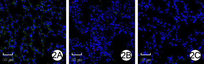
术后24 h, 3组小鼠肺组织中血管内皮细胞特异性标志物CD31均呈阳性表达。单纯CLP组小鼠肺组织中8-OHdG阳性细胞荧光强度较正常对照组显著升高, CLP+ADSC外泌体组小鼠肺组织中8-OHdG阳性细胞荧光强度较单纯CLP组明显下降。见图 2。
图 2.
术后24 h 3组脓毒症小鼠的肺组织中CD31和8-羟基脱氧鸟苷(8-OHdG)荧光共染检测其氧化性损伤情况 激光扫描共聚集显微镜×40, 图中标尺为50 μm。2A.正常对照组小鼠肺组织中CD31呈阳性表达, 8-OHdG阳性细胞少;2B.单纯CLP组小鼠肺组织中CD31呈阳性表达, 8-OHdG阳性细胞较图 2A明显增加;2C.CLP+脂肪间充质干细胞外泌体组小鼠肺组织中CD31呈阳性表达, 8-OHdG阳性细胞较图 2B明显减少
注:CLP为盲肠结扎穿孔;绿色荧光为CD31阳性表达, 红色荧光为8-OHdG阳性表达, 蓝色荧光为细胞核阳性表达
2.3. 人ADSC来源外泌体对PMVEC的影响
2.3.1. PMVEC的鉴定及其吞噬ADSC来源外泌体情况
第3代PMVEC呈鹅卵石样生长, 免疫荧光法检测显示细胞呈CD31阳性表达(图 3A), 流式细胞术检测结果显示CD31阳性细胞占比为99.5%(图 3B)。共培养12 h, ADSC来源外泌体成功被PMVEC吞噬, 进入胞质(图 3C)。
图 3.
第3代肺血管内皮细胞(PMVEC)鉴定及吞噬人脂肪间充质干细胞(ADSC)来源外泌体情况。3A.内皮细胞特异性标志物CD31呈阳性表达 FITC-4', 6-二脒基-2-苯基吲哚×40, 图中标尺为150 μm;3B.流式细胞术检测显示CD31阳性细胞占比为99.5%;3C.ADSC来源外泌体被PMVEC吞噬, 分布于细胞质中 PKH26-4', 6-二脒基-2-苯基吲哚×200, 图中标尺为100 μm
注:图 3A中绿色为CD31阳性表达, 蓝色为细胞核阳性表达;图 3B中FITC为异硫氰酸荧光素, 图中线段指平均荧光强度, P2为流式细胞仪设定的门, 即在FITC的P2门里阳性细胞占总细胞数的99.5%;图 3C中蓝色为细胞核阳性表达, 红色为外泌体阳性表达

2.3.2. PMVEC活性氧含量
第3代PMVEC培养24 h后, 与空白对照组比较, 单纯巨噬细胞上清液组PMVEC的活性氧荧光强度明显增加(P < 0.01);与单纯巨噬细胞上清液组比较, 巨噬细胞上清液+ADSC外泌体组PMVEC的活性氧荧光强度明显降低(P < 0.01), 见表 1。
表 1.
3组第3代肺血管内皮细胞培养24 h后活性氧含量和通透性比较(x ± s)
| 组别 | 样本数 | 活性氧 | 通透性(ng/mL) |
| 注:ADSC为脂肪间充质干细胞;活性氧含量以荧光强度反映, 通透性以牛血清白蛋白质量浓度反映;F值、P值为3组间各指标总体比较所得;t1值、P1值为单纯巨噬细胞上清液组与空白对照组各指标比较所得;t2值、P2值为巨噬细胞上清液+ADSC外泌体组与单纯巨噬细胞上清液组各指标比较所得 | |||
| 空白对照组 | 3 | 2 227±90 | 70±10 |
| 单纯巨噬细胞上清液组 | 3 | 3 650±128 | 193±32 |
| 巨噬细胞上清液+ADSC外泌体组 | 3 | 2 808±281 | 121±28 |
| F值 | 44.54 | 18.25 | |
| P值 | < 0.001 | < 0.005 | |
| t1值 | 15.73 | 6.34 | |
| P1值 | < 0.001 | 0.003 | |
| t2值 | 4.72 | 2.93 | |
| P2值 | 0.009 | 0.043 | |
2.3.3. PMVEC氧化性损伤情况
第3代PMVEC培养24 h, 与空白对照组相比, 单纯巨噬细胞上清液组PMVEC的8-OHdG阳性细胞荧光强度明显增强;巨噬细胞上清液+ADSC外泌体组PMVEC的8-OHdG阳性细胞荧光强度介于空白对照组和单纯巨噬细胞上清液组之间, 见图 4。
图 4.
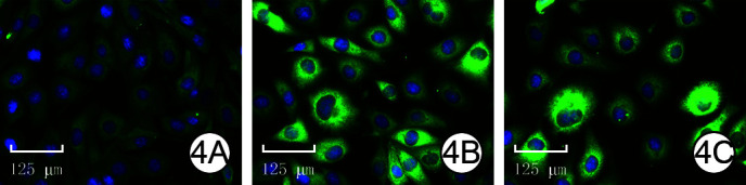
免疫荧光法检测第3代肺血管内皮细胞(PMVEC)的8-羟基脱氧鸟苷(8-OHdG)表达情况 Alexa Fluor-4', 6-二脒基-2-苯基吲哚×40, 图中标尺为125 μm。4A.空白对照组细胞8-OHdG低表达;4B.单纯巨噬细胞上清液组8-OHdG表达较图 4A明显增加;4C.巨噬细胞上清液+脂肪间充质干细胞外泌体组细胞8-OHdG表达介于图 4A与图 4B之间
2.3.4. PMVEC通透性情况
第3代PMVEC培养24 h, 与空白对照组比较, 单纯巨噬细胞上清液组的PMVEC单分子层细胞通透性明显增加(P < 0.01);与单纯巨噬细胞上清液组比较, 巨噬细胞上清液+ADSC外泌体组的PMVEC单分子层细胞通透性明显降低(P < 0.05), 见表 1。
3. 讨论
脓毒症是由感染引起的SIRS, 是严重创(烧)伤等危重患者常见的并发症。脓毒症时, 机体炎症反应及氧化应激是肺组织受损的重要原因[11, 29]。ALI时, 肺泡巨噬细胞首先被激活, 同时募集大量炎症细胞, 随之巨噬细胞产生大量的活性氧, 最终造成细胞和组织损伤, 甚至导致MODS。因此, 抑制炎症反应及减少细胞氧化应激损伤, 是减轻脓毒症MODS的重要治疗策略[29-30]。有研究报道, ADSC外泌体可以调控巨噬细胞, 减轻巨噬细胞引起的炎症反应进而发挥其保护作用[31-40]。ADSC来源的外泌体可以调控Notch信号通路, 减轻LPS诱导的炎症和脓毒症反应[41]。本研究观察到, ADSC来源的外泌体可以减轻CLP诱导的脓毒症小鼠血清中炎症因子的表达, 减轻肺组织炎症细胞浸润。
除巨噬细胞浸润外, PMVEC损伤在脓毒症肺损伤发病机制中也起关键作用。脓毒症发生时, 大量白细胞浸润肺组织, 局部过度炎症反应造成肺微血管病变, 促进内皮细胞活性氧累积。过量的活性氧破坏脂质、蛋白质和DNA, 并促进内皮细胞不可逆氧化应激损伤, 造成PMVEC凋亡和内皮屏障的破坏。内皮屏障破坏后会加剧炎症细胞浸润和活性氧累积, 进一步破坏肺组织结构。越来越多的研究表明, 间充质干细胞外泌体在多种疾病模型中可减轻细胞氧化应激损伤, 发挥保护作用。本课题组前期研究表明, ADSC来源外泌体可以通过调控巨噬细胞核因子E2相关因子/血红素加氧酶1轴, 减轻LPS诱导的巨噬细胞活性氧累积, 在脓毒症肺损伤中发挥保护作用。作为活性氧的另一主要来源, PMVEC在脓毒症肺损伤中同样可以产生大量氧化自由基, 破坏肺血管的完整性。本研究以PMVEC为靶细胞进行了相关的研究, 进一步阐明人ADSC来源外泌体减轻脓毒症诱导的ALI的分子机制。本研究通过CLP构建小鼠脓毒症模型, 进一步通过尾静脉注射人ADSC来源的外泌体来观察外泌体的保护作用。组织病理切片结果显示, 外泌体处理可以明显改善CLP诱导的肺脏结构破坏和炎症细胞浸润。8-OHdG和CD31免疫荧光染色显示ADSC来源外泌体处理后可以明显改善PMVEC氧化应激损伤。本研究通过经LPS刺激的巨噬细胞培养上清液刺激PMVEC, 模拟在体氧化应激损伤, 观察ADSC来源外泌体对PMVEC的保护作用, 结果显示ADSC来源外泌体可以明显减轻巨噬细胞培养上清诱导的活性氧累积和氧化应激损伤, 同时降低PMVEC通透性。
综上所述, CLP诱导脓毒症小鼠发生ALI时, 肺组织水肿明显, 炎症细胞浸润明显增加且细胞坏死增多, 炎症细胞浸润增加, 同时DNA氧化损伤的指标8-OHdG水平显著增加。人ADSC外泌体处理后, 脓毒症小鼠肺水肿症状明显减轻, 炎症细胞和红细胞浸润明显减少, 细胞坏死情况明显改善, 且炎症细胞浸润明显减轻、8-OHdG表达显著降低。体外培养的PMVEC经ADSC来源外泌体处理后, 细胞内活性氧含量以及8-OHdG表达显著降低, 内皮通透性明显减少, 从而达到减轻PMVEC的损伤及肺脏的保护作用。本研究为脓毒症诱导的ALI的防治提供了一条新的途径, 但在CLP引起的ALI过程中, 氧化性损伤以及活性氧的产生和清除、细胞通透性调控过程非常复杂, 其调控机制有待于进一步深入研究。
Funding Statement
国家自然科学基金青年科学基金项目(81601680)
Youth Science Foundation Project of National Natural Science Foundation of China (81601680)
Footnotes
利益冲突 所有作者均声明不存在利益冲突
作者贡献声明 蔡维霞:负责本研究酝酿和设计实验及文章撰写、整理和提交;沈括:负责本研究的脓毒症动物模型构建及数据整理和分析;曹涛:负责实验实施、采集数据;王璟:负责临床标本的收集和汇总及肺血管内皮细胞培养;赵明:负责脂肪间充质干细胞外泌体的提取和鉴定;王克甲:负责统计分析;张月:负责巨噬细胞培养;胡大海:提供研究思路、数据处理方法等研究过程中的指导工作;韩军涛:对文章的知识性内容作批评性审阅和支持性贡献;陶克:负责研究相关人员的工作分配及本研究的实验思路、数据处理方法等研究过程中的指导工作
References
- 1.Rudd KE, Johnson SC, Agesa KM, et al. Global, regional, and national sepsis incidence and mortality, 1990-2017: analysis for the Global Burden of Disease Study. Lancet. 2020;395(10219):200–211. doi: 10.1016/S0140-6736(19)32989-7. [DOI] [PMC free article] [PubMed] [Google Scholar]
- 2.Young P, Mackle D, Bellomo R, et al. Conservative oxygen therapy for mechanically ventilated adults with sepsis: a post hoc analysis of data from the intensive care unit randomized trial comparing two approaches to oxygen therapy (ICU-ROX) Intensive Care Med. 2020;46(1):17–26. doi: 10.1007/s00134-019-05857-x. [DOI] [PMC free article] [PubMed] [Google Scholar]
- 3.Stanski NL, Wong HR. Prognostic and predictive enrichment in sepsis. Nat Rev Nephrol. 2020;16(1):20–31. doi: 10.1038/s41581-019-0199-3. [DOI] [PMC free article] [PubMed] [Google Scholar]
- 4.Komorowski M. Clinical management of sepsis can be improved by artificial intelligence: yes. Intensive Care Med. 2020;46(2):375–377. doi: 10.1007/s00134-019-05898-2. [DOI] [PubMed] [Google Scholar]
- 5.黎 鳌. 黎鳌烧伤学. 上海: 上海科学技术出版社; 2001. [Google Scholar]
- 6.黄 巧冰. 内皮细胞屏障与烧伤后血管通透性的关系及机制. 中华烧伤杂志. 2007;23(5):324–326. doi: 10.3760/cma.j.issn.1009-2587.2007.05.002. [DOI] [PubMed] [Google Scholar]
- 7.Maniatis NA, Kotanidou A, Catravas JD, et al. Endothelial pathomechanisms in acute lung injury. Vascul Pharmacol. 2008;49(4/6):119–133. doi: 10.1016/j.vph.2008.06.009. [DOI] [PMC free article] [PubMed] [Google Scholar]
- 8.Delano MJ, Ward PA. Sepsis-induced immune dysfunction: can immune therapies reduce mortality? J Clin Invest. 2016;126(1):23–31. doi: 10.1172/JCI82224. [DOI] [PMC free article] [PubMed] [Google Scholar]
- 9.Douglas SD. Monocyte and macrophage reactions. Environ Health Perspect. 1974;9:281–282. doi: 10.1289/ehp.749281. [DOI] [PMC free article] [PubMed] [Google Scholar]
- 10.邹 宪宝, 李 新宇. 脓毒症致肺血管内皮细胞损伤机制的研究进展. 现代生物医学进展. 2009;9(22):4384–4386. [Google Scholar]
- 11.吴 丽丽, 梁 群, 曹 雪丹. 中医药保护脓毒症肺血管内皮细胞损伤的机制研究进展. 中国中医急症. 2021;30(1):172–175. doi: 10.3969/j.issn.1004-745X.2021.01.050. [DOI] [Google Scholar]
- 12.柴 云飞, 雷 黎明, 李 鹏, et al. 血管内皮细胞损伤与脓毒症休克的相关性. 实用医学杂志. 2017;33(15):2529–2532. doi: 10.3969/j.issn.1006-5725.2017.15.027. [DOI] [Google Scholar]
- 13.Pu CM, Liu CW, Liang CJ, et al. Adipose-derived stem cells protect skin flaps against ischemia/reperfusion injury via IL-6 expression. J Invest Dermatol. 2017;137(6):1353–1362. doi: 10.1016/j.jid.2016.12.030. [DOI] [PubMed] [Google Scholar]
- 14.Joffre J, Hellman J, Ince C, et al. Endothelial responses in sepsis. Am J Respir Crit Care Med. 2020;202(3):361–370. doi: 10.1164/rccm.201910-1911TR. [DOI] [PubMed] [Google Scholar]
- 15.Colbert JF, Schmidt EP. Endothelial and microcirculatory function and dysfunction in sepsis. Clin Chest Med. 2016;37(2):263–275. doi: 10.1016/j.ccm.2016.01.009. [DOI] [PMC free article] [PubMed] [Google Scholar]
- 16.Hamidzadeh K, Christensen SM, Dalby E, et al. Macrophages and the recovery from acute and chronic inflammation. Annu Rev Physiol. 2017;79:567–592. doi: 10.1146/annurev-physiol-022516-034348. [DOI] [PMC free article] [PubMed] [Google Scholar]
- 17.Yang C, Luo L, Bai X, et al. Highly-expressed micoRNA-21 in adipose derived stem cell exosomes can enhance the migration and proliferation of the HaCaT cells by increasing the MMP-9 expression through the PI3K/AKT pathway. Arch Biochem Biophys. 2020;681:108259. doi: 10.1016/j.abb.2020.108259. [DOI] [PubMed] [Google Scholar]
- 18.Zhang W, Bai X, Zhao B, et al. Cell-free therapy based on adipose tissue stem cell-derived exosomes promotes wound healing via the PI3K/Akt signaling pathway. Exp Cell Res. 2018;370(2):333–342. doi: 10.1016/j.yexcr.2018.06.035. [DOI] [PubMed] [Google Scholar]
- 19.Wei P, Zhong C, Yang X, et al. Exosomes derived from human amniotic epithelial cells accelerate diabetic wound healing via PI3K-AKT-mTOR-mediated promotion in angiogenesis and fibroblast function[J/OL]. Burns Trauma, 2020, 8: tkaa020[2022-03-01]. https://pubmed.ncbi.nlm.nih.gov/32923490/. DOI: 10.1093/burnst/tkaa020.
- 20.Hu P, Yang Q, Wang Q, et al. Mesenchymal stromal cells-exosomes: a promising cell-free therapeutic tool for wound healing and cutaneous regeneration[J/OL]. Burns Trauma, 2019, 7: 38[2022-03-02]. https://pubmed.ncbi.nlm.nih.gov/31890717/. DOI: 10.1186/s41038-019-0178-8.
- 21.Hwang I, Jo K, Shin KC, et al. GABA-stimulated adipose-derived stem cells suppress subcutaneous adipose inflammation in obesity. Proc Natl Acad Sci U S A. 2019;116(24):11936–11945. doi: 10.1073/pnas.1822067116. [DOI] [PMC free article] [PubMed] [Google Scholar]
- 22.Domingues CC, Kundu N, Kropotova Y, et al. Antioxidant-upregulated mesenchymal stem cells reduce inflammation and improve fatty liver disease in diet-induced obesity. Stem Cell Res Ther. 2019;10(1):280. doi: 10.1186/s13287-019-1393-8. [DOI] [PMC free article] [PubMed] [Google Scholar]
- 23.Xie J, Jones TJ, Feng D, et al. Human adipose-derived stem cells suppress elastase-induced murine abdominal aortic inflammation and aneurysm expansion through paracrine factors. Cell Transplant. 2017;26(2):173–189. doi: 10.3727/096368916X692212. [DOI] [PMC free article] [PubMed] [Google Scholar]
- 24.Tian M, Ticer T, Wang Q, et al. Adipose-derived biogenic nanoparticles for suppression of inflammation. Small. 2020;16(10):e1904064. doi: 10.1002/smll.201904064. [DOI] [PubMed] [Google Scholar]
- 25.Hong P, Yang H, Wu Y, et al. The functions and clinical application potential of exosomes derived from adipose mesenchymal stem cells: a comprehensive review. Stem Cell Res Ther. 2019;10(1):242. doi: 10.1186/s13287-019-1358-y. [DOI] [PMC free article] [PubMed] [Google Scholar]
- 26.沈 括, 王 许杰, 刘 开拓, et al. 人脂肪间充质干细胞外泌体对小鼠RAW264.7细胞的炎症反应和小鼠全层皮肤缺损创面愈合的影响. 中华烧伤与创面修复杂志. 2022;38(3):215–226. doi: 10.3760/cma.j.cn501120-20201116-00477. [DOI] [PMC free article] [PubMed] [Google Scholar]
- 27.Rittirsch D, Huber-Lang MS, Flierl MA, et al. Immunodesign of experimental sepsis by cecal ligation and puncture. Nat Protoc. 2009;4(1):31–36. doi: 10.1038/nprot.2008.214. [DOI] [PMC free article] [PubMed] [Google Scholar]
- 28.胡 国栋, 陈 英华, 刘 爱华, et al. 一种改良的肺微血管内皮细胞培养方法. 南方医科大学学报. 2012;32(8):1151–1153. doi: 10.3969/j.issn.1673-4254.2012.08.17. [DOI] [PubMed] [Google Scholar]
- 29.Qiu P, Liu Y, Zhang J. Review: the role and mechanisms of macrophage autophagy in sepsis. Inflammation. 2019;42(1):6–19. doi: 10.1007/s10753-018-0890-8. [DOI] [PubMed] [Google Scholar]
- 30.Venet F, Monneret G. Advances in the understanding and treatment of sepsis-induced immunosuppression. Nat Rev Nephrol. 2018;14(2):121–137. doi: 10.1038/nrneph.2017.165. [DOI] [PubMed] [Google Scholar]
- 31.Gotts JE, Matthay MA. Sepsis: pathophysiology and clinical management. BMJ. 2016;353:i1585. doi: 10.1136/bmj.i1585. [DOI] [PubMed] [Google Scholar]
- 32.Marshall JC. Sepsis definitions: a work in progress. Crit Care Clin. 2018;34(1):1–14. doi: 10.1016/j.ccc.2017.08.004. [DOI] [PubMed] [Google Scholar]
- 33.Feehan KT, Gilroy DW. Is resolution the end of inflammation? Trends Mol Med. 2019;25(3):198–214. doi: 10.1016/j.molmed.2019.01.006. [DOI] [PubMed] [Google Scholar]
- 34.Joo HJ, Kim JH, Hong SJ. Adipose tissue-derived stem cells for myocardial regeneration. Korean Circ J. 2017;47(2):151–159. doi: 10.4070/kcj.2016.0207. [DOI] [PMC free article] [PubMed] [Google Scholar]
- 35.Glass GE, Ferretti P. Adipose-derived stem cells in aesthetic surgery. Aesthet Surg J. 2019;39(4):423–438. doi: 10.1093/asj/sjy160. [DOI] [PubMed] [Google Scholar]
- 36.Moreno-Manzano V, Mellado-López M, Morera-Esteve MJ, et al. Human adipose-derived mesenchymal stem cells accelerate decellularized neobladder regeneration. Regen Biomater. 2020;7(2):161–169. doi: 10.1093/rb/rbz049. [DOI] [PMC free article] [PubMed] [Google Scholar]
- 37.Bandeira F, Goh TW, Setiawan M, et al. Cellular therapy of corneal epithelial defect by adipose mesenchymal stem cell-derived epithelial progenitors. Stem Cell Res Ther. 2020;11(1):14. doi: 10.1186/s13287-019-1533-1. [DOI] [PMC free article] [PubMed] [Google Scholar]
- 38.Guo J, Hu H, Gorecka J, et al. Adipose-derived mesenchymal stem cells accelerate diabetic wound healing in a similar fashion as bone marrow-derived cells. Am J Physiol Cell Physiol. 2018;315(6):C885–C896. doi: 10.1152/ajpcell.00120.2018. [DOI] [PMC free article] [PubMed] [Google Scholar]
- 39.Lai TC, Lee TL, Chang YC, et al. MicroRNA-221/222 mediates ADSC-exosome-induced cardioprotection against ischemia/reperfusion by targeting PUMA and ETS-1. Front Cell Dev Biol. 2020;8:569150. doi: 10.3389/fcell.2020.569150. [DOI] [PMC free article] [PubMed] [Google Scholar]
- 40.Shen K, Jia Y, Wang X, et al. Exosomes from adipose-derived stem cells alleviate the inflammation and oxidative stress via regulating Nrf2/HO-1 axis in macrophages. Free Radic Biol Med. 2021;165:54–66. doi: 10.1016/j.freeradbiomed.2021.01.023. [DOI] [PubMed] [Google Scholar]
- 41.Shi Y, Wang Y, Li Q, et al. Immunoregulatory mechanisms of mesenchymal stem and stromal cells in inflammatory diseases. Nat Rev Nephrol. 2018;14(8):493–507. doi: 10.1038/s41581-018-0023-5. [DOI] [PubMed] [Google Scholar]