



Abstract
α-Synuclein (α-syn) is a presynaptic protein that is widely implicated in the pathophysiology of Parkinson’s disease, a neurodegenerative disorder characterized by a progressive loss of dopaminergic neurons and the formation of Lewy bodies. Evidence suggests that α-syn could be imported into the nucleus and subsequently disrupt normal neuronal function. The existence of α-syn in the nucleus provides the possibility of interaction with DNA leading to gene transcript regulation. Thus, CD spectra were used to determine the specific DNA sequence with which α-syn is most likely to interact. Our results indicated that α-syn was prone to preferentially interact with the GC-box-like sequence in vitro at a ratio of 2:1 or less (α-syn: the GC-box-like sequence).
Keywords: α-Synuclein, GC-box-like sequence, Circular dichroism, DNA
Introduction
Parkinson’s disease (PD) is the most common movement neurodegenerative disorder and is characterized by a loss of dopaminergic neurons in the substantia nigra pars compacta. Pathological analysis of PD brain tissue reveals intracytoplasmic, perikaryal inclusions, known as Lewy bodies (LBs), as well as similar inclusions in neuronal processes, termed Lewy neuritis (Goedert 2001; von Bohlen Und Halbach 2004; Lee and Trojanowski 2006). The major component of LBs and Lewy neuritis is α-synuclein (α-syn) (Goedert 2001; von Bohlen Und Halbach 2004). As its name implies, α-syn was initially localized to the presynaptic nerve terminals and nucleus (Maroteaux et al. 1988). Subsequent studies indicated that the nuclear localization of this protein had been noted in a variety of experimental systems, including α-syn transgenic Drosophila (Takahashi et al. 2003), mice (Masliah et al. 2000; Goers et al. 2003), and cultured cells (McLean et al. 2000; Specht et al. 2005).
It has been reported that nucleic acids can interact with different amyloid peptides (β-amyloid, prion peptides) and modulate their aggregation (Ahn et al. 2000; Kampers et al. 1996; Nandi et al. 2002; Nandi 1998; Cordeiro et al. 2001; Gabus et al. 2001). These interactions induce structural changes (from an α-helical form into a β-sheet isoform) in prion peptides by forming stable complexes, which can catalyze and modulate further polymerization (Nandi et al. 2002; Cordeiro et al. 2001). Dmitry et al. have reported that double-stranded DNA (dsDNA), either linear or supercoiled, can interact with wild-type α-syn, leading to a significant stimulation of α-syn assembly into mature fibrils characterized by a reduced lag phase (Cherny et al. 2004). Hegde’s results also demonstrated that double-stranded oligonucleotides induced partial folding of α-syn and promoted its aggregation (Hegde et al. 2010). Therefore, DNA can induce the aggregation of α-syn in some pathological conditions, but can α-syn affect the DNA in turn?
Recent studies have shown that α-syn possesses DNA binding properties (Padmaraju et al. 2011). Subsequent studies from Eirene and Boyer have indicated that α-syn protein in the nucleus could disturb the normal functions of neurons. (Kontopoulos et al. 2006; Boyer and Dreyer 2007). Our previous work also demonstrated that α-syn could accumulate in the nucleus and promote neurotoxicity by accelerating the cell cycle (Ma et al. 2013). Thus, the existence of α-syn in the nucleus allows the regulation of transcription or other functions associated with gene expression. However, it was unknown which specific DNA sequence α-syn preferentially interact with.
In this study, a series of DNA sequences were synthesized, and the circular dichroism (CD) spectra were obtained to investigate the specific DNA sequence with which α-syn is most likely to interact. Our results indicated that α-syn favored the GC-box-like sequence in vitro at a ratio of 2:1 or less (α-syn:the GC-box-like sequence).
Material and Method
Single-Stranded DNA Synthesis
A series of DNA sequences (Table 1) were synthesized and dissolved in CD buffer (0.2 M KC1, 10 % glycerol, 10 mM Tris (pH 8.0) with <0.05 mM of EDTA and DTT). The final concentration was 100 μM.
Table 1.
The DNA sequence
| Gene | Sequence |
|---|---|
| A30 | aaaaaaaaaaaaaaaaaaaaaaaaaaaaaa |
| T30 | tttttttttttttttttttttttttttttt |
| C30 | cccccccccccccccccccccccccccccc |
| AT10 | atatatatatatatatatat |
| AC10 | acacacacacacacacacac |
| AG10 | agagagagagagagagagag |
| GC10 | gcgcgcgcgcgcgcgcgcgc |
| GT10 | gtgtgtgtgtgtgtgtgtgt |
| TC10 | tctctctctctctctctctc |
| GC box like | gggcgggggcgggggcgggggcgggggcgg |
| CAAT box like | gccaatgccaatgccaatgccaatgccaat |
Protein Expression in Pichia Pastoris
The α-syn-GFP gene was cloned into pPIC9 K expression vectors and sequenced. Then, 5–10 μg of the constructed plasmid pPIC9 K-α-syn-GFP was linearized by SacI digestion and transformed into Pichia pastoris. The cells were spread on YPD agar plates (1 % BactoTM yeast extract, 2 % BactoTM peptone, 1.5 % BactoTM agar, and 2 % dextrose) containing 4 mg/ml of G418 and incubated at 30 °C for 3–4 days. Six colonies were randomly picked and cultivated in shake flasks (scaled up from the above described small tube expression) for downstream purification; 100 units/ml of penicillin and 100 μg/ml of streptomycin were added to suppress bacterial contamination. The supernatant was clarified by centrifugation at 3,000 rpm at 4 °C for 10 min prior to protein purification.
Protein Purification
Anion exchange chromatography was used for protein purification. The column was equilibrated with 50 mM Tris–Cl, pH 7.5 (10 CV). The above dialyzed sample was loaded onto the column followed by washing with 50 mM Tris–Cl, pH 7.5 (8 CV). The bound protein was eluted with 100 and 200 mM sodium chloride in 50 mM Tris–Cl, pH 7.5, into eight fractions. The purification fractions were analyzed using 12 % NuPAGE SDS gel. Protein concentrations were determined using the Pierce BCA protein assay kit (Thermo Scientific).
Western Blot Analysis
α-Syn-GFP protein was solubilized in SDS sample buffer, separated by SDS-PAGE gel, and then transferred to a PVDF membrane (Millipore). The membrane was blocked with 3 % BSA (Sigma) and incubated with the primary antibody anti-alpha-synuclein (Santa Cruz), followed by horseradish peroxidase (HRP)-conjugated secondary antibody, and detected with the Enhanced Chemiluminescence (ECL) plus detection system (Molecular Device, Lmax).
Circular Dichroism Spectra
For CD spectroscopy, near UV (250–350 nm) CD spectra were recorded using a Jasco J600 spectropolarimeter purged with nitrogen gas. Near UV CD spectra were recorded in 1-cm path length cells. All spectra were recorded in 50 mM Tris-HCI, 5 mM MgCl2, 100 mM NaCI, pH 8.0, and corrected by subtraction of the buffer spectrum. Far UV CD spectra were recorded for α-syn and for 1:1, 1:2, 1:3, and 1:4 complexes with DNA at concentrations of 5, 10, 15, and 20 μM. Spectra of α-syn were recorded at equivalent concentrations and subtracted to obtain the CD spectrum of the bound DNA.
Results
Expression and Purification of the α-Syn-GFP Protein
The purity of the α-syn-GFP protein using anion exchange chromatography is shown in Fig. 1a. The main bands of each lane have the same molecular weights (45 kD) and represent the α-syn-GFP protein. The final concentration of the purified α-syn-GFP protein is 800 μg/ml. Further WB results (Fig. 1b) showed that the commercial α-syn antibody could recognize and bind a protein with the same molecular weight as α-syn-GFP.
Fig. 1.
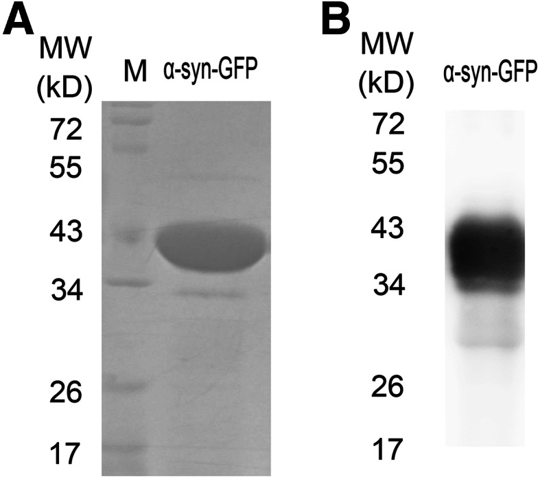
The expression and purification of α-syn-GFP protein. a The separated α-syn-GFP protein using anion exchange chromatography was subjected to SDS-PAGE gel electrophoresis and visualized via staining with Bromophenol Blue. Lane 1 markers (in kDa from the highest to lowest): 72, 55, 43, 34, 26, and 17. Lane 2 α-syn-GFP protein. b Western blot analysis for α-syn-GFP was performed. A mouse monoclonal antibody specific for α-syn (Santa Cruz) was used at a dilution of 1:500
α-Syn-GFP Protein is Prone to Interaction with the GC-Box-Like Sequence
CD spectra are highly sensitive to the conformation and environment of optically active chromophores and are a valuable probe of secondary structure in proteins and DNA. Considering the localization of α-syn protein in the nucleus, we have employed CD spectra to look for the specific DNA sequence with which α-syn is likely to interact. Among a series of synthesized DNA sequences which were designed using different combinations of the four types of bases, DNA rich in the bases AG or TC were found to be the DNA with which α-syn-GFP protein is most likely to interact (Fig. 2a). Thus, both the CAAT-box-like sequence and the GC-box-like sequence were further synthesized based on this result, and the CAAT-box-like sequence showed no interaction with the α-syn-GFP protein. Finally, the GC-box-like sequence was found to be the sequence that α-syn-GFP protein preferentially interact with (Fig. 2b).
Fig. 2.

Circular dichroism spectra show α-syn binding. a The binding assay between the purified α-syn-GFP protein and the DNA sequences rich in bases AG or TC. b The binding assay between α-syn and the CCAT-box-like sequence or GC-box-like sequence. Four scans were taken and averaged from 410 to 230 nm, and the data were collected every 0.5 nm. Representative spectra are shown, corresponding to protein: DNA ratios of 1:1. These experiments were carried out in 50 mM Tris-HCI, 5 mM MgCl2, 100 mM NaCI, pH 8.0 at 20 °C in a 4-mm path length cell
Quantitative Analysis of the Ratio of α-Syn-GFP Protein Interaction with the GC-Box-Like Sequence
To explore the ratio of α-syn-GFP protein interaction with the GC-box-like sequence, mixtures with differing ratios of α-syn-GFP protein and GC-box-like sequence were prepared. The status of α-syn-GFP protein interactions with the GC-box-like sequence is reflected by the difference in peak intensity at wavelengths from 250 to 310 nm. As shown in Fig. 3a, the peak intensity at 270 nm gradually increased with the increasing α-syn-GFP protein concentrations but stopped when the concentration of the α-syn-GFP protein reached 10 μM (α-syn-GFP:GC-box-like sequence, 2:1). The peak intensity at 250–310 nm was identical for the following sequence of ratios of α-syn-GFP protein and the GC-box-like sequence: 2:1, 3:1, and 4:1. Thus, the ratio of α-syn-GFP protein interaction with the GC-box-like sequence obtained in our study is 2:1 or less.
Fig. 3.

Circular dichroism spectra showing α-syn binding. a Result from the binding assay between the purified α-syn-GFP protein and the GC-box-like DNA sequence. b The binding assay results between the commercial α-syn protein and the GC-box-like DNA sequence. c The binding assay results between the commercial GFP protein and the GC-box-like DNA sequence. Four scans were taken and averaged from 410 to 230 nm, and the data were collected every 0.5 nm. Representative spectra are shown, corresponding to protein: DNA ratios of a 0.5:1, 1:1, 2:1, 3:1, and 4:1; b 1:1, 2:1, 3:1, and 4:1; and c 1:1, 2:1, 3:1, and 4:1. These experiments were carried out in 50 mM Tris-HCI, 5 mM MgCl2, 100 mM NaCI, pH 8.0 at 20 °C in a 4-mm path length cell
Quantitative Analysis of the Ratio of α-Syn Protein Interaction with the GC-Box-Like Sequence
The commercial α-syn protein without fusion GFP was purchased from Sigma and used in the CD spectra assays to verify the ratio of α-syn protein interaction with the GC-box-like sequence. The commercial GFP protein was also purchased from Abcam and used in the CD spectra assay to eliminate the possible effects of GFP protein itself on the α-syn protein. As shown in Fig. 3b, the commercial α-syn protein interacted with the GC-box-like sequence with affinity similar to that of α-syn-GFP protein. Additionally, the acting ratio assay at the peak intensity from 250 nm to 310 nm remained unchanged up to an α-syn protein concentration of 10 μM (α-syn:GC-box-like sequence, 2:1). The similar results were found in Fig. 3c, with the increasing of GFP protein concentration; the CD spectra at the peak intensity from 250 to 310 nm only showed slight changes. Thus, these results indicate that regardless of GFP, α-syn per se was prone to interaction with the GC-box-like sequence at a ratio of 2:1 or less.
Discussion
α-syn is a presynaptic protein widely implicated in the pathophysiology of PD (Spillantini et al. 1997; Dawson and Dawson 2003; Recchia et al. 2004) and has been reported to enter the nucleus and interfere with normal neuronal function (Kontopoulos et al. 2006; Boyer and Dreyer 2007). The existence of α-syn in the nucleus allows for α-syn to interact with DNA leading to regulation of gene expression. Thus, we employed CD spectra analysis to determine with which DNA sequence α-syn is most likely to interact. Our results indicated that α-syn preferentially interacted with the GC-box-like sequence in vitro at a ratio of 2:1 or less (α-syn:GC-box-like sequence).
Circular dichroism (CD) has found widespread use for predicting the secondary structure of proteins using a number of modeling techniques (Compton and Johnson 1986; Johnson 1990; Lees et al. 2006). DNA is polymorphic in nature and exists in different conformations in cells under physiological conditions (Rich 1993). Recent research has sought to correlate the structural parameters of DNA with their CD spectrum (Johnson et al. 1981; Basham et al. 1995; Scarlett et al. 2004), and some success in assigning quaternary structures to nucleic acids has been demonstrated (e.g., in the case of triplex DNA and G-quartet mediated structures) (Gray et al. 1995; Hardin et al. 1991). The overlap of the UV absorption bands of nucleic acids and proteins complicates the CD studies of protein–DNA interactions because the contributions from both components are observed. In practice, CD spectra between 250 and 300 nm are dominated by nucleic acids, as the contribution arising from the aromatic chromophores of proteins are weak in comparison and generally fewer in number (Cary and Kneale 2009). Thus, the peak intensity at 250–300 nm was chosen as the main outcome measure in this study.
To discover additional DNA sequences which the α-syn protein may interact with different combinations of DNA with four types of bases were synthesized and employed in our CD assay. It was found that the α-syn protein preferentially interacted with the DNA rich in the bases AG or TC. The CAAT-box-like sequence was synthesized based on this result. However, our results were disappointing in that the CD spectra peak remained unchanged when the CAAT-box-like sequence was added. Only the addition of the GC-box-like sequence caused a significant change in the CD spectra. Thus, the GC-box-like sequence demonstrated to be the sequence of α-syn protein preferential interaction with. We then examined the ratio of α-syn protein interaction with GC-box-like sequence and demonstrated an acting ratio of 2:1 or less (α-syn protein:GC-box-like sequence, 2:1).
Considering the α-syn-GFP fusion expression vectors used in our previous study, α-syn-GFP fusion protein was expressed in Pichia pastoris, purified by anion exchange chromatography, and used in the present study. To exclude the possible confounding effect of GFP protein, we acquired both the commercial α-syn protein without fusion GFP from Sigma and the commercial GFP protein, which we employed in parallel. GFP protein showed no effect; instead, α-syn demonstrated preferential interaction with the GC-box-like sequence at a ratio of 2:1 or less.
In summary, our results indicate that α-syn preferentially interacts with the GC-box-like sequence in vitro at a ratio of 2:1 or less (α-syn: GC-box-like sequence).
Acknowledgments
This work was supported by the National Natural Science Foundation of China Grants (No. 81274122, 81301073, 81073078,30973887, 31100127, Key Program No. U832008), Special Purpose for New Drug Development (No. 2012ZX09301002-004), Studies on Structure and function of Bioactive Substances from the Natural Medicines (No. IRT1007), the Specialized Research Fund for the Doctoral Program of Higher Education of China (No. 20121106120056), Yunnan Natural Science Foundation (No.2013FZ132; No.2011FB116), Beijing Key Laboratory of New Drug Mechanisms and Pharmacological Evaluation Study (No. BZ0150), the PUMC Youth Fund and the Fundamental Research Funds for the Central Universities (No. 3332013082), and Graduate Innovation Fund 2012 of Peking Union Medical College (No. 2012-1007-003).
Conflict of interest
The authors have declared that no competing interests exist.
Footnotes
Kai-Li Ma and Lian-Kun Song have contributed equally to this work.
References
- Ahn BW, Song DU, Jung YD, Chay KO, Chung MA, Yang SY, Shin BA (2000) Detection of beta-amyloid peptide aggregation using DNA electrophoresis. Anal Biochem 284(2):401–405 [DOI] [PubMed] [Google Scholar]
- Basham B, Schroth GP, Ho PS (1995) An A-DNA triplet code: thermodynamic rules for predicting A- and B-DNA. Proc Natl Acad Sci USA 92(14):6464–6468 [DOI] [PMC free article] [PubMed] [Google Scholar]
- Boyer F, Dreyer JL (2007) Alpha-synuclein in the nucleus accumbens induces changes in cocaine behaviour in rats. Eur J Neurosci 26(10):2764–2776 [DOI] [PubMed] [Google Scholar]
- Cary PD, Kneale GG (2009) Circular dichroism for the analysis of protein-DNA interactions. Methods Mol Biol 543:613–624 [DOI] [PubMed] [Google Scholar]
- Cherny D, Hoyer W, Subramaniam V, Jovin TM (2004) Double-stranded DNA stimulates the fibrillation of alpha-synuclein in vitro and is associated with the mature fibrils: an electron microscopy study. J Mol Biol 344(4):929–938 [DOI] [PubMed] [Google Scholar]
- Compton LA, Johnson WC Jr (1986) Analysis of protein circular dichroism spectra for secondary structure using a simple matrix multiplication. Anal Biochem 155(1):155–167 [DOI] [PubMed] [Google Scholar]
- Cordeiro Y, Machado F, Juliano L, Juliano MA, Brentani RR, Foguel D, Silva JL (2001) DNA converts cellular prion protein into the beta-sheet conformation and inhibits prion peptide aggregation. J Biol Chem 276(52):49400–49409 [DOI] [PubMed] [Google Scholar]
- Dawson TM, Dawson VL (2003) Molecular pathways of neurodegeneration in Parkinson’s disease. Science 302(5646):819–822 [DOI] [PubMed] [Google Scholar]
- Gabus C, Derrington E, Leblanc P, Chnaiderman J, Dormont D, Swietnicki W, Morillas M, Surewicz WK, Marc D, Nandi P, Darlix JL (2001) The prion protein has RNA binding and chaperoning properties characteristic of nucleocapsid protein NCP7 of HIV-1. J Biol Chem 276(22):19301–19309 [DOI] [PubMed] [Google Scholar]
- Goedert M (2001) Alpha-synuclein and neurodegenerative diseases. Nat Rev Neurosci 2(7):492–501 [DOI] [PubMed] [Google Scholar]
- Goers J, Manning-Bog AB, McCormack AL, Millett IS, Doniach S, Di Monte DA, Uversky VN, Fink AL (2003) Nuclear localization of alpha-synuclein and its interaction with histones. Biochemistry 42(28):8465–8471 [DOI] [PubMed] [Google Scholar]
- Gray DM, Hung SH, Johnson KH (1995) Absorption and circular dichroism spectroscopy of nucleic acid duplexes and triplexes. Methods Enzymol 246:19–34 [DOI] [PubMed] [Google Scholar]
- Hardin CC, Henderson E, Watson T, Prosser JK (1991) Monovalent cation induced structural transitions in telomeric DNAs: G-DNA folding intermediates. Biochemistry 30(18):4460–4472 [DOI] [PubMed] [Google Scholar]
- Hegde ML, Vasudevaraju P, Rao KJ (2010) DNA induced folding/fibrillation of alpha-synuclein: new insights in Parkinson’s disease. Front Biosci 15:418–436 [DOI] [PubMed] [Google Scholar]
- Johnson WC Jr (1990) Protein secondary structure and circular dichroism: a practical guide. Proteins 7(3):205–214 [DOI] [PubMed] [Google Scholar]
- Johnson BB, Dahl KS, Tinoco I Jr, Ivanov VI, Zhurkin VB (1981) Correlations between deoxyribonucleic acid structural parameters and calculated circular dichroism spectra. Biochemistry 20(1):73–78 [DOI] [PubMed] [Google Scholar]
- Kampers T, Friedhoff P, Biernat J, Mandelkow EM, Mandelkow E (1996) RNA stimulates aggregation of microtubule-associated protein tau into Alzheimer-like paired helical filaments. FEBS Lett 399(3):344–349 [DOI] [PubMed] [Google Scholar]
- Kontopoulos E, Parvin JD, Feany MB (2006) Alpha-synuclein acts in the nucleus to inhibit histone acetylation and promote neurotoxicity. Hum Mol Genet 15(20):3012–3023 [DOI] [PubMed] [Google Scholar]
- Lee VM, Trojanowski JQ (2006) Mechanisms of Parkinson’s disease linked to pathological alpha-synuclein: new targets for drug discovery. Neuron 52(1):33–38 [DOI] [PubMed] [Google Scholar]
- Lees JG, Miles AJ, Wien F, Wallace BA (2006) A reference database for circular dichroism spectroscopy covering fold and secondary structure space. Bioinformatics 22(16):1955–1962 [DOI] [PubMed] [Google Scholar]
- Ma KL, Song LK, Yuan YH, Zhang Y, Han N, Gao K, Chen NH (2013) The nuclear accumulation of alpha-synuclein is mediated by importin alpha and promotes neurotoxicity by accelerating the cell cycle. Neuropharmacology. doi:10.1016/j.neuropharm.2013.07.035 [DOI] [PubMed]
- Maroteaux L, Campanelli JT, Scheller RH (1988) Synuclein: a neuron-specific protein localized to the nucleus and presynaptic nerve terminal. J Neurosci 8(8):2804–2815 [DOI] [PMC free article] [PubMed] [Google Scholar]
- Masliah E, Rockenstein E, Veinbergs I, Mallory M, Hashimoto M, Takeda A, Sagara Y, Sisk A, Mucke L (2000) Dopaminergic loss and inclusion body formation in alpha-synuclein mice: implications for neurodegenerative disorders. Science 287(5456):1265–1269 [DOI] [PubMed] [Google Scholar]
- McLean PJ, Ribich S, Hyman BT (2000) Subcellular localization of alpha-synuclein in primary neuronal cultures: effect of missense mutations. J Neural Transm Suppl 58:53–63 [DOI] [PubMed] [Google Scholar]
- Nandi PK (1998) Polymerization of human prion peptide HuPrP 106-126 to amyloid in nucleic acid solution. Arch Virol 143(7):1251–1263 [DOI] [PubMed] [Google Scholar]
- Nandi PK, Leclerc E, Nicole JC, Takahashi M (2002) DNA-induced partial unfolding of prion protein leads to its polymerisation to amyloid. J Mol Biol 322(1):153–161 [DOI] [PubMed] [Google Scholar]
- Padmaraju V, Bhaskar JJ, Prasada Rao UJ, Salimath PV, Rao KS (2011) Role of advanced glycation on aggregation and DNA binding properties of alpha-synuclein. J Alzheimers Dis 24(Suppl 2):211–221 [DOI] [PubMed] [Google Scholar]
- Recchia A, Debetto P, Negro A, Guidolin D, Skaper SD, Giusti P (2004) Alpha-synuclein and Parkinson’s disease. Faseb J 18(6):617–626 [DOI] [PubMed] [Google Scholar]
- Rich A (1993) DNA comes in many forms. Gene 135(1–2):99–109 [DOI] [PubMed] [Google Scholar]
- Scarlett GP, Elgar SJ, Cary PD, Noble AM, Orford RL, Kneale GG, Guille MJ (2004) Intact RNA-binding domains are necessary for structure-specific DNA binding and transcription control by CBTF122 during Xenopus development. J Biol Chem 279(50):52447–52455 [DOI] [PubMed] [Google Scholar]
- Specht CG, Tigaret CM, Rast GF, Thalhammer A, Rudhard Y, Schoepfer R (2005) Subcellular localisation of recombinant alpha- and gamma-synuclein. Mol Cell Neurosci 28(2):326–334 [DOI] [PubMed] [Google Scholar]
- Spillantini MG, Schmidt ML, Lee VM, Trojanowski JQ, Jakes R, Goedert M (1997) Alpha-synuclein in Lewy bodies. Nature 388(6645):839–840 [DOI] [PubMed] [Google Scholar]
- Takahashi M, Kanuka H, Fujiwara H, Koyama A, Hasegawa M, Miura M, Iwatsubo T (2003) Phosphorylation of alpha-synuclein characteristic of synucleinopathy lesions is recapitulated in alpha-synuclein transgenic Drosophila. Neurosci Lett 336(3):155–158 [DOI] [PubMed] [Google Scholar]
- von Bohlen Und Halbach O (2004) Synucleins and their relationship to Parkinson’s disease. Cell Tissue Res 318(1):163–174 [DOI] [PubMed] [Google Scholar]