



Abstract
The chemokine receptor CXCR4 has been reported to be aberrantly expressed in human cancers and has also been shown to participate in the development of cancer metastasis. The present study was carried out to assess immunohistochemically the pattern of CXCR4 expression in patients with metastatic prostate cancer. We analyzed whether there may be an association between CXCR4 expression and prognosis. Fifty‐two patients who received hormonal therapy were enrolled. Specimens were obtained from transperineal needle biopsy before treatment, and were stained with antihuman CXCR4 antibody. We also evaluated the pathological grade, extent of bony metastasis, clinical response to hormonal therapy, and patient prognosis. CXCR4 was detected in 94.2% patients. Its expression showed no association with pathological grade, extent of bony metastasis, or clinical response to hormonal therapy. Patients with a high expression of CXCR4 in tumors had poorer cancer‐specific survival than those with low expression of CXCR4. CXCR4 expression is a useful prognostic factor for patients with metastatic prostate cancer treated with androgen‐withdrawal therapy. (Cancer Sci 2008; 99: 539–542)
Most prostate cancer‐related deaths are not the result of primary tumor growth but, rather, are caused by the spread of the cancer to other organs. Approximately 80% of patients with untreated prostate cancer respond to androgen‐withdrawal therapy; however, disease recurrence occurs frequently after progression to hormone‐refractory status, in which androgen‐independent growth of the tumor is observed. Better prediction of the progression and growth potential of prostate cancer is needed urgently.
Chemokines, a superfamily of small cytokine‐like proteins, induce cytoskeletal rearrangement through binding the corresponding G‐protein‐coupled receptors, adhesion to endothelial cells, and directional migration.( 1 , 2 ) For example, it has been shown that CXCL12–CXCR4 interactions may play a significant role in the metastasis of prostate cancer to bone.( 3 ) CXCR4 is a seven‐domain transmembrane chemokine receptor expressed predominantly on lymphocytes where it activates chemotaxis. CXCL12 is the only physiological ligand for CXCR4, and is expressed by osteoblasts and bone marrow stromal cells. It has also been demonstrated that normal breast tissue expresses low amounts of CXCR4, whereas neoplastic breast tissue expresses higher levels of CXCR4.( 4 ) Recently, CXCL12 and CXCR4 have been implicated in the pathogenesis and progression of various cancers. For example, there are a few studies reporting that CXCR4 expression predicts poor prognosis in lung cancer,( 5 ) melanoma,( 6 ) esophageal cancer,( 7 ) and ovarian cancer.( 8 )
We hypothesized that there may be an association between CXCR4 expression and prognostic factors. In the present study, CXCR4 was stained immunohistochemically using transperineal needle biopsy specimens from patients with metastatic prostate cancer, and we compared its expression to cancer‐specific survival and other prognostic factors.
Materials and Methods
Data collection. Fifty‐two patients with untreated metastatic prostate cancer (TxNxM1) who received hormonal therapy between 1986 and 1999 were included in the present study. All patients underwent surgical or medical castration plus diethylstilbestrol diphosphate or chlormadinone acetate administration. The age distribution ranged from 54 to 87 years (mean ± SD: 73.4 ± 7.7 years). All specimens were primary cancer tissue and were obtained by transperineal needle biopsy before any treatment. This study was undertaken with the approval and institutional overview of the institutional review board at the University of Toyama.
Immunohistochemical staining of CXCR4 in prostate cancer. Immunohistochemical staining was carried out using the goat antihuman CXCR4 polyclonal antibody fusin (A‐17) (SC‐6191; Santa Cruz Biotechnology, Santa Cruz, CA, USA). In brief, formalin‐fixed, paraffin‐embedded specimens were deparaffinized in xylene and dehydrated with ethanol. Endogenous peroxidase was blocked with 0.1% hydrogen peroxide–methanol for 30 min at room temperature. After washing with distilled water, the specimens were incubated in a microwave oven (95°C, 750 W; MF‐2; Nissin, Tokyo, Japan) in target retrieval solution (Dako, Glostrup, Denmark) for 10 min and then washed with distilled water. Nonspecific binding was blocked by treatment with a special blocking reagent (Dako) for 15 min. Anti‐CXCR4 antibody was applied at a dilution of 1:100, and the sections were then incubated in a moist chamber overnight at 4°C. After washing the specimens with Tris‐buffered saline, they were then incubated with a peroxidase‐conjugated antigoat IgG polymer (Histofine for goat; Nichirei, Tokyo, Japan). CXCR4 antigen was visualized after the specimens were treated with diaminobenzidine (Vector Laboratories, Burlingame, CA, USA) for 5 min at room temperature. The nuclei were counterstained using Meyer's hematoxylin. Negative control sections were prepared by incubation with normal mouse IgG (Vector Laboratories) at a dilution of 1:100. Immunohistochemical detection of CXCR4 expression was carried out for specimens from patients with prostate cancer and bony metastasis. The evaluations were recorded as the percentage of positively stained tumor cells in each of three intensity categories. A consensus judgment was adopted as to the proper immunohistochemical score of the tumors based on the strength of CXCR4 expression: negative, weak staining, moderate staining, or strong staining. In the present study, as in a previous study, the distribution of positive cells was also recorded to portray the diffuse or focal nature of the positive cells: sporadic (positive cells <5%); focal (positive cells >11% but less than 50%); or diffuse (positive cells >50%).( 9 ) Samples with immunohistochemical scores of negative, weak, or moderate staining with sporadic to focal distributions were considered to have ‘low’ expression whereas diffuse distributions were considered to have ‘high’ expression for CXCR4 antibodies. In each category, the percentage of positively stained tumor cells was assessed by scoring at least 1000 adjacent cells in the area with the highest density of CXCR4‐positive cells (Fig. 1). The pathological tumor grade was evaluated according to the method of Gleason.( 10 ) The extent of bony metastasis (EOD) was classified according to the method described by Soloway.( 11 ) The clinical response of serum prostate‐specific antigen (PSA) was estimated 3 months after the beginning of hormonal therapy. A complete response (CR) was considered normalization of the PSA level (<4 ng/mL), a partial response (PR) was a greater than 50% decrease from the pretreatment level, progressive disease (PD) was a greater than 25% increase from the pretreatment level, and no change (NC) was that between a partial response and progressive disease. In terms of the clinical response to hormonal therapy, a non‐responder was defined as either new or worsened bone metastasis, with a greater than 25% increase in local or soft‐tissue disease.
Figure 1.
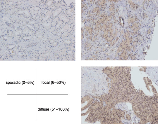
CXCR4 expression in prostate cancer (with bone metastasis) (×200). The evaluations were recorded as the percentage of positively stained tumor cells in each of three intensity categories: sporadic, 0–5%; focal, 6–50%; and diffuse, 51–100%.
Statistical analysis. All statistical analyses were carried out with the Stat‐View program, version 5.0 (Abacus Concepts, Berkeley, CA, USA). Cause‐specific survival was calculated by the Kaplan–Meier method. According to the Kaplan–Meier plots, the log‐rank statistic was used for evaluating statistical significance in the analyses. Statistical significance was estimated using Student's t‐test and the Cox–Mantel method. For multivariate analyses, we used a Cox proportional hazard model.
Results
The clinical and pathological features of 52 patients are shown in Table 1. The percentage of stained tumor cells in each of the three intensity categories was 5.8% (sporadic; n = 3), 59.6% (focal; n = 31), and 34.6% (diffuse; n = 18). No statistically significant differences were found in CXCR4 expression according to the pretreatment PSA level (P = 0.8914). CXCR4 expression showed no association with pathological grade (Gleason score; P = 0.8812), clinical response to hormonal therapy (P =0.3447), or extent of bony metastasis (P =0.9443). However, pathological grade (P =0.0025), clinical response to hormonal therapy (P =0.0257), and CXCR4 expression (P =0.0159) exhibited correlations with cause‐specific survival (Fig. 2). The results of multivariate analysis to determine the relative importance of the prognostic factors were found to be statistically significant for pathological grade (P = 0.0040, 95% confidence interval [CI] 0.216–0.747) and CXCR4 expression (P =0.0329, 95% CI 0.284–0.948). In the present analysis, CXCR4 expression was found to be one of the prognostic factors (Table 2).
Table 1.
CXCR4 expression and clinicopathological features
| Feature | All cases | CXCR4 expression | P‐value (low vs high) | ||
|---|---|---|---|---|---|
| Low | High | ||||
| Sporadic | Focal | Diffuse | |||
| Mean age ± SD (years) | 73.4 ± 7.7 | 77.7 ± 13.6 | 73.2 ± 7.18 | 73.5 ± 7.9 | 0.9692 |
| Mean pretreatment PSA ± SD (ng/mL) | 412.7 ± 726.9 | 21.4 ± 23.1 | 443.0 ± 752.0 | 436.0 ± 747.1 | 0.8914 |
| Mean Gleason score ± SD | 8.10 ± 1.40 | 7.3 ± 2.1 | 8.1 ± 1.5 | 8.06 ± 1.31 | 0.8812 |
| Clinical response to hormonal therapy † (n) | |||||
| CR | 32 | 2 | 21 | 9 | 0.3447 |
| PR + NC + PD | 20 | 1 | 10 | 9 | |
| EOD (n) | |||||
| Low (0–2) | 32 | 3 | 20 | 11 | 0.9443 |
| High (3–4) | 20 | 0 | 11 | 7 | |
Changes in prostate‐specific antigen level after 3 months. CR, complete response; EOD, extent of bony metastasis; NC, no change; PD, progressive disease; PE, partial response.
Figure 2.
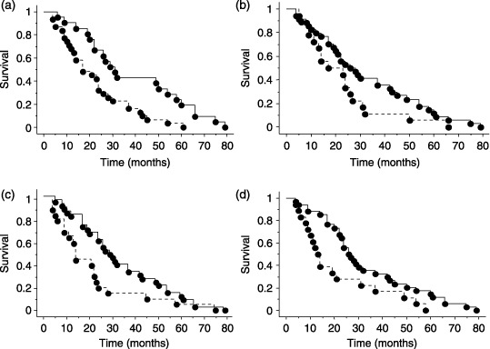
Kaplan–Meier cause‐specific survival curves for patients with metastatic prostate cancers relative to clinicopathological features. (a) Cancer‐specific survival according to pretreatment Gleason score. Solid and dotted lines represent Gleason score <8 (n = 21) and = 8 (n = 31), respectively; P = 0.0025. (b) Cancer‐specific survival according to extent of disease (EOD). Solid and dotted lines represent EOD 0–2 (n = 32) and EOD 3–4 (n = 20), respectively; P = 0.0891. (c) Cancer‐specific survival according to clinical response to hormonal therapy. Solid and dotted lines represent complete response (n = 32), and partial response, no change, and progressive disease (n = 20), respectively; P = 0.0257. (d) Cancer‐specific survival according to CXCR4 expression. Solid and dotted lines represent CXCR4 expression of low (sporadic and focal, n = 34) and high (diffuse, n = 18), respectively; P = 0.0159.
Table 2.
Multivariate analysis of prognostic factors in relation to cause‐specific survival
| Prognostic factor | P‐value |
|---|---|
| Pretreatment Gleason score | 0.0040 |
| Clinical response to hormonal therapy | 0.0551 |
| CXCR4 expression | 0.0329 |
Discussion
Many studies on the CXCL12–CXCR4 ligand–receptor system have been carried out mainly in the field of immunology and infection, such as hematopoiesis, lymphocyte homing, and HIV infection. In 2001, Muller et al. showed that CXCR4 is expressed more highly in breast cancer tissue than in normal breast tissue, and CXCL12 is expressed in many organs in which breast cancer metastasis is often found, such as lymph nodes, bone marrow, and lungs, but is not expressed in the kidney where metastasis hardly occurs.(4) In addition, in vivo studies have shown that injecting the antibody that neutralizes CXCR4 activity leads to inhibition of metastasis to the bone marrow and lungs.( 4 ) These findings indicate that the CXCL12–CXCR4 ligand–receptor system plays a critical role in determining the metastatic destination of cancer cells. Similar experimental results have also been reported in other types of cancer such as esophageal,( 7 ) malignant melanoma,( 12 ) ovarian,( 13 ) and lung cancers;( 14 ) however, such studies are limited in prostate cancer.
A previous study revealed that in the three prostate cancer cell lines LNCaP, PC3, and DU‐145, CXCR4 expression was detected at both the mRNA and protein levels.( 15 ) In contrast, in normal human prostate epithelial cells, no detectable level of CXCR4 expression was found. However, CXCL12 was not expressed in these three prostate cancer cell lines; therefore, although the autocrine action of the CXCR12–CXCR4 ligand–receptor system is supposedly present in neuroblastoma,( 16 ) prostate cancer cells might be affected not by endogenous CXCL12, but by exogenous CXCL12. The preoperative PSA level is one of the strongest predictors of recurrence in prostate cancer.( 17 ) Meanwhile, a previous study did not reveal a statistically significant association between CXCR4 expression and patient status, for example, with preoperative PSA level, T category, Gleason sum, metastasis (lymph node, bone, lung), and patient prognosis.( 18 ) In the present study, we used immunohistochemical staining of transperineal needle biopsy specimens from patients with metastatic prostate cancer treated with androgen‐withdrawal therapy. We also evaluated the relationship between CXCR4 expression and prognosis, and other clinicopathological factors. Although a statistically significant association was noted between CXCR4 expression and cancer‐specific survival in this study, correlations with pretreatment PSA level, Gleason score, clinical response to hormonal therapy, and extent of bony metastasis, conventionally suggested to be related to survival, were not noted. The reason why none of these factors had correlations with CXCR4 expression might be because the subjects in the present study all had prostate cancer with bone metastasis (in a sense, biased object). So the mean value of each parameter (EOD, Gleason score and clinical response to hormonal therapy) had no relationship with CXCR4 expression.
The present results suggest that CXCR4 was detected in most patients (94.2%) with metastatic prostate cancer or specimens. According to a previous study, Mochizuki et al. reported that positive CXCR4 protein was found in 57.1% of clinical prostate cancer samples, and that the positive expression of CXCR4 protein was an independent and superior predictor of bone metastasis to the Gleason score.( 18 ) Previous studies reported that CXCR4 expression predicted poor prognosis in 61 patients with completely resected non‐small cell lung cancer,( 5 ) in esophageal cancer,( 7 ) and in epithelial ovarian cancer.( 8 ) Recent clinical studies have verified that cytoplasmic CXCR4 expression is associated with tumor aggressiveness and has prognostic value.( 19 , 20 ) To the best of our knowledge, this is the first report demonstrating the prognostic role of CXCR4 in prostate cancer.
CXCR4 expression appears to be an independent prognostic factor in prostate cancer with bone metastasis. In our study, subjects were a limited patient group (stage D2 only) who had received androgen‐withdrawal therapy, but the data showed that CXCR4 expression predicts poor prognosis, supporting the inhibition of CXCR4 as a possible therapeutic target.
Experiments in vitro revealed that anti‐CXCR4 antibody inhibited the migration and invasion of a prostate cancer cell line.( 3 ) In prostate cancer, Darash‐Yanaha et al. showed that high expression levels of CXCR4 correlated with the presence of metastatic disease.( 21 ) These findings suggest that CXCR4 expression in prostate cancer tissues is closely associated with cancer progression or metastasis and is a useful tool in predicting metastasis in prostate cancer patients.
Therapeutic strategies are classified mainly into two categories: the application of neutralizing antibody against CXCR4 and specific CXCR4 antagonists. It was first reported that lung metastasis of human breast cancer is suppressed by the administration of antihuman CXCR4 monoclonal antibody in an SCID mouse model.( 4 ) The antibody also inhibits lung metastasis of murine B16 melanoma( 22 ) and bone metastasis of human prostate cancer in a murine model.( 14 ) However, currently, CXCR4 antagonists are thought to be a promising therapeutic approach for cancer metastasis. AMD3100 is very effective against HIV‐1 and HIV‐2, based on its inhibition of viral replication, and is the most potent and selective CXCR4 antagonist ever discovered.( 23 ) Administration of AMD3100 effectively suppresses the growth of glioblastoma cells transplanted intracranially into mice, and also increases apoptosis of the cells.( 24 ) Another CXCR4 antagonist, T140, reduced pulmonary metastasis of human breast cancer cells in SCID mice.( 25 ) In any case, both AMD3100‐ and T140‐derived CXCR4 antagonists appear to have activity in animal tumor models, providing the rationale for future clinical trials of these agents in patients with various cancers.( 26 , 27 )
Thus, identifying CXCR4 as a potential therapeutic target is attractive in not only early stages but also in advanced (metastatic) cases. Clinical experiments with larger samples and further study will be needed to verify the results of our study.
References
- 1. Zlotnik A, Yoshie O. Chemokines. a new classification system and their role in immunity. Immunity 2000; 12: 121–7. [DOI] [PubMed] [Google Scholar]
- 2. Butcher EC, Williams M, Youngman K, Rott L, Briskin M. Lymphocyte trafficking and regional immunity. Adv Immunol 1999; 72: 209–53. [DOI] [PubMed] [Google Scholar]
- 3. Taichman RS, Cooper C, Keller ET, Pienta KJ, Taichman NS, McCauley LK. Use of stromal cell‐derived factor‐1/CXCR4 pathway in prostate cancer metastasis to bone. Cancer Res 2002; 62: 1832–7. [PubMed] [Google Scholar]
- 4. Muller A, Homey B, Soto H et al . Involvement of chemokine receptors in breast cancer metastasis. Nature 2001; 410: 50–6. [DOI] [PubMed] [Google Scholar]
- 5. Spano JP, Andre F, Morat L, Sabatier L et al . Chemokine receptor CXCR4 and early‐stage non‐small cell lung cancer: pattern of expression and correlation with outcome. Ann Oncol 2004; 15: 613–17. [DOI] [PubMed] [Google Scholar]
- 6. Scala S, Ottaiano A, Ascierto PA et al . Expression of CXCR4 predicts poor prognosis in patients with malignant melanoma. Clin Cancer Res 2005; 11: 1835–41. [DOI] [PubMed] [Google Scholar]
- 7. Koishi K, Yoshikawa R, Tsujimura T et al . Persistent CXCR4 expression after preoperative chemoradiotherapy predicts early recurrence and poor prognosis in esophageal cancer. World J Gastroenterol 2006; 12: 7585–90. [DOI] [PMC free article] [PubMed] [Google Scholar]
- 8. Jiang YP, Wu XH, Shi B, Wu WX, Yin GR. Expression of chemokine CXCL12 and its receptor CXCR4 in human epithelial ovarian cancer: an independent prognostic factor for tumor progression. Gynecol Oncol 2006; 103: 226–33. [DOI] [PubMed] [Google Scholar]
- 9. Oda Y, Yamamoto H, Tamiya S et al . CXCR4 and VEGF expression in the primary site and the metastatic site of human osteosarcoma: analysis within a group of patients, all of whom developed lung metastasis. Mod Pathol 2006; 19: 738–45. [DOI] [PubMed] [Google Scholar]
- 10. Gleason DF. Histologic grading of prostate cancer: a perspective. Hum Pathol 1992; 23: 273–9. [DOI] [PubMed] [Google Scholar]
- 11. Soloway MS. The important prognostic factors in advanced prostate cancer. Cancer 1990; 66: 1017–21. [DOI] [PubMed] [Google Scholar]
- 12. Payne AS, Cornelius LA. The role of chemokines in melanoma tumor growth and metastasis. J Invest Dermatol 2002; 118: 915–22. [DOI] [PubMed] [Google Scholar]
- 13. Scotton CJ, Wilson JL, Scott K et al . Multiple actions of the chemokine CXCL12 on epithelial tumor cells in human ovarian cancer. Cancer Res 2002; 62: 5930–8. [PubMed] [Google Scholar]
- 14. Kijima T, Maulik G, Ma PC et al . Regulation of cellular proliferation, cytoskeletal function, and signal transduction through CXCR4 and c‐Kit in small cell lung cancer cells. Cancer Res 2002; 62: 6304–11. [PubMed] [Google Scholar]
- 15. Sun YX, Wang J, Shelburne CE et al . Expression of CXCR4 and CXCL12 (SDF‐1) in human prostate cancers (PCa) in vivo . J Cell Biochem 2003; 89: 462–73. [DOI] [PubMed] [Google Scholar]
- 16. Geminder H, Sagi‐Assif O, Goldberg L et al . A possible role for CXCR4 and its ligand, the CXC chemokine stromal cell‐derived factor‐1, in the development of bone marrow metastases in neuroblastoma. J Immunol 2001; 167: 4747–57. [DOI] [PubMed] [Google Scholar]
- 17. Catalona WJ, Smith DS. Cancer recurrence and survival rates after anatomic radical retropubic prostatectomy for prostate cancer: intermediate‐term results. J Urol 1998; 160: 2428–34. [DOI] [PubMed] [Google Scholar]
- 18. Mochizuki H, Matsubara A, Teishima J et al . Interaction of ligand‐receptor system between stromal‐cell‐derived factor‐1 and CXC chemokine receptor 4 in human prostate cancer: a possible predictor of metastasis. Biochem Biophys Res Commun 2004; 30: 656–63. [DOI] [PubMed] [Google Scholar]
- 19. Hart CA, Brown M, Bagley S, Sharrard M, Clarke NW. Invasive characteristics of human prostatic epithelial cells: understanding the metastatic process. Br J Cancer 2005; 92: 503–12. [DOI] [PMC free article] [PubMed] [Google Scholar]
- 20. Salvucci O, Bouchard A, Baccarelli A et al . The role of CXCR4 receptor expression in breast cancer: a large tissue microarray study. Breast Cancer Res Treat 2006; 97: 275–83. [DOI] [PubMed] [Google Scholar]
- 21. Darash‐Yanaha M, Pikarsky E, Abramovitch R et al . Role of high expression levels of CXCR4 in tumor growth, vascularization, and metastasis. FASEB J 2004; 18: 1240–2. [DOI] [PubMed] [Google Scholar]
- 22. Murakami T, Maki W, Cardones AR et al . Expression of CXC chemokine receptor‐4 enhances the pulmonary metastatic potential of murine B16 melanoma cells. Cancer Res 2002; 62: 7328–34. [PubMed] [Google Scholar]
- 23. De Clercq E. Potential clinical applications of the CXCR4 antagonist Bicyclam AMD3100. Mini Rev Med Chem 2005; 5: 805–24. [DOI] [PubMed] [Google Scholar]
- 24. Rubin JB, Kung AL, Klein RS et al . A small‐molecule antagonist of CXCR4 inhibits intracranial growth of primary brain tumors. Proc Natl Acad Sci USA 2003; 100: 13 513–18. [DOI] [PMC free article] [PubMed] [Google Scholar]
- 25. Tamamura H, Hori A, Kanzaki N et al . T140 analogs as CXCR4 antagonists identified as anti‐metastatic agents in the treatment of breast cancer. FEBS Lett 2003; 28: 79–83. [DOI] [PubMed] [Google Scholar]
- 26. Smith MC, Luker KE, Garbow JR et al . CXCR4 regulates growth of both primary and metastatic breast cancer. Cancer Res 2004; 64: 8604–12. [DOI] [PubMed] [Google Scholar]
- 27. Takenaga M, Tamamura H, Hiramatsu K et al . A single treatment with microcapsules containing a CXCR4 antagonist suppresses pulmonary metastasis of murine melanoma. Biochem Biophys Res Commun 2004; 320: 226–32. [DOI] [PubMed] [Google Scholar]