


Figure 1.
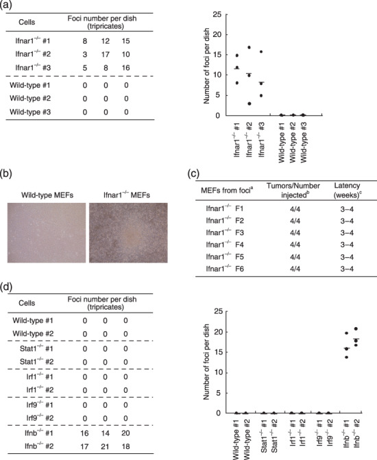
Spontaneous transformation of Ifnar1−/– MEF. (a) Primary wild‐type or Ifnar1−/– MEF (3 × 10⁶ cells) were plated on 10‐cm culture dishes, and after 4‐ to 6‐week cell culture, numbers of foci formed in the dish were counted. Assay was done with three different MEF in three independent dishes. Average of the numbers of foci observed in the three dishes are shown as a horizontal line in the graph (right). (b) Appearance of a cell colony observed after 50‐day culture. (c) Six cell clones (F1–F6) were isolated from separate foci that formed after long‐term cell culture, were inoculated in nude mice for tumorigenicity. aMEF genotypes; bnumber of tumors arising per number of sites injected; cperiod until visible tumor formation. (d) Primary wild‐type or other mutant MEF lacking signaling molecules downstream of the IFNAR were plated on 10‐cm culture dishes, and were examined for the formation of colonies during the long‐term culture, as described in (a). IFNAR, IFN‐α/β receptor; MEF, mouse embryonic fibroblasts.