



Abstract
Deficient mismatch repair (dMMR) results in microsatellite instability (MSI), a pronounced mutator phenotype. High‐frequency MSI (MSI‐H)/dMMR is gaining increasing interest as a biomarker for advanced cancer patients to determine their eligibility for immune checkpoint inhibitors (ICIs). Various methods based on next‐generation sequencing (NGS) have been developed to assess the MSI status. Comprehensive genomic profiling (CGP) testing can precisely ascertain the MSI status as well as genomic alterations in a single NGS test. The MSI status can be also ascertained through the liquid biopsy‐based CGP assays. MSI‐H has thus been identified in various classes of tumors, resulting in a greater adoption of immunotherapy, which is hypothesized to be effective against malignancies that possess a substantial number of mutations and/or neoantigens. NGS‐based studies have also characterized MSI‐driven carcinogenesis, including significant rates of fusion kinases in colorectal cancers (CRCs) with MSI‐H that are targets for therapeutic kinase inhibitors, particularly in MLH1‐methylated CRCs with wild‐type KRAS/BRAF. NTRK fusion is linked to the colorectal serrated neoplasia pathway. Recent advances in investigations of MSI‐H malignancies have resulted in the development of novel diagnostic or therapeutic techniques, such as a synthetic lethal therapy that targets the Werner gene. DNA sensing in cancer cells is required for antitumor immunity induced by dMMR, opening up novel avenues and biomarkers for immunotherapy. Therefore, clinical relevance exists for analyses of MSI and MSI‐H‐associated genomic alterations in malignancy. In this article, we provide an update on MSI‐driven carcinogenesis, with an emphasis on unique landscapes of diagnostic and immunotherapeutic strategies.
Keywords: DNA mismatch repair, fusion kinases, immunotherapy, microsatellite instability, next‐generation sequencing
High‐frequency microsatellite instability (MSI‐H)/deficient mismatch repair (dMMR) is gaining increasing interest as a biomarker for patients with advanced cancer to determine their eligibility for immune checkpoint inhibitors (ICIs). This review provides an update on MSI‐driven carcinogenesis, with an emphasis on unique landscapes of diagnostic and immunotherapeutic strategies.

1. INTRODUCTION
Deficient mismatch repair (dMMR) results in microsatellite instability (MSI), a pronounced mutator phenotype (Figure 1). High‐frequency MSI (MSI‐H)/dMMR is gaining increasing interest as a biomarker for advanced cancer patients to determine their eligibility for immune checkpoint inhibitors (ICIs). In this article, we provide an update on MSI‐driven carcinogenesis, with an emphasis on unique landscapes of diagnostic and immunotherapeutic strategies.
FIGURE 1.
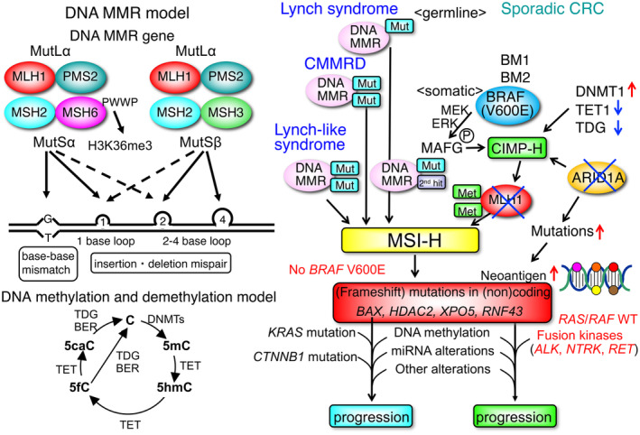
DNA mismatch repair (MMR) model, DNA methylation and demethylation model, and pathogenesis of high‐frequency microsatellite instability (MSI‐H) colorectal cancers (CRCs). Upper left: DNA MMR model based on DNA mismatch. Lower left: DNA methylation and demethylation model. 5caC, 5‐carboxylcytosine; 5fC, 5‐formylcytosine; 5hmC, 5‐hydroxy methylcytosine; 5mC, 5‐methylcytosine; BER, base excision repair; DNMT, DNA methyltransferase; TDG, thymine DNA glycosylase; TET, Tet methylcytosine dioxygenase. Right: a model of pathogenesis of MSI‐H CRCs based on hereditary and sporadic cases. CMMRD, constitutional mismatch repair deficiency; MAFG, MAF BZIP transcription factor G.
2. ANALYSES OF MSI
2.1. Next‐generation sequencing (NGS) analyses of MSI in diverse tumor types
MSI status is now routinely assessed in various types of advanced malignancies using polymerase chain reaction and/or immunohistochemistry of DNA mismatch repair (MMR) proteins 1 , 2 , 3 , 4 , 5 (Figure 2). NGS enables a comprehensive profile of genomic alterations as well as MSI status in a single test. 6 To determine MSI status in NGS analysis, various algorithms using targeted, whole‐exome, or whole‐genome sequencing data have been devised 1 , 7 , 8 , 9 , 10 (Figure 2). MSI status can be determined in the real world utilizing FDA‐approved tumor profiling NGS tests like Foundation Medicine's FoundationOne CDx and MSK‐IMPACT.
FIGURE 2.
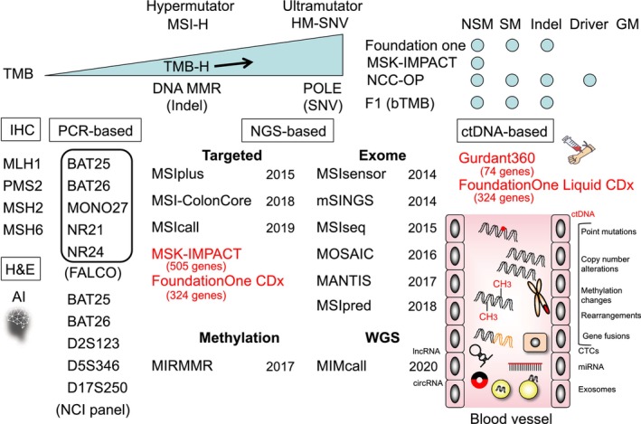
High‐frequency microsatellite instability (MSI‐H) and TMB and evaluation of the MSI status. Upper left: an association of MSI‐H with TMB. HM‐SNV, hypermutated‐single nucleotide variant. Upper right: evaluation of TMB. GM, germline mutations; NCC‐OP, National Cancer Center‐OncoPanel; NSM, nonsynonymous mutations; SM, synonymous mutations. Lower: the MSI status is evaluated by immunohistochemistry, PCR‐based assays, next‐generation sequencing (NGS)‐based techniques, and ctDNA‐based techniques.
2.2. Microsatellite instability evaluation by liquid biopsy
Reportedly, circulating tumor DNA (ctDNA)‐based NGS can ascertain the MSI status in a wide range of cancer types, including gastrointestinal (GI) malignancies, in close agreement with tissue‐based sequencing 11 , 12 (Figure 2). Recently, the clinical validity of MSI evaluation using plasma was demonstrated in advanced GI malignancies enrolled in the SCRUM‐Japan GOZILA substudy. 13 The positive, negative, and overall percent agreement in 658 patients with advanced GI malignancies that received tissue and plasma evaluation of MSI were 71.4%, 99.1%, and 98.2%, respectively. Importantly, the positive percent agreement was 100.0% in individuals with a ctDNA proportion of 1.0% or higher in their plasma samples. Thus, ctDNA genotyping could determine MSI status with good concordance to validated tissue‐based testing of MSI, particularly in patients whose cancers shed an abundance of ctDNA. 14
Microsatellite instability status can be determined in the real world with FDA‐approved liquid biopsy tests such as Foundation Medicine's FoundationOne Liquid CDx and Guardant360 CDx (Figure 2). ctDNA‐based MSI testing with Guardant360 was greatly concordant with tissue‐based MSI analysis, allowing for a more accurate assessment of MSI status in conjunction with CGP. This improves immunotherapy accessibility for patients diagnosed with advanced cancer who do not have a tissue‐based CGP test. 12
2.3. Microsatellite instability prediction using artificial intelligence (AI) based on tumor histomorphology
High‐frequency MSI/dMMR is gaining increasing interest as a biomarker for ICI eligibility in advanced malignancies. AI technology is being used to forecast MSI/dMMR status based on histomorphological traits on hematoxylin–eosin slides. A systematic review found that AI‐based systems exhibited exceptional performance, as evidenced by the highest standard of 0.972 in colorectal cancer (CRC). 15 The performance of AI‐based systems was comparatively subpar as evidenced by the highest standards of 0.81 for gastric cancer (GC) and 0.82 for endometrial cancer. Cases identified as MSI‐H by AI‐based systems, nevertheless, require direct molecular confirmation via PCR‐based testing or NGS due to substantial limitations. 15 , 16
3. HIGH‐FREQUENCY MSI‐DRIVEN CARCINOGENESIS
3.1. BRAF V600E mutations in CRC
In the setting of CpG island methylator phenotype–high (CIMP‐H), the majority of sporadic MSI‐H cases evolve by epigenetic silencing of MLH1 via promoter hypermethylation 17 , 18 (Figure 1). A minority of sporadic MSI‐H tumors that did not have MLH1 methylation had somatic mutations in DNA MMR genes, indicating an alternate pathway to DNA MMR inactivation (Figure 1). The latter malignancies were linked to KRAS mutations rather than BRAF mutations.
BRAF V600E mutations are strongly correlated with MLH1 promoter‐methylated sporadic CRCs with MSI‐H 19 (Figure 1). Therefore, Lynch syndrome (LS) can be essentially ruled out in patients who are positive for the BRAF V600E variants and/or MLH1 promoter methylation. 19 As a diagnostic process for LS, the Japanese Society for Cancer of the Colon and Rectum (JSCCR) guidelines recommend tests for the BRAF V600E variants in CRC cases with MSI‐H or loss of MLH1 and PMS2 expression. 19 After that, MLH1 promoter methylation testing is recommended for those with BRAF V600E wild type. 19
Moreover, CRCs with BRAF V600E mutations can be divided into two groups based on gene expression: BRAF mutant 1 (BM1) or BRAF mutant 2 (BM2) 20 (Figure 1). BM1 is distinguished by the activation of the KRAS/AKT pathway, the dysregulation of mTOR/4EBP, the activation of epithelial to mesenchymal transition (EMT), and immune infiltration. In contrast, BM2 exhibits dysregulation of the cell cycle checkpoints. BM1 is additionally distinguished by elevated levels of phosphorylated AKT and 4EBP1, whereas BM2 exhibits decreased cyclin D1 levels and elevated CDK1 levels.
Among the 218 BRAF‐mutant CRCs, 69 were categorized as BM1 and 149 as BM2. MSI‐H enrichment in BM2 was marginally greater than in BM1 (43% vs. 65% in BM2). Four subtypes comprise the CRC Subtyping Consortium (CRCSC) subtype consensus: consensus molecular subtype (CMS) CMS1 (defined by immune patterns and MSI‐H), CMS2 (CIN and activation of WNT), CMS3 (metabolic pattern), and CMS4 (EMT) 21 (Figure 3). A minority of BRAF‐mutant patients were identified in CMS4 (17%), CMS3 (5%), and CMS2 (2%), while CMS1 accounted for the overwhelming majority (70%) of BRAF‐mutant patients. Interestingly, CMS1 BRAF mutants were found to be distributed in both BM1 and BM2, whereas all BRAF‐mutated CMS4 were BM1. Additional research is required to elucidate the clinical significance of this BRAF classification, particularly with regard to diverse targeted therapy combinations. Individuals with the BM1 or CMS4 subtype, which are both associated with increased MAP3K8 expression, may benefit more from triplet therapy as opposed to doublet therapy.
FIGURE 3.
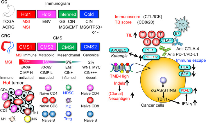
Immune microenvironment and immunotherapy in cancer. Upper left: classification of gastric cancer (GC) based on immunogram. ACRG, Asian Cancer Research Group; EMT, epithelial to mesenchymal transition; GS, genomically stable. Lower left: classification of colorectal cancer (CRC) based on CMS. Right: The immune response against high‐microsatellite instability (MSI‐H) cancers can be interfered with immune evasion mechanisms, such as inactivation of HLA and B2M. APOBEC3 alterations, APOBEC3 overexpression, and kataegis play roles in the overexpression of PD‐L1/2. cGAS, cyclic GMP‐AMP synthase; CTL/ICK, tumor‐infiltrating T cell/immune checkpoint; DC, dendritic cell; TBK1, TANK binding kinase 1; TIL, tumor‐infiltrating lymphocyte.
3.2. ARID1A deficiency and a CIMP
If MLH1 is disrupted in the presence of CIMP‐H, the tumor transforms into MSI‐H; conversely, if MLH1 is not silenced, the tumor progresses along the CIN pathway and transforms into CIMP‐H MSS CIN malignancies. 22 BRAF V600E mutations were nearly ubiquitous in CIMP‐H ascending colon tumors but were absent in CIMP‐H GCs. Moreover, BRAF V600E mutations were not found in a subset of CIMP‐H CRCs with comparable DNA methylation signatures. Therefore, BRAF V600E mutation is probably one of the minor mechanisms by which CIMP is induced (Figure 1). On the contrary, CIMP‐H could potentially create a favorable milieu for the BRAF V600E mutation by inhibiting pathways implicated in apoptosis and senescence induced by oncogenes. 23
CpG island methylator phenotype is thought to be caused by genetic abnormalities in the tricarboxylic acid cycle (TCA) cycle, that is, IDH1 or IDH2 mutations in glioblastoma multiforme (GBM) and acute myelogenous leukemia (AML). 24 TCA cycle gene mutations are uncommon in CIMP‐H GCs and CRCs, in contrast to GBM and AML. Even in AML, IDH mutations can only explain roughly half of the instances of CIMP‐H. The cause of CIMP‐H cases in which IDH mutations are absent is still unknown.
ARID1A is a member of a core component of the SWI/SNF chromatin remodeling complexes. Regarding the relationship between ARID1A and MSI‐H, ARID1A promotes MMR and reportedly recruits MSH2 to chromatin during DNA replication. 25 ARID1A mutation is linked to CIMP and EBV infection in GCs, as well as CRCs and uterine endometrial malignancies. Loss of function of ARID1A was strongly associated with aberrant DNA methylation induction. 26 In 293FT cell lines and GES1 gastric epithelial cell lines, aberrant DNA methylation at genomic regions containing acquired and pre‐existing H3K27me3 was induced by ARID1A knockout. Thus, aberrant DNA methylation induced by ARID1A mutation is one of the prospective mechanisms by which CIMP is induced (Figure 1).
3.3. Frequent fusion kinases in MLH1 ‐methylated CRCs with MSI‐H
Colorectal cancers with MLH1 promoter methylation (called MM) displayed clinicopathological characteristics different to CRCs with germline mutations (LS or LS‐associated) or Lynch‐like somatic MMR alterations (Figure 1). Frequent mutations in BRAF/RNF43 as well as more insertions and deletions were found in MLH1‐methylated sporadic MSI‐H CRCs than in Lynch‐like or LS‐associated MSI‐H cancers. 27 Importantly, 11 fusion kinases were only found in MLH1‐methylated MSI‐H CRCs with wild‐type KRAS/BRAF and were linked to a short post‐relapse survival 27 (Figure 1). Sato et al. created a simple approach for identifying MLH1‐methylated cancers and tested it on a validation cohort of 28 CRCs with MSI‐H, detecting 16 MLH1‐methylated cancers and two fusion kinases. 27 By employing the novel approach for identifying MLH1‐methylated tumors, MSI‐H CRC patients can be efficiently categorized as LS or sporadic cases that are potential candidates for fusion kinase carriers, which are therapeutic targets of kinase inhibitors.
Through the utilization of variant‐specific PCR, 5′/3′‐end expression imbalance, and RNA‐based NGS, actionable gene rearrangements in malignancies with MSI were comprehensively analyzed. 28 A total of 58/471 (12.3%) CRCs, 4/69 (5.8%) GCs, and 3/65 (4.6%) endometrial malignancies exhibited gene fusions (8 ALK; 24 NTRK1; 2 NTRK2; 19 NTRK3; 12 RET). Gene rearrangements occurred most frequently in 53 (26%) of 204 CRCs with wild‐type KRAS/NRAS/BRAF. Analysis of gene rearrangements of tyrosine kinase is especially viable in MSI‐H CRCs with wild‐type KRAS/NRAS/BRAF, although these events also occur to a moderate degree in other types of malignancies harboring MSI‐H.
3.4. NTRK fusions in the colorectal serrated neoplasia pathway
Fusions of NTRK gene are promising tissue‐agnostic therapeutic targets in a variety of cancers. Immunohistochemical positivity of TRK was found in 19% (11/58) of MLH1‐methylated CRCs with MSI‐H, 17% (4/23) of sessile serrated lesions with dysplasia, and 4% (5/132) of sessile serrated lesions but not in any of the hyperplastic polyps, traditional serrated adenomas, conventional adenomas, or MSS CRCs 29 (Figure 4). The 11 MSI‐H CRCs with TRK positivity all had CIMP‐H, methylated MLH1, wild‐type KRAS/BRAF, and NTRK1 or NTRK3 fusion. Therefore, NTRK‐rearranged CRCs exclusively develop via the colorectal serrated neoplasia pathway and can originate from KRAS/BRAF wild‐type non‐dysplastic SSLs before reaching complete development of CRCs with MSI‐H/CIMP‐H (Figure 4).
FIGURE 4.
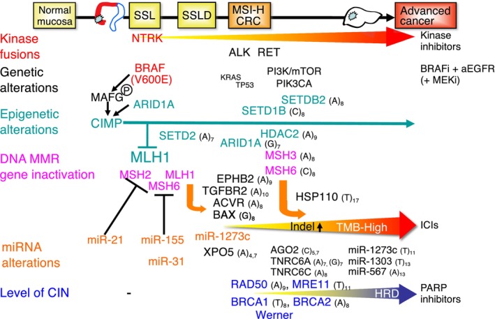
Carcinogenesis of sporadic high‐microsatellite instability (MSI‐H) colorectal cancers (CRCs). A model for MSI‐H CRC progression is presented based on genetic, epigenetic, and miRNA alterations with target therapies. Mutations that promote cancer cell growth are considered to be the driving force of MSI‐H carcinogenesis and are referred to as driver mutations or Real Common Target genes. SSL, sessile serrated lesions; SSLD, serrated lesions with dysplasia.
3.5. Identification of Werner (WRN) in MSI‐H tumors as a synthetic lethal therapeutic target
Four research groups independently determined, through the use of distinct functional screening techniques, that MSI‐H malignancies rely on the WRN helicase to maintain genome stability and ensure cellular survival. 30 , 31 , 32 , 33 , 34 Expanded TA‐dinucleotide repeats were identified as prevalent sites of DNA breakage in MSI cancer cells when WRN was deficient. 35 These findings uncovered the mechanism underlying the synthetic lethality that exists between MSI and WRN deficiency and identified a novel biomarker for MSI (Figure 4).
The propensity of TA repeats to aggregate into cruciform structures elucidates the reason why downregulation of WRN results in preferential disruption of these DNA sequences in MSI cells. This provides a mechanistic foundation for the synthetic–lethal interaction involving TA repeats, which has been identified as a promising approach to precision cancer therapy. 36
In MSI CRC cells, WRN depletion significantly increased the expression of TP53 and PUMA, its apoptotic downstream target. 37 WRN depletion‐induced apoptosis was eliminated by TP53 or PUMA deletion in MSI CRC cells. Notably, the TP53 locus remains wild type in most MSI CRCs. 38 These findings suggest that TP53/PUMA‐mediated apoptosis plays a crucial role in susceptibility to WRN depletion for MSI CRC, and it endorses WRN as a viable treatment target for MSI CRCs with wild‐type TP53.
4. UNIQUE LANDSCAPES OF IMMUNOTHERAPEUTIC STRATEGIES
4.1. Gastric cancer is classified immunologically using an immunogram
Because cancer–immune system interactions are complex, biomarker combinations will be needed to forecast each patient's response to ICI therapy and devise approaches to surmount resistance to therapy (Figure 3). A comprehensive framework called “Immunogram” has been proposed in order to encompass nearly all pertinent immunological variables (Figure 3). A method for converting transcriptomic data to immunogram scores was devised by Kobayashi et al. 39 Axis 1 (innate immunity), 2 (priming and activation), 3 (T cells), 4 (interferon gamma response), 5 (inhibitory molecules), 6 (inhibitory Treg cells), 7 (inhibitory myeloid‐derived suppressor cells (MDSCs)), 8 (recognition of tumor cells), 9 (proliferation), and 10 (glycolysis) are the 10 molecular profiles comprising this immunogram.
Four subtypes of immune signatures of GCs were identified by immunogram analysis: Hot1, Hot2, Intermediate, and Cold (Figure 3). An impaired T cell signature was observed in Hot tumors, whereas an exclusion signature was noted in cold tumors. 40 The T cell dysfunction was more pronounced in Hot1 tumors as opposed to Hot2 tumors. Survival was superior in Hot2 subtypes in their cohort as well as TCGA cohort. Moreover, metabolic and cell adhesion pathways were distinct between Hot 1 and Hot 2 malignancies. 41 Hence, it is imperative to incorporate active metabolism modulation into treatment strategies for Hot 1 tumors.
While these immunological subtypes and TCGA molecular subtypes shared some degree of overlap, histological or molecular subtyping alone did not provide sufficient information to predict intratumoral immune responses in GC (Figure 3). Thus, molecular and immunological classifications complement each other in predicting ICI therapy responses and have the potential to be biomarkers for the treatment of GCs.
4.2. Enhancement of mutability and ICI‐induced antitumor immunity by ARID1A deficiency
ARID1A is a gene that experiences frequent mutations in cancer. 42 , 43 ARID1A inactivation, on the other hand, impeded MMR and increased mutagenesis. A correlation was observed between ARID1A deficiency and the MSI‐H genomic signature (preponderant C>T mutation pattern), and an increased burden of tumor mutations across a range of human cancer types. Thus, ARID1A deficiency relates to cancer's defective MMR (dMMR) and mutator phenotype, and it may interact favorably with ICI therapy (Figure 1).
4.3. The accumulation of gene mutations results in decreased expression of HLA‐ABC genes, which contributes to immune evasion in CRC
Understanding antitumor immunity is critical for effective cancer immune treatment (Figure 3). The accumulation of gene mutations leads to a decrease in HLA‐ABC gene expression and could result in immune evasion in CRC (Figure 3). The impact of defective mutations in the HLA‐ABC genes on tumor immunity was investigated by Kawazu et al. 44 A total of 114 MSI‐H CRC samples were subjected to long‐read sequencing to identify mutations in the HLA‐ABC loci. There were 101 truncating mutations and 61 allele losses found in 57 and 21 tumors, respectively. Immunological subclassification also identified a subtype of tumor characterized by reduced lymphocyte infiltration, which was partially attributable to nongenetic alterations in the HLA‐ABC genes (Figure 5). This group of tumors consistently showed the highest TMB, implying that the immunogenic effects of tumor mutations were mitigated by alterations that reduced immunoreactivity. Various genetic and epigenetic changes, including RFX5 frameshift mutations and PSMB8 promoter methylation, contributed to a lower HLA‐ABC gene expression (Figure 5). Lastly, patients afflicted with these tumors had a shorter survival period than those with other forms of tumors. The insights gained from these results regarding the immune evasion mechanisms of MSI‐H CRC contribute to the advancement and patient selection of cancer immunotherapy.
FIGURE 5.
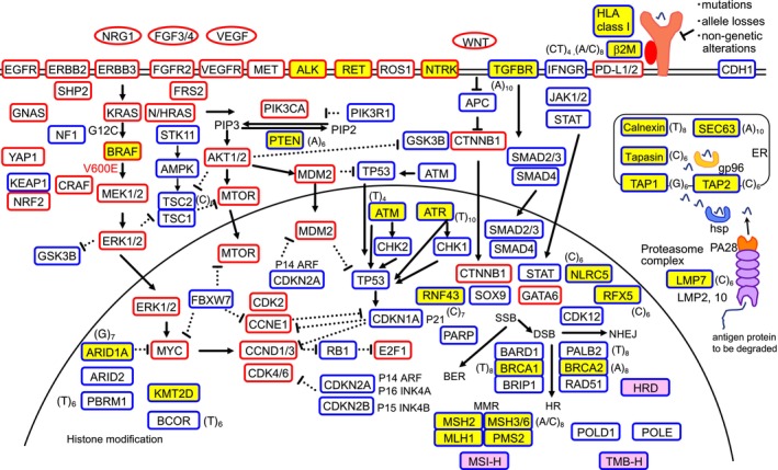
Representative genes altered in high‐microsatellite instability (MSI‐H) cancers. Coding microsatellites in cancer‐associated genes are potential targets of frameshift mutations in MSI‐H cancers. A number of cancer‐associated genes mutated in MSI‐H cancers have been reported. The relevance of each mutation has not necessarily been proven. Because MSI‐H cancers accumulate many mutations, disruption of cell growth and/or survival regulation can be accomplished in different cancers by mutations in different genes of the same signaling pathways.
4.4. The majority of beta 2 microglobulin ( B2M )‐deficient/mutant CRCs benefit clinically from ICI therapy and are MSI‐H
As ICI therapy becomes standard for advanced MSI‐H CRC, ICI response and resistance predictors have become increasingly significant (Figure 3). In melanoma, loss of expression‐associated B2M mutations have been linked to acquired immunotherapy resistance. 45 B2M mutations have also been linked to primary resistance in patients with immunotherapy‐naive CRC. 46 , 47 Middha et al. 48 investigated the association of mutations of B2M with expression of B2M as well as MHC class I, tumor‐infiltrating lymphocytes (TILs), molecular correlates, and immunotherapy response in CRC. B2M mutations were identified in 24% (44/182) of immunotherapy‐naive MSI‐H CRCs; in 93% of these cases, B2M loss was observed, although loss of MHC class I was not typically observed. After controlling for MSI status, B2M mutations were not related to expression of MHC class I, TIL level, PD‐L1 expression, or KRAS or BRAF mutation. Among the 13 patients having B2M‐mutated CRC, partial response, stable disease, and pseudoprogression were observed in five, six, and one patient, respectively. Accordingly, the fact that 11 (85%) of 13 CRCs harboring B2M mutations exhibited some clinical benefit from ICIs indicates that B2M mutations do not serve as a predictor of primary ICI resistance in CRC. Despite loss of B2M, recognition of neoantigen was functional, as evidenced by abundance of TILs in B2M‐defective MSI‐H CRC. Thus, loss of B2M in CRC with MSI‐H may not be enough to result in primary immunotherapy resistance. To determine whether the proportion and period of response differ by status of B2M mutations, larger, prospective studies are required.
4.5. The common frameshift mutation landscape of MSI‐H malignancies supports immunoediting in tumor progression
On cancer cells, the immune system is capable of identifying and eliminating cancer cells, particularly those that contain abundant mutation‐induced neoantigens. Neoantigens of this nature are prevalent in MSI‐H malignancies. Ballhausen et al. established a method for quantifying frameshift mutations in CRC and endometrial cancer with MSI‐H. 49 Counterselection of cell clones having marked immunogenic frameshift peptides is suggested by the negative correlation between the frequency of frameshift mutations and the expected immunogenicity of the resulting peptides (Figure 3). The aforementioned correlation was not observed in tumors harboring B2M mutations. Conversely, coding microsatellite mutation patterns were found to be associated with HLA‐A*02:01 status. Significantly, specific outlier mutations that were associated with frameshift peptides that have been functionally verified as immunogenic were prevalent in MSI‐H malignancies. This suggests that these mutations may have played a driving role in the progression of MSI tumors.
Vaccine candidates that are nascent due to shared mutations hold great potential in the prevention of MSI‐H malignancies 49 (Figure 6). A first phase I/IIa trial established the safety and immunological efficacy of a vaccine containing frameshift peptides (https://clinicaltrials.gov/show/NCT01461148). The landscapes of frameshift mutations in MSI‐H tumors show that frameshift mutations that yield highly immunogenic neoantigens are being negatively selected. This demonstrates the viability of immunoediting in human nonviral malignancies. Frameshift peptide‐based vaccination strategies, which target to fortify the host's immune response against driver mutation‐derived neoantigens, may account for the host's natural immune surveillance during the MSI‐H tumorigenesis. 49 The findings suggest that the condition of the tumor microenvironment and the composition of neoantigens hold promise as novel prognostic biomarkers that may have therapeutic implications for advanced CRC.
FIGURE 6.
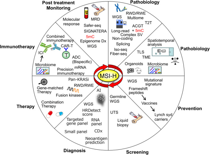
Overview of current and future pathobiology of high‐microsatellite instability (MSI‐H) and precision medicine. MSI‐H‐driven tumorigenesis, focusing on a novel landscape for diagnostic and therapeutic approach, is shown. Abbreviations in this figure are summarized with brief explanations in Table 1.
4.6. Antitumor immunity requires DNA sensing in tumor cells with dMMR
It has been hypothesized that elevated neoantigen levels in hypermutated malignancies with dMMR are the primary cause of the elevated objective response rate observed in therapy targeting PD‐1. On the other hand, the precise mechanism underlying drug resistance remains poorly comprehended. The contribution of tumor cell‐ and host‐derived cGAS‐STING pathways to tumor control has been documented in the context of DNA damage‐inducing therapies. 50 Nevertheless, mounting evidence indicates that activation of tumor cell‐intrinsic cGAS/STING may facilitate tumor initiation, progression, and metastasis in the absence of treatment. Lu et al. 50 demonstrated that the cGAS‐STING pathway in tumor cells is crucial for inhibiting tumor progression in a dMMR setting. They demonstrated that tumor cells with defective MLH1 accumulate cytosolic DNA and release IFN‐γ in a cGAS‐STING‐dependent way, causing tumors with defective MLH1 to grow slowly and be very sensitive to immune checkpoint blockade (Figure 3). Moreover, MLH1 reportedly controls nuclease activity of Exo1 in DNA repair, and MLH1 loss leads to hyperexcision of DNA and replication protein A exhaustion, resulting in chromosomal instability as well as cytosolic accumulation of DNA. Then, the cGAS‐STING pathway is activated and facilitates immunotherapy. 51
dMLH1 tumors effectively induced priming of T cell and lost resistance to immune checkpoint therapy in neoantigen‐fixed models, irrespective of TMB. Consequently, the absence of cGAS/STING in cancer cells resulted in reduced T cell infiltration and confers resistance to immune checkpoint inhibition. Downregulation of cGAS/STING was clinically associated with an unfavorable prognosis in human dMMR malignancies. DNA sensing in tumor cells with dMMR is crucial for antitumor immunity, and it provides novel biomarkers as well as mechanisms for immunotherapy targeting dMMR cancer.
4.7. Immune checkpoint inhibitor therapy in cancer neoadjuvant and adjuvant treatment
Anti‐PD‐1, PD‐L1, and CTLA‐4 monoclonal antibodies have shown remarkable efficacy against a variety of malignancies. MSI‐H/dMMR tumors have been identified as notable indicators for responsiveness to ICIs. The presence of cancer‐associated (clonal) neoantigens, dense immune cell infiltration, and a greater mutational burden are characteristics of these biomarkers that elicit a robust immune response. Pembrolizumab and nivolumab, with or without ipilimumab, are suggested therapeutic options for chemotherapy‐resistant patients with metastatic MSI‐H/dMMR CRCs. The FDA has granted full approval to pembrolizumab for the treatment of patients with unresectable/metastatic MSI‐H/dMMR solid tumors that have advanced following prior treatment.
4.8. Pembrolizumab as first‐line treatment for patients with MSI‐H/dMMR mCRC
For patients having mCRC with MSI‐H/dMMR, the FDA approved pembrolizumab as the first‐line therapy on the basis of the findings of the trial KEYNOTE‐177, in which patients were assigned randomly to receive standard chemotherapy or pembrolizumab. Diaz et al. 52 recently reported the KEYNOTE‐177 study's final overall survival analysis. While pembrolizumab demonstrated persistent antitumor activity and fewer treatment‐related side events when compared with chemotherapy, there was no statistically significant distinction in overall survival between the two treatment cohorts. Pembrolizumab is supported as an efficacious first‐line therapy in patients having mCRC with MSI‐H/dMMR by these findings. Yoshino et al.'s 53 findings provide additional evidence in favor of pembrolizumab as first‐line treatment for Asian patients having mCRC with MSI‐H/dMMR.
5. CONCLUSIONS
We provided an update on MSI‐driven carcinogenesis, with an emphasis on unique landscapes of diagnostic and immunotherapeutic strategies. As shown in Figure 6 and Table 1, the current and future innovations in research of MSI‐H pathogenesis will lead to the development of novel diagnostic, therapeutic, and preventive strategies in clinical settings.
TABLE 1.
Abbreviations used in Figure 6 with brief explanations.
| Abbreviations | Full spelling | Brief explanations |
|---|---|---|
| ADC | Antibody–drug conjugate | A substance made up of a monoclonal antibody chemically linked to a drug |
| CDx | Companion diagnostics | A medical device which provides information that is essential for the safe and effective use of a corresponding drug or biological product |
| Fiber‐seq | Fiber sequencing | A method for analyzing regions with open chromatin that takes advantage of PacBio's ability to call methylated bases |
| HRDetect | Homologous recombination deficiency | A WGS‐based classifier designed to predict BRCA1/2 deficiency based on six mutational signatures |
| Iso‐seq | Iso‐sequencing | Sequencing full‐length cDNA using PacBio SMRT sequencing technology |
| MRD | Minimal residual disease | Cancer cells remaining after treatment that cannot be detected by those same scans or tests |
| RWD | Real‐world data | Data relating to patient health status and/or the delivery of health care routinely collected from a variety of sources |
| RWE | Real‐world evidence | The clinical evidence about the usage and potential benefits or risks of a medical product derived from analysis of RWD |
| Safer‐seq | Safer‐sequencing | A method for detecting rare mutations in blood in a highly efficient manner |
| SIGNATERA | A sensitive ctDNA test for colorectal cancer that can identify relapse sooner than standard tools | |
| SV | Structural variation | A genetic alteration of more than 50 base pairs, including deletions, duplications, insertions, inversions, and translocations, as well as complex rearrangements |
| T2T | Telomere‐to‐telomere | The first complete, gapless sequence of a human genome |
| TLS | Tertiary lymphoid structures | Ectopic lymphocyte aggregates that form at sites of chronic inflammation, including cancers, in nonlymphoid tissues |
| TME | Tumor microenvironment | The normal cells, molecules, and blood vessels that surround and feed a tumor cell |
| UTS | Universal tumor screening | A strategy to identify high‐risk individuals by testing all colorectal tumors for molecular features suggestive of Lynch syndrome |
| WGS | Whole genome sequencing | A comprehensive method for analyzing entire genomes |
AUTHOR CONTRIBUTIONS
Hiroyuki Yamamoto: Conceptualization; visualization; writing – original draft; writing – review and editing. Yoshiyuki Watanabe: Data curation; visualization; writing – review and editing. Hiroyuki Arai: Data curation; visualization; writing – review and editing. Kumiko Umemoto: Data curation; visualization; writing – review and editing. Keisuke Tateishi: Supervision; writing – review and editing. Yu Sunakawa: Supervision; writing – review and editing.
FUNDING INFORMATION
This work was supported by JSPS KAKENHI Grant Number 22H02975.
CONFLICT OF INTEREST STATEMENT
The authors declare no conflict of interest.
ETHICS STATEMENTS
Approval of the research protocol by an Institutional Reviewer Board: N/A.
Informed Consent: N/A.
Registry and the Registration No. of the study/trial: N/A.
Animal Studies: N/A.
ACKNOWLEDGMENTS
Not applicable.
Yamamoto H, Watanabe Y, Arai H, Umemoto K, Tateishi K, Sunakawa Y. Microsatellite instability: A 2024 update. Cancer Sci. 2024;115:1738‐1748. doi: 10.1111/cas.16160
REFERENCES
- 1. Ruiz‐Bañobre J, Goel A. DNA mismatch repair deficiency and immune checkpoint inhibitors in gastrointestinal cancers. Gastroenterology. 2019;156:890‐903. [DOI] [PMC free article] [PubMed] [Google Scholar]
- 2. Ganesh K, Stadler ZK, Cercek A, et al. Immunotherapy in colorectal cancer: rationale, challenges and potential. Nat Rev Gastroenterol Hepatol. 2019;16:361‐375. [DOI] [PMC free article] [PubMed] [Google Scholar]
- 3. Yamamoto H, Imai K. Microsatellite instability: an update. Arch Toxicol. 2015;89:899‐921. [DOI] [PubMed] [Google Scholar]
- 4. Yamamoto H, Imai K. An updated review of microsatellite instability in the era of next‐generation sequencing and precision medicine. Semin Oncol. 2019;46:261‐270. [DOI] [PubMed] [Google Scholar]
- 5. Yamamoto H, Watanabe Y, Maehata T, Imai K, Itoh F. Microsatellite instability in cancer: a novel landscape for diagnostic and therapeutic approach. Arch Toxicol. 2020;94:3349‐3357. [DOI] [PubMed] [Google Scholar]
- 6. Nakamura Y, Kawazoe A, Lordick F, Janjigian YY, Shitara K. Biomarker‐targeted therapies for advanced‐stage gastric and gastro‐oesophageal junction cancers: an emerging paradigm. Nat Rev Clin Oncol. 2021;18:473‐487. [DOI] [PubMed] [Google Scholar]
- 7. Foltz SM, Liang WW, Xie M, Ding L. MIRMMR: binary classification of microsatellite instability using methylation and mutations. Bioinformatics. 2017;33:3799‐3801. [DOI] [PMC free article] [PubMed] [Google Scholar]
- 8. Wang C, Liang C. MSIpred: a python package for tumor microsatellite instability classification from tumor mutation annotation data using a support vector machine. Sci Rep. 2018;8:17546. [DOI] [PMC free article] [PubMed] [Google Scholar]
- 9. Hirotsu Y, Nagakubo Y, Amemiya K, Oyama T, Mochizuki H, Omata M. Microsatellite instability status is determined by targeted sequencing with MSIcall in 25 cancer types. Clin Chim Acta. 2020;502:207‐213. [DOI] [PubMed] [Google Scholar]
- 10. Fujimoto A, Fujita M, Hasegawa T, et al. Comprehensive analysis of indels in whole‐genome microsatellite regions and microsatellite instability across 21 cancer types. Genome Res. 2020;30:334‐346. [DOI] [PMC free article] [PubMed] [Google Scholar]
- 11. Mayrhofer M, De Laere B, Whitington T, et al. Cell‐free DNA profiling of metastatic prostate cancer reveals microsatellite instability, structural rearrangements and clonal hematopoiesis. Genome Med. 2018;10:85. [DOI] [PMC free article] [PubMed] [Google Scholar]
- 12. Willis J, Lefterova MI, Artyomenko A, et al. Validation of microsatellite instability detection using a comprehensive plasma‐based genotyping panel. Clin Cancer Res. 2019;25:7035‐7045. [DOI] [PubMed] [Google Scholar]
- 13. Nakamura Y, Okamoto W, Denda T, et al. Clinical validity of plasma‐based genotyping for microsatellite instability assessment in advanced GI cancers: SCRUM‐Japan GOZILA substudy. JCO Precis Oncol. 2022;6:e2100383. [DOI] [PMC free article] [PubMed] [Google Scholar]
- 14. Nakamura Y, Shitara K. Development of circulating tumour DNA analysis for gastrointestinal cancers. ESMO Open. 2020;5:e000600. [DOI] [PMC free article] [PubMed] [Google Scholar]
- 15. Park JH, Kim EY, Luchini C, et al. Artificial intelligence for predicting microsatellite instability based on tumor histomorphology: a systematic review. Int J Mol Sci. 2022;23:2462. [DOI] [PMC free article] [PubMed] [Google Scholar]
- 16. Echle A, Ghaffari Laleh N, Quirke P, et al. Artificial intelligence for detection of microsatellite instability in colorectal cancer‐a multicentric analysis of a pre‐screening tool for clinical application. ESMO Open. 2022;7:100400. [DOI] [PMC free article] [PubMed] [Google Scholar]
- 17. Herman JG, Umar A, Polyak K, et al. Incidence and functional consequences of hMLH1 promoter hypermethylation in colorectal carcinoma. Proc Natl Acad Sci USA. 1998;95:6870‐6875. [DOI] [PMC free article] [PubMed] [Google Scholar]
- 18. Leung SY, Yuen ST, Chung LP, Chu KM, Chan AS, Ho JC. hMLH1 promoter methylation and lack of hMLH1 expression in sporadic gastric carcinomas with high‐frequency microsatellite instability. Cancer Res. 1999;59:159‐164. [PubMed] [Google Scholar]
- 19. Tomita N, Ishida H, Tanakaya K, et al. Japanese Society for Cancer of the Colon and Rectum (JSCCR) guidelines 2020 for the clinical practice of hereditary colorectal cancer. Int J Clin Oncol. 2021;26:1353‐1419. [DOI] [PMC free article] [PubMed] [Google Scholar]
- 20. Barras D, Missiaglia E, Wirapati P, et al. BRAF V600E mutant colorectal cancer subtypes based on gene expression. Clin Cancer Res. 2017;23:104‐115. [DOI] [PubMed] [Google Scholar]
- 21. Guinney J, Dienstmann R, Wang X, et al. The consensus molecular subtypes of colorectal cancer. Nat Med. 2015;21:1350‐1356. [DOI] [PMC free article] [PubMed] [Google Scholar]
- 22. Fang M, Ou J, Hutchinson L, Green MR. The BRAF oncoprotein functions through the transcriptional repressor MAFG to mediate the CpG Island methylator phenotype. Mol Cell. 2014;55:904‐915. [DOI] [PMC free article] [PubMed] [Google Scholar]
- 23. Hinoue T, Weisenberger DJ, Pan F, et al. Analysis of the association between CIMP and BRAF in colorectal cancer by DNA methylation profiling. PLoS One. 2009;4:e8357. [DOI] [PMC free article] [PubMed] [Google Scholar]
- 24. Kelly AD, Kroeger H, Yamazaki J, et al. A CpG island methylator phenotype in acute myeloid leukemia independent of IDH mutations and associated with a favorable outcome. Leukemia. 2017;31:2011‐2019. [DOI] [PMC free article] [PubMed] [Google Scholar]
- 25. Shen J, Ju Z, Zhao W, et al. ARID1A deficiency promotes mutability and potentiates therapeutic antitumor immunity unleashed by immune checkpoint blockade. Nat Med. 2018;24:556‐562. [DOI] [PMC free article] [PubMed] [Google Scholar]
- 26. Yamada H, Takeshima H, Fujiki R, et al. ARID1A loss‐of‐function induces CpG island methylator phenotype. Cancer Lett. 2022;532:215587. [DOI] [PubMed] [Google Scholar]
- 27. Sato K, Kawazu M, Yamamoto Y, et al. Fusion kinases identified by genomic analyses of sporadic microsatellite instability‐high colorectal cancers. Clin Cancer Res. 2019;25:378‐389. [DOI] [PubMed] [Google Scholar]
- 28. Mulkidjan RS, Saitova ES, Preobrazhenskaya EV, et al. ALK, ROS1, RET and NTRK1–3 gene fusions in colorectal and non‐colorectal microsatellite‐unstable cancers. Int J Mol Sci. 2023;24:13610. [DOI] [PMC free article] [PubMed] [Google Scholar]
- 29. Kim JH, Hong JH, Choi YL, et al. NTRK oncogenic fusions are exclusively associated with the serrated neoplasia pathway in the colorectum and begin to occur in sessile serrated lesions. J Pathol. 2021;255:399‐411. [DOI] [PubMed] [Google Scholar]
- 30. van Wietmarschen N, Nathan WJ, Nussenzweig A. The WRN helicase: resolving a new target in microsatellite unstable cancers. Curr Opin Genet Dev. 2021;71:34‐38. [DOI] [PubMed] [Google Scholar]
- 31. Chan EM, Shibue T, McFarland JM, et al. WRN helicase is a synthetic lethal target in microsatellite unstable cancers. Nature. 2019;568:551‐556. [DOI] [PMC free article] [PubMed] [Google Scholar]
- 32. Lieb S, Blaha‐Ostermann S, Kamper E, et al. Werner syndrome helicase is a selective vulnerability of microsatellite instability‐high tumor cells. elife. 2019;8:e43333. [DOI] [PMC free article] [PubMed] [Google Scholar]
- 33. Kategaya L, Perumal SK, Hager JH, Belmont LD. Werner syndrome helicase is required for the survival of cancer cells with microsatellite instability. iScience. 2019;13:488‐497. [DOI] [PMC free article] [PubMed] [Google Scholar]
- 34. Behan FM, Iorio F, Picco G, et al. Prioritization of cancer therapeutic targets using CRISPR‐Cas9 screens. Nature. 2019;568:511‐516. [DOI] [PubMed] [Google Scholar]
- 35. van Wietmarschen N, Sridharan S, Nathan WJ, et al. Repeat expansions confer WRN dependence in microsatellite‐unstable cancers. Nature. 2020;586:292‐298. [DOI] [PMC free article] [PubMed] [Google Scholar]
- 36. Mengoli V, Ceppi I, Sanchez A, et al. WRN helicase and mismatch repair complexes independently and synergistically disrupt cruciform DNA structures. EMBO J. 2023;42:e111998. [DOI] [PMC free article] [PubMed] [Google Scholar]
- 37. Hao S, Tong J, Jha A, et al. Synthetical lethality of Werner helicase and mismatch repair deficiency is mediated by p53 and PUMA in colon cancer. Proc Natl Acad Sci USA. 2022;119:e2211775119. [DOI] [PMC free article] [PubMed] [Google Scholar]
- 38. Rampino N, Yamamoto H, Ionov Y, et al. Somatic framesfhift mutations in the BAX gene in colon cancers of the microsatellite mutator phenotype. Science. 1997;275:967‐969. [DOI] [PubMed] [Google Scholar]
- 39. Kobayashi Y, Kushihara Y, Saito N, Yamaguchi S, Kakimi K. A novel scoring method based on RNA‐Seq immunograms describing individual cancer‐immunity interactions. Cancer Sci. 2020;111:4031‐4040. [DOI] [PMC free article] [PubMed] [Google Scholar]
- 40. Sato Y, Wada I, Odaira K, et al. Integrative immunogenomic analysis of gastric cancer dictates novel immunological classification and the functional status of tumor‐infiltrating cells. Clin Transl Immunol. 2020;9:e1194. [DOI] [PMC free article] [PubMed] [Google Scholar]
- 41. Saito N, Kobayashi Y, Nagaoka K, et al. Two distinct phenotypes of immunologically hot gastric cancer subtypes. Biochem Biophys Rep. 2021;28:101167. [DOI] [PMC free article] [PubMed] [Google Scholar]
- 42. Wilson BG, Roberts CW. SWI/SNF nucleosome remodellers and cancer. Nat Rev Cancer. 2011;11:481‐492. [DOI] [PubMed] [Google Scholar]
- 43. Wu JN, Roberts CW. ARID1A mutations in cancer: another epigenetic tumor suppressor? Cancer Discov. 2013;3:35‐43. [DOI] [PMC free article] [PubMed] [Google Scholar]
- 44. Kawazu M, Ueno T, Saeki K, et al. HLA class I analysis provides insight into the genetic and epigenetic background of immune evasion in colorectal cancer with high microsatellite instability. Gastroenterology. 2022;162:799‐812. [DOI] [PubMed] [Google Scholar]
- 45. Sade‐Feldman M, Jiao YJ, Chen JH, et al. Resistance to checkpoint blockade therapy through inactivation of antigen presentation. Nat Commun. 2017;8:1136. [DOI] [PMC free article] [PubMed] [Google Scholar]
- 46. Janikovits J, Müller M, Krzykalla J, et al. High numbers of PDCD1(PD‐1)‐positive Tcells and B2M mutations in microsatellite‐unstable colorectalc ancer. Onco Targets Ther. 2017;7:e1390640. [DOI] [PMC free article] [PubMed] [Google Scholar]
- 47. Grasso CS, Giannakis M, Wells DK, et al. Genetic mechanisms of immune evasion in colorectal cancer. Cancer Discov. 2018;8:730‐749. [DOI] [PMC free article] [PubMed] [Google Scholar]
- 48. Middha S, Yaeger R, Shia J, et al. Majority of B2M‐mutant and ‐deficient colorectal carcinomas achieve clinical benefit from immune checkpoint inhibitor therapy and are microsatellite instability‐high. JCO Precis Oncol. 2019;3:PO.18.00321. [DOI] [PMC free article] [PubMed] [Google Scholar]
- 49. Ballhausen A, Przybilla MJ, Jendrusch M, et al. The shared frameshift mutation landscape of microsatellite‐unstable cancers suggests immunoediting during tumor evolution. Nat Commun. 2020;11:4740. [DOI] [PMC free article] [PubMed] [Google Scholar]
- 50. Lu C, Guan J, Lu S, et al. DNA sensing in mismatch repair‐deficient tumor cells is essential for anti‐tumor immunity. Cancer Cell. 2021;39:96‐108. [DOI] [PMC free article] [PubMed] [Google Scholar]
- 51. Guan J, Lu C, Jin Q, et al. MLH1 deficiency‐triggered DNA hyperexcision by exonuclease 1 activates the cGAS‐STING pathway. Cancer Cell. 2021;39:109‐121. [DOI] [PMC free article] [PubMed] [Google Scholar]
- 52. Diaz LA Jr, Shiu KK, Kim TW, et al. Pembrolizumab versus chemotherapy for microsatellite instability‐high or mismatch repair‐deficient metastatic colorectal cancer (KEYNOTE‐177): final analysis of a randomised, open‐label, phase 3 study. Lancet Oncol. 2022;23:659‐670. [DOI] [PMC free article] [PubMed] [Google Scholar]
- 53. Yoshino T, Andre T, Kim TW, et al. Pembrolizumab in Asian patients with microsatellite‐instability‐high/mismatch‐repair‐deficient colorectal cancer. Cancer Sci. 2023;114:1026‐1036. [DOI] [PMC free article] [PubMed] [Google Scholar]