


Fig. 2 |. Experimental approaches for measuring chromatin accessibility and nucleosome positioning.
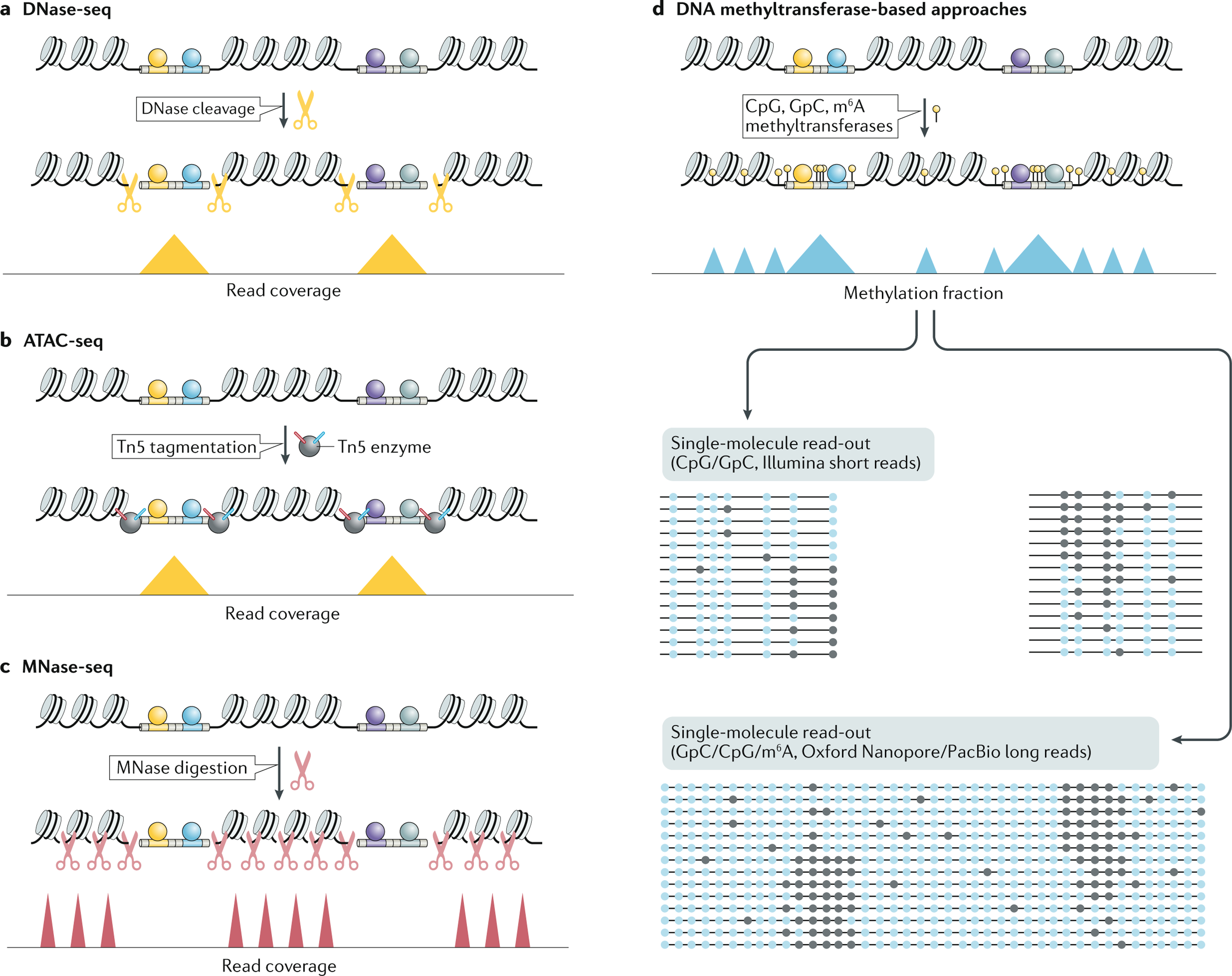
a | In deoxyribonuclease I (DNase I) hypersensitive site sequencing (DNase-seq), the DNase I enzyme (represented as yellow scissors) is used to preferentially cleave accessible chromatin, generating fragments that can then be amplified into sequencing libraries. b | In Assay for Transposase-Accessible Chromatin using sequencing (ATAC-seq), a hyperactive version of the Tn5 transposase (represented by the dark grey circle) is used to preferentially insert into accessible chromatin while simultaneously attaching adapters (represented by the red and blue lines on the Tn5 transposase) to the resulting fragments that can be used to directly amplify sequencing libraries. Both DNase-seq and ATAC-seq generate peaks in read coverage over accessible regions in the genome. c | In micrococcal nuclease sequencing (MNase-seq), the MNase enzyme (represented as red scissors) is used to digest DNA that is not protected by bound proteins, leaving intact fragments protected by protein occupancy (primarily nucleosomes). These fragments are then amplified, resulting in increased read coverage over positioned nucleosomes. d | DNA methyltransferase-based approaches rely on the labelling of accessible DNA with DNA methylation modifications (represented by drawing pins), which can either be sequenced using Illumina platforms following bisulfite conversion or via long-read sequencing platforms that directly read the modified bases (unmodified and modified bases are represented as light and dark blue circles, respectively). These single-molecule chromatin accessibility profiling approaches tend to provide a simultaneous read-out of both nucleosome positioning and accessible chromatin regions. Accessible chromatin regions represent themselves as higher peaks due the fact that they have more nucleotides that are accessible to the methyltransferases and are therefore more frequently methylated, compared with the internucleosomal sequences that are thus not methylated in every single-molecule read. In all four panels, bound transcription factors (TFs) are visualized via coloured circles on the accessible chromatin.