



Abstract
Background.
Antiretroviral treatment has enabled people living with HIV infection to have a near-normal lifespan. With longevity come opportunities for engaging in risky behavior, including initiation of excessive drinking. Given that both HIV infection and alcohol use disorder (AUD) can disrupt brain white matter integrity, we questioned whether HIV infection, even if successfully treated, or AUD alone results in signs of accelerated white matter aging and whether HIV+AUD comorbidity further accelerates brain aging.
Methods.
Longitudinal MRI-FLAIR data were acquired over 15 years in 179 controls, 204 AUD participants, 70 with HIV, and 75 comorbid for HIV+AUD. White matter hyperintensity (WMH) volumes were quantified and localized and their functional relevance was examined with cognitive and motor testing.
Results.
The three diagnostic groups each had larger WMH volumes than controls. Although all four groups exhibited accelerating volume increases with aging, only the HIV groups showed faster WMH enlargement than controls; the comorbid group showed faster acceleration than the HIV-only group. Sex and HIV infection length, but not viral suppression status, moderated acceleration. Correlations emerged between WMH volumes and Attention/Working Memory and Executive Function scores of the AUD and HIV groups, and between WMH volumes and Motor Skills in the three diagnostic groups.
Conclusions.
Even treated HIV can show accelerated aging, possibly from treatment sequelae or legacy effects, and notably from AUD comorbidity. WMH volumes may be especially relevant for tracking HIV and AUD brain health because each condition is associated with liability for hypertensive processes, for which WMHs are considered a marker.
Keywords: HIV infection, alcohol, brain, white matter hyperintensity, aging, cognition
INTRODUCTION
Currently, an estimated 38.4 million people worldwide are aging with the human immunodeficiency virus (HIV)(1), and 51% of people living with HIV (PLWH) in the U.S. are 50 years and older(2). Antiretroviral therapy (ART) has reduced the risk for opportunistic infections(3) and has considerably improved prognosis and life expectancy in PLWH(4–6). Brain white matter injuries such as progressive multifocal leukoencephalopathy(7–9), HIV-related encephalopathy(10), and ART-triggered immune responses (i.e., immune reconstitution inflammatory syndrome, IRIS)(11–13) have all declined. Yet, quantitative neuroimaging studies provide initial cross-sectional(14) and longitudinal(15) (but see16) evidence for greater age-related declines in cortical volume of largely treated HIV groups than in uninfected control groups. Thus, even in ART-treated, virally-suppressed HIV, infection or pharmacotreatment legacy effects may contribute to accelerated aging(17–19).
Accompanying expanded longevity are opportunities to develop comorbidities, thereby complicating an understanding of aging in PLWH. Indeed, a large-scale longitudinal study reported more comorbidities in PLWH than in uninfected participants who were followed biennially for about 6 years. At their initial examination, participants, who were ages 50–55 years old, had nearly all been treated successfully indexed by low viral loads. Greater numbers of comorbidities unrelated to HIV infection in PLWH increased their risk of death and number of disability-adjusted life-years(20) and highlight the need to consider common comorbidities in studies of aging with HIV.
Evidence for quadratic or even exponential increases in brain structural features, such as lateral ventricular volumes(21) or presence of white matter hyperintensities (WMH),(22, 23) occurs with advancing age in neurologically healthy men and women. The possibility of acceleration of normal aging in PLWH is consistent with the hypothesis that early disease or insult, even when overcome, can re-emerge as a factor to accelerate normal aging(24). Accelerated aging in HIV alone and in combination with common comorbidities, notably alcohol use disorder (AUD)(25, 26), may also herald signs of premature cognitive and motor decline(15, 27–29) and HIV Associated Neurocognitive Disorder(30, 31), especially as PLWH reach ages at risk for dementia. For brain structure, factors accelerating gray matter and white matter degradation are HIV+AUD comorbidity and aging(15, 32). Several studies noted a compounded adverse effect of HIV+AUD comorbidity on white matter diffusivity metrics(33, 34). Additionally, WMH, visible in periventricular (PVWMH) and deep (DWMH) white matter regions, occur in HIV, AUD, and normal aging(18, 35–37), and tracking the trajectories of these neuroradiological signs may serve to identify modifiable risk factors and comorbidity targets for prevention.
WMH is a neuroimaging feature of cerebral small vessel disease commonly found in normal aging(38–43), HIV infection(44, 45) (46–48), and AUD(49, 50). As in the general population, age, hypertension, and diabetes(36, 51–55) are major risk factors(35) contributing to WMH burden, which is also associated with worse cognitive performance [general population(56–58); PLWH on ART(35, 52, 54, 59, 60)]. Relations between WMH and cognitive and motor performance may be mediated by hypertension(61, 62) or other factors(63, 64), including education(14, 65), ART(52, 66, 67), hepatitis C virus (HCV) comorbidity(68), and WMH location(69–72).
Quantitative measurement of the size and location of WMH(73) in AUD, HIV, and HIV+AUD comorbidity has the potential of leading to a mechanistic understanding of this anomalous tissue as people age with these conditions(41). Accordingly, we focused on WMH volumetry to test the following hypotheses in people with AUD, HIV, and HIV+AUD and in an unaffected comparison group examined contemporaneously: 1) MRI data acquired longitudinally would reveal significantly larger WMH volumes and faster enlargement in the three diagnostic groups than in control participants; 2) high viral load, high blood pressure, and heavy alcohol use would be associated with faster acceleration of WMH volumes; and 3) greater WMH volumes would be predictive of poorer cognitive and motor performance regardless of diagnosis. Exploratory analysis examined potential sex differences in presence and progression of WMH volume enlargement.
METHOD
Participants
Data collection proceeded from 12 October 2006 to 25 October 2021 of our four study groups: 179 control, 204 AUD, 70 HIV, 75 HIV+AUD (Supplemental Figure 1; Table 1). Participants were recruited from local alcohol and drug recovery centers, HIV clinics, post card mailings, recruitment flyers, and word of mouth. Most of these participants were also represented in our earlier longitudinal studies(15, 74). The current study focus was on the detection and quantification of WMHs quantified from 1,056 FLAIR images; participants were aged 18.5 to 86.1 years old at study entry.
Table 1.
Study entry demographics of the study groups at baseline: mean (SD) or frequency count.
| Control | AUD | HIV† | HIV+AUD | lm for Control vs. each diagnostic group | ||
|---|---|---|---|---|---|---|
|
| ||||||
| Age (yrs) | 47.77 | 49.74 | 53.75 | 51.63 | C<H, H+A | |
| (16.41) | (10.86) | (9.13) | (9.32) | |||
| male/female n= | 99/80 | 151/53 | 49/21 | 50/25 | ||
| Education (yrs) | 16.25 | 13.27 | 13.49 | 13.11 | C>A, H, H+A | |
| (2.29) | (2.46) | (2.76) | (2.00) | |||
| n= | 165 | 196 | 70 | 75 | ||
| Socioeconomic status (lower score=higher status) | 24.67 | 41.47 | 38.97 | 43.57 | C<A, H, H+A | |
| (11.75) | (15.01) | (15.15) | (12.52) | |||
| n= | 177 | 203 | 70 | 75 | ||
| Body Mass Index (BMI) | 25.54 | 27.01 | 26.03 | 26.23 | C<A | |
| (4.20) | (4.36) | (4.28) | (4.18) | |||
| n= | 143 | 186 | 70 | 74 | ||
| Diastolic blood pressure (dBP) | 75.39 | 78.72 | 76.23 | 78.52 | C<A | |
| (10.03) | (11.30) | (10.99) | (9.09) | |||
| n= | 87 | 161 | 60 | 56 | ||
| Systolic blood pressure (sBP) | 126.91 | 129.33 | 125.80 | 128.88 | n.s. | |
| (17.57) | (17.00) | (15.44) | (14.92) | |||
| n= | 87 | 161 | 60 | 56 | ||
| Mean test composite Z-score†† (lower scores=worse performance) | 0.05 | −0.75 | −0.75 | −1.06 | C>A, H, H+A | |
| (0.63) | (0.81) | (0.89) | (0.89) | |||
| n= | 100 | 160 | 70 | 72 | ||
| Global Assessment of Functioning (GAF) | 85.21 | 66.54 | 70.99 | 65.11 | C>A, H, H+A | |
| (6.55) | (10.35) | (10.86) | (9.25) | |||
| n= | 147 | 196 | 70 | 73 | ||
| Falls in past year | 1.00 | 1.49 | 0.97 | 1.09 | n.s. | |
| (1.16) | (1.92) | (1.13) | (1.44) | |||
| n= | 35 | 63 | 30 | 32 | ||
| Beck Depression Inventory II (BDI) | 2.20 | 8.64 | 9.88 | 10.52 | C<A, H, H+A | |
| (3.45) | (8.20) | (8.02) | (8.92) | |||
| n= | 140 | 175 | 69 | 73 | ||
| Age at AUD diagnosis | — | 25.67 | — | — | — | |
| (9.56) | ||||||
| n= | 187 | |||||
| Length of HIV diagnosis | — | — | 18.44 | 17.09 | HIV length: n.s. | |
| (8.97) | (8.09) | |||||
| n= | 64 | 70 | ||||
| Viral load (count/mL) | Median= | — | — | 48 | 47 | n.s. |
| SD= | (20079.57) | (44418.00) | ||||
| n= | 62 | 67 | ||||
| 10 Viral load log−10 (count/mL) | Mean= | — | — | 2.03 | 2.23 | n.s. |
| Median= | (1.67) | (1.67) | ||||
| n= | 62 | 67 | ||||
| CD4 (count/mL) | Mean= | — | — | 597.34 | 552.80 | n.s. |
| SD= | (252.95) | (331.19) | ||||
| n= | 67 | 70 | ||||
| CD8 (count/mL) | Mean= | — | — | 1016.21 | 952.76 | n.s. |
| SD= | (487.26) | (461.11) | ||||
| n= | 67 | 70 | ||||
| CD4/CD8 (count/mL) | Mean= | — | — | 0.67 | 0.70 | n.s. |
| SD= | (0.30) | (0.50) | ||||
| n= | 67 | 70 | ||||
| CD4 nadir | — | — | 173.00 | 195.32 | n.s. | |
| (142.53) | (163.50) | |||||
| n= | 54 | 53 | ||||
| AIDS yes/no | — | — | 35/29 | 41/31 | chi2=.07, p=.79 | |
| HCV yes/no | 1/92 | 33/124 | 20/46 | 33/40 | chi2=48.77, p=.00001 | |
| Age at nicotine diagnosis | 19.19 | 19.40 | 17.22 | 17.04 | n.s. | |
| (3.61) | (8.39) | (6.52) | (5.12) | |||
| n= | 21 | 125 | 27 | 42 | ||
| Smoker: never/past or current | 121/21 | 64/132 | 39/27 | 24/44 | chi2=101.12, p=.00001 | |
| Race: Black/White/Asian/other | 26/106/37/9 | 69/94/7/34 | 25/34/0/11 | 46/13/0/16 | chi2=117.96, p=.00001 | |
After obtaining written informed consent for study participation, approved by the SRI International and Stanford University School of Medicine Institutional Review Boards, all participants underwent Structured Clinical Interviews for DSM-IV revised (75) to determine diagnosis for alcohol dependence and DSM-5 diagnosis for AUD.(76) Interviews included structured health questionnaires, a semi-structured timeline follow-back interview to quantify lifetime alcohol consumption.(77, 78) Principal exclusion criteria at study entry were history of schizophrenia, bipolar disorder, and neurological disorders other than AUD. Substance use dependence (current or in remission) for illicit drugs was common and involved cocaine, amphetamines, opiates, and cannabis (although now legal in California). Hematological evaluation determined HIV and hepatitis C presence and load. All participants also completed screening to ensure MRI safety. Starting each study day, participants underwent a breathalyzer to test for recent alcohol consumption.
Neuroimaging acquisition and analysis
Protocols and specific parameters are presented in supplemental material. Briefly, scanning was conducted at SRI International on a GE Discovery MR750 system. Detection and localization of WMHs used three MRI acquisition protocols: T1-weighted (T1-w) MRI for anatomical localization, T2-weighted MRI for skull stripping, and FLAIR (FLuid-Attenuated Inversion Recovery) imaging for estimates of WMH volumes.
WMH analysis was accomplished with the “UBO Detector,” a cluster-based, fully automated pipeline for extracting and calculating WMH on a voxel basis(79, 80); see Supplemental Material. This procedure yielded voxel maps of total, periventricular (PVWMH), and deep (DWMH) WMHs (Figure 1) and WMH locations by four lobar regions. Analysis required that FLAIR and T1-w data be transformed into MNI space prior to non-rigid transformation into standard SRI atlas space. This was necessary for accurate placement of anatomical locations to enable comparisons across individuals and across imaging modalities on a voxel-wise basis without the need for further correction for intracranial volume.
Figure 1.
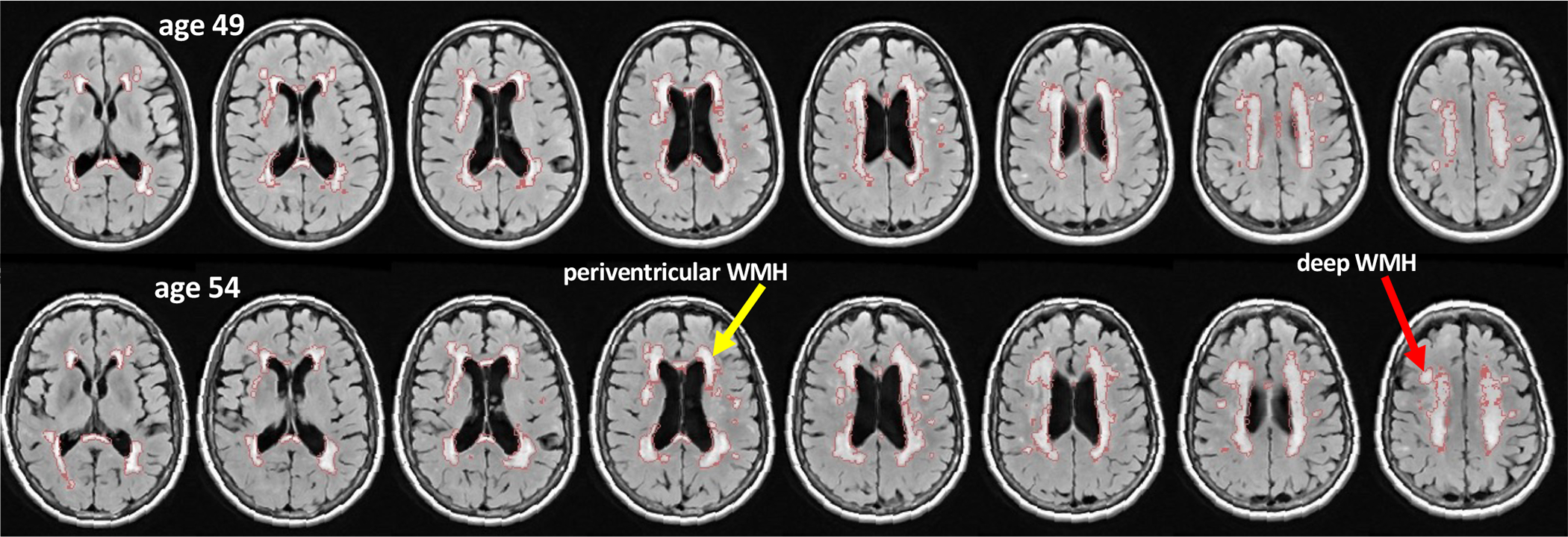
Axial images from FLAIR images acquired in an HIV infected woman examined when she was age 49 and then 54 years old. Note the enlargement of the WMHs delineated in red outlining. Examples of periventricular WMH (yellow arrows) and deep WMH tissue (red arrows) are indicated.
Functional measures
The neuropsychological test battery used cognitive and motor tests (Supplemental Table 1) to calculate neuropsychological composite scores(30, 31) representing five functional domains: Executive Functioning, Attention/Working Memory, Verbal and Visual Learning, Verbal and Visual Memory, and Motor Skills. To create a composite, scores of each test were transformed into age, education, and sex corrected Z-scores based on scores from 102 cognitively-intact, volunteers (47 women; 47.9±14.9 years; 50 White, 25 Black, 27 other race; 15.8±2.4 years educated; 27.1±12.9 SES; 90 right handed)(81). Composite scores for each domain equaled the average, demographically-adjusted Z-scores comprising that domain. A summary composite score was the mean of the five domain composite scores(82).
Sitting blood pressure was collected twice. The mean of each of the two diastolic and systolic measures was used as a correlate of WMH metrics.
Statistical analysis
Statistical analysis was performed using R 3.5.1 (R Core Team, 2019) on WMH volumes obtained with the UBO analysis procedure. The primary statistic was a linear mixed effects model (lmer) separately predicting each WMH volume as a function of group (control, AUD, HIV, and HIV+AUD)+age. Because of the heteroscedasticity of WMH volumes across individuals, their values were square-root transformed before statistical analysis. Interactions with age tested potential group differences in rates of change over the exam range. To account for differences in demographic characteristics, statistical models included diagnosis, age (and age2), sex, race, socioeconomic status (SES), years of education, BMI, systolic and diastolic blood pressure, and depressive symptoms (Beck Depression Inventory score) in multiple regression to test whether any of these variables could account for group differences observed in WMH volumes or their interactions with age. Secondary analyses examined the influence on the WMH volumes of HIV-related factors in the HIV groups and four principal drugs of abuse—cocaine, amphetamine, opiates, and cannabis—within each patient group. The model outputs produced t and p values for each diagnostic group relative to the controls. To compare HIV to HIV+AUD, the model was rerun on the two HIV groups with HIV as the index level. Further lmer calculations included sex in the model. Correlations examined relations between regional WMH volumes and cognitive and motor, hematological, and blood pressure scores within each diagnostic group. Bonferroni correction was applied to correct for multiple comparisons in each primary analysis.
RESULTS
The mainstay of the statistical test results is presented in the tables and figures.
Tests of diagnostic differences in WMH volumes and evidence for accelerated aging
For all four study groups, age-squared regression rather than linear age models best fit the relations between regional WMH volumes and age (Figure 2). Thus, WMH volumes were larger in older than younger participants in all four study groups, including those in the control group screened to meet health criteria. Tests of group differences were based on multiple regression using lmer accounting for sex, race, SES, education, BMI, BDI, and systolic and diastolic blood pressure. The results indicated that the three diagnostic groups—AUD, HIV, and HIV+AUD—had significantly larger total, PVWMH, and DWMH volumes than the control group, with one exception: the DWMH volume difference was not significant between the control and comorbid group (Table 2, left).
Figure 2.
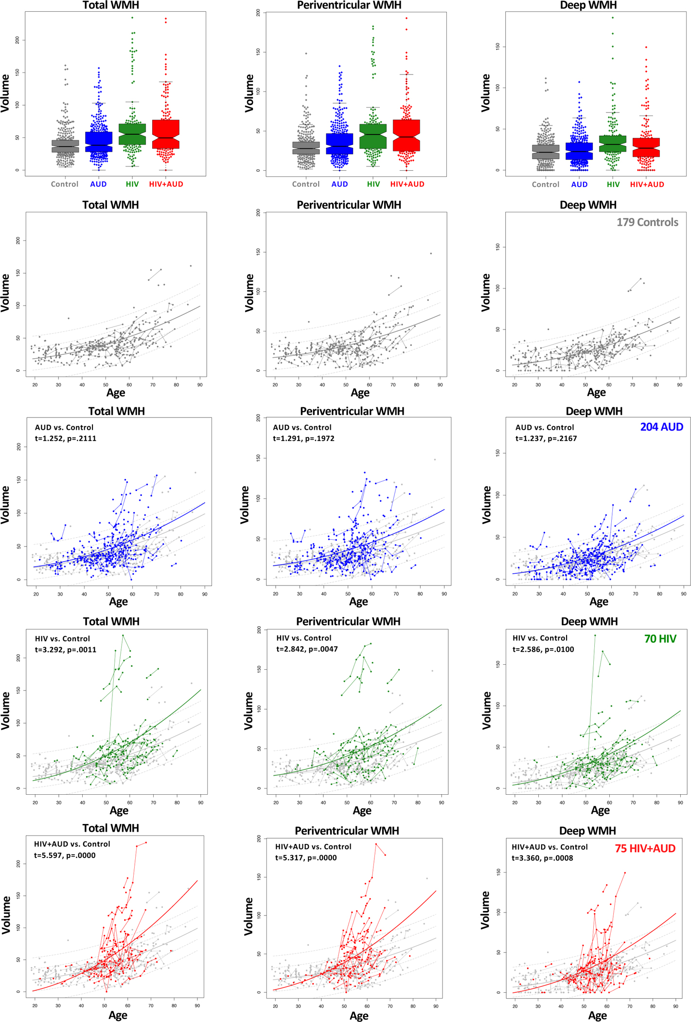
Top row: Box plots and data points of volumes of the Total (left column), PVWMH (middle column), and DWMH (right column) volumes for each participant’s data for all visits. Controls (gray), AUD (blue), HIV (green), and HIV+AUD (red). Bottom 12 spaghetti plots: Total, PVWMH, and DWMH volumes for individual from each study group. Data and regression lines of the three diagnostic groups are plotted on the control regression lines for comparison. The diagnostic group-by-control interaction results are provided in the upper left corner of the plots. Although all three diagnostic groups had larger WMH volumes in each region, only the two HIV groups showed significant interactions with controls over age, indicating age accelerations.
Table 2.
Diagnostic and sex differences in volume and interaction with aging.
| Group difference | Group × age2 interaction | |||
|---|---|---|---|---|
|
|
|
|||
| t | p | t | p | |
|
| ||||
| AUD vs. control | ||||
| Total WMH | 3.291 | 0.001 | 0.091 | 0.928 |
| Periventricular WMH | 3.130 | 0.002 | 0.308 | 0.758 |
| Deep WMH | 2.606 | 0.010 | 0.339 | 0.735 |
| HIV vs. control | ||||
| Total WMH | 3.054 | 0.003 | 2.124 | 0.034 |
| Periventricular WMH | 3.492 | 0.001 | 2.138 | 0.033 |
| Deep WMH | 2.235 | 0.027 | 1.667 | 0.097 |
| HIV+AUD vs. control | ||||
| Total WMH | 2.132 | 0.034 | 2.887 | 0.004 |
| Periventricular WMH | 2.426 | 0.016 | 2.729 | 0.007 |
| Deep WMH | 1.125 | 0.262 | 1.602 | 0.110 |
| HIV+AUD vs. HIV | ||||
| Total WMH | −0.100 | 0.921 | 1.503 | 0.134 |
| Periventricular WMH | −0.419 | 0.676 | 2.518 | 0.012 |
| Deep WMH | −0.311 | 0.756 | 0.688 | 0.492 |
Group differences resulting from multiple regression using lmer accounting for sex, race (Asian, Black, other), SES, education, BMI, BDI, systolic and diastolic blood pressure
Bold font indicates p≤.05 without correction for multiple comparisons.
The next analyses sought accelerated aging differences by testing for interactions between diagnostic group and the age-squared functions. The age-related accelerations in the AUD group were not statistically greater than those in the control group. By contrast, significant group by age interactions were identified in both HIV groups relative to controls in PVWMH and total WMH volumes (Table 2, right). A further comparison of aging interactions focused on the two HIV groups and yielded a significantly greater accelerated aging of PVWMH volumes in the HIV+AUD group relative to the HIV only group (Figure 3).
Figure 3.
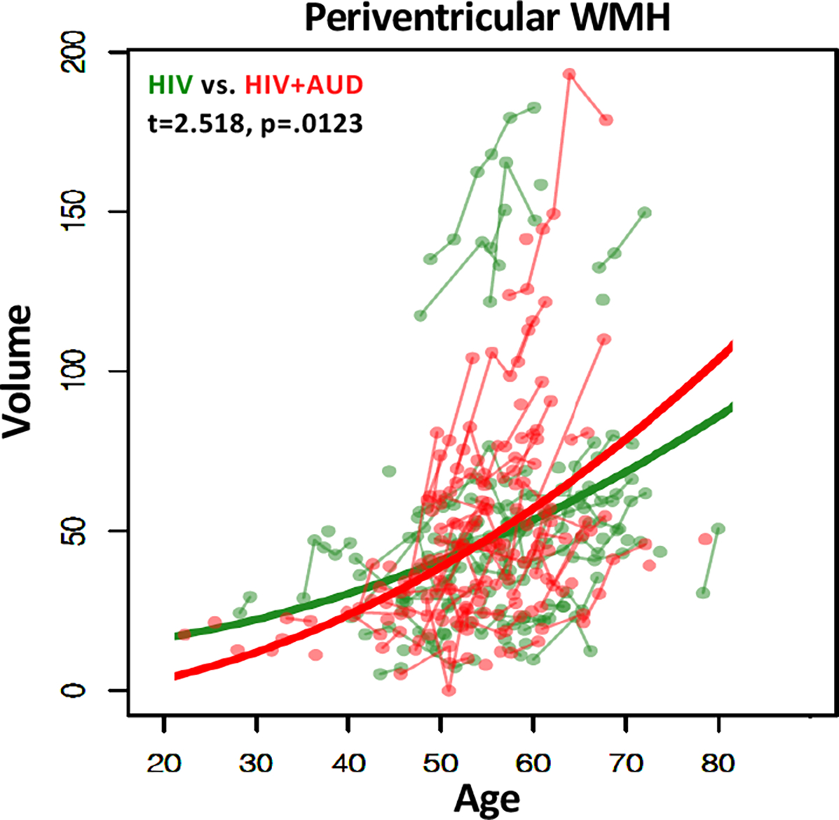
Longitudinal data and regression lines for PVWMH volumes of individuals with HIV (green) and HIV+AUD (red). The HIV+AUD group shows a faster volume acceleration over age than the HIV group.
Of the factors intended to adjust for demographic differences among the groups, sex, SES, race, and blood pressure emerged as significant contributors in a few instances but did not remove the primary group or interaction effects noted above. Specifically, for the comparisons between AUD and controls, the significant effects were for age and diagnosis for all three WMH measures but not accelerated age effects greater than those in the controls. For the HIV comparisons, age and diagnosis effects were significant as were the effects of sex (larger relative WMH volumes in women than men) and SES (larger volumes in people with poorer SES); interactions endured for total and PVWM but not DWMH volumes. For the HIV+AUD comparisons, significant age and diagnosis effects endured for total and PVWMH volume despite additional significant effects of sex (larger relative WMH volumes in women than men), race (greater burden in Black and Asian participants), and systolic blood pressure; interactions endured for total and PVWM but not DWMH volumes. Finally, for the HIV only vs. HIV+AUD comparisons, the age-by diagnosis interaction remained significant for PVWMH, where the volumes accelerated faster over age in the comorbid group than in HIV-only group.
Disease and drug variables potentially influencing WMH volumes
Factors relevant to AUD were age of AUD onset and to HIV infection were estimated age of infection, ART history with reference to history of efavirenz, CD4 nadir and count, CD8 count, CD4/CD8, viral suppression, and HCV infection. Specifically, more years with an HIV infection diagnosis in the HIV+AUD group (total r=.378, p=0013; PVWMH r=.270, p=.024; DWMH r=.392, p=8e-04) and older age at initial AUD diagnosis in the AUD group correlated significantly with larger WMH volumes (total r=.282, p=.0001; PVWMH r=.265, p=.0003; DWMH r=.278, all p’s=.0001). Individuals with versus without an HIV-related disease conditions—CD4 cell count nadir, CD4 cell count, history of an AIDS-defining event, HCV, and nicotine diagnosis—did not differ in regional WMH volumes (p-values=.0899 to .9377, data not shown). We also examined the potential effect of ART type, focusing on treatments that included efavirenz, which are considered especially neurotoxic(83), but found that history of efavirenz use did not remove evidence for age acceleration of the PVWMH volumes of the PLWH with versus without AUD (t=−0.229, p=.819).
Regarding the role of viral suppression on WMH volumes, the HIV groups were divided according to individuals with viral loads < 50 counts/mL (47 HIV and 46 HIV+AUD, considered virally suppressed) and those with viral loads ≥ 50 counts/mL (15 HIV and 21 HIV+AUD). In both HIV groups, those considered virally suppressed had significantly higher CD4 cell counts (HIV t=3.411, p=.0014; HIV+AUD t=4.29, p=.0001) and CD4/CD8 ratios (HIV t=5.10, p<.0001; HIV+AUD t=3.82, p=.0003). Despite these differences, virally suppressed versus non-suppressed HIV groups did not differ in the expected direction for any regional WMH volume. Although PVWMH volumes of the comorbid group were larger than those of the HIV-only group, the difference was not significant (t=1.744, p=.0852); unexpectedly, the non-suppressed HIV group had a lesser DWMH burden than the virally-suppressed group (t=2.92, p=.0051).
We also sought differences within each diagnostic group based on presence vs. absence of HCV infection. None was significant: HIV: total WMH t=0.947, p=.351; PVWMH t=1.162, p=.253; DWMH t=.435, p=.666. HIV+AUD: total WMH t=1.205, p=.233; PVWMH t=1.727, p=.090; DWMH t=−.113, p=.910.
Finally, the influence on the WMH volumes of recent use (that is, over the past month) of any of four principal drugs of abuse—cocaine, amphetamine, opiates, and cannabis—was tested within each patient group. In no case were our findings of group differences or age-accelerated changes in WMH burden accounted for by such use of any of these drugs. This result held whether the lmer was conducted for each drug alone (p=.163 to .944) or whether all drugs were entered into a single analysis for each diagnostic comparison (p=.083 to .948).
Group differences on test composite scores and correlations with WMH volumes
Each diagnostic group achieved five neuropsychological test composite scores and a composite mean score that were significantly lower than those of the control group (p≤.001, Figure 4). Performance on multiple composites across the four study groups correlated with larger WMH volumes, and most correlations noted next met correction for multiple comparisons with directional testing (p≤.017, Table 3; Figure 5). Specifically, in the AUD group, Attention/Working Memory, Executive Function, Motor Skills, and mean domain scores correlated with larger total, PVWMH, and DWMH volumes. In the HIV group, the strongest relations were between lower scores on the Attention/Working Memory and the Motor Skills composites and larger total and DWMH volumes. In the HIV+AUD group, Motor Skills and mean composite scores correlated with larger total and PVWMH volumes. The relations endured despite controlling for age and blood pressure.
Figure 4.
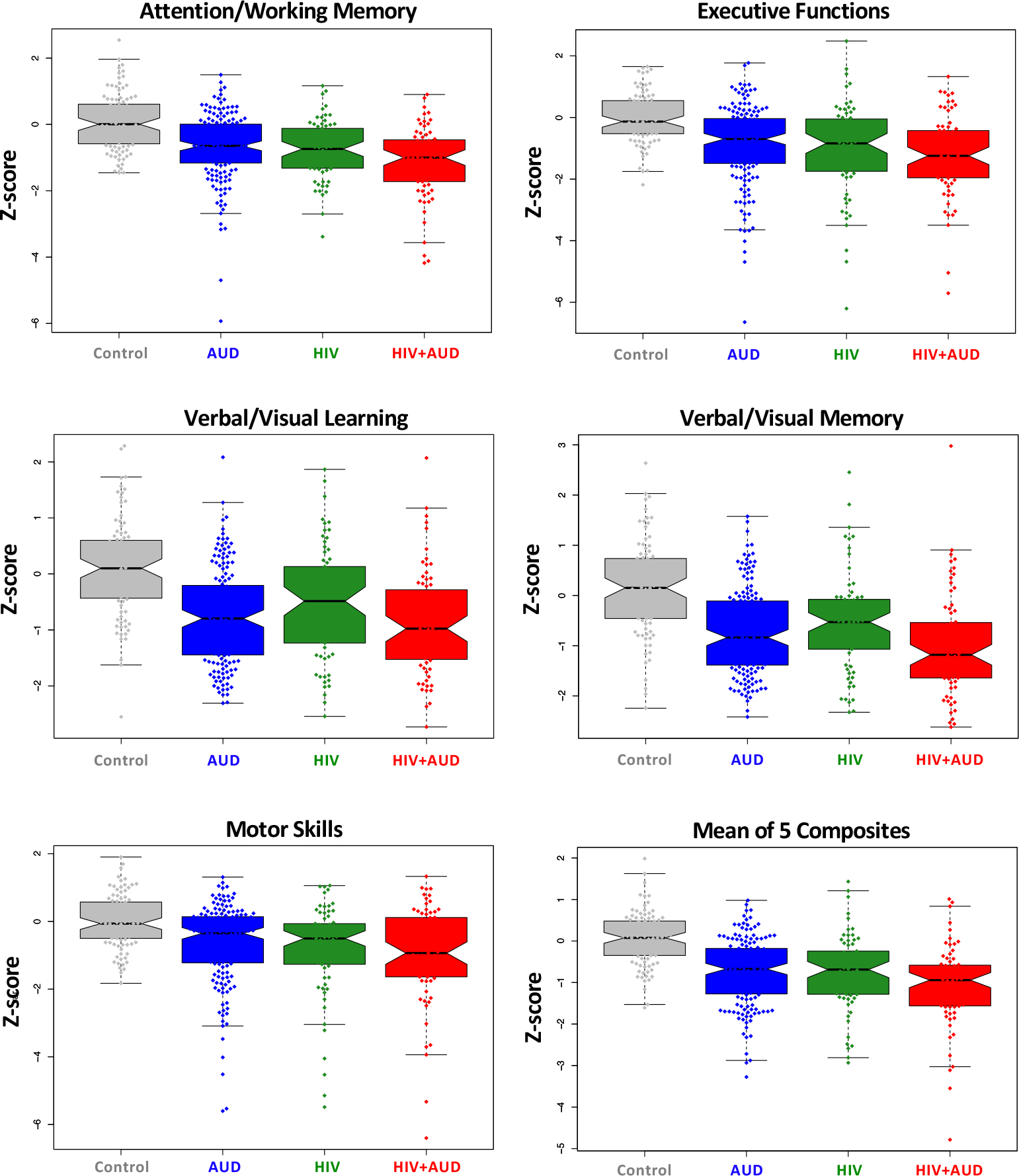
Box plots overlaid on the scatterplots of Z-scores for the five domain composite scores and the mean domain scores, each of which was age, sex, and education adjusted on the controls and acquired at initial study time. The four study groups are color coded.
Table 3.
Results of lm analyses testing relations between diagnostic and other study variables and regional WMH volumes.
| Total WMH | PVWMH | DWMH | ||||
|---|---|---|---|---|---|---|
|
| ||||||
| AUD | t | p | t | p | t | p |
|
|
|
|
||||
| Attention/Working Memory | −3.641 | 0.000 | −3.928 | 0.000 | −2.088 | 0.038 |
| Executive Functions | −3.619 | 0.000 | −3.687 | 0.000 | −2.781 | 0.006 |
| Verbal/Visual Learning | −1.035 | 0.302 | −0.961 | 0.338 | −0.808 | 0.420 |
| Verbal/Visual Memory | −1.909 | 0.058 | −1.693 | 0.092 | −1.580 | 0.116 |
| Motor Skills | −3.279 | 0.001 | −3.224 | 0.002 | −2.490 | 0.014 |
| Mean | −3.747 | 0.000 | −3.769 | 0.000 | −2.683 | 0.008 |
|
| ||||||
| HIV | t | p | t | p | t | p |
|
|
|
|
||||
| Attention/Working Memory | −2.996 | 0.004 | −2.423 | 0.018 | −3.704 | 0.000 |
| Executive Functions | −1.533 | 0.130 | −1.572 | 0.121 | −1.408 | 0.164 |
| Verbal/Visual Learning | −1.042 | 0.301 | −0.694 | 0.490 | −1.491 | 0.140 |
| Verbal/Visual Memory | −0.954 | 0.343 | −0.671 | 0.504 | −1.272 | 0.208 |
| Motor Skills | −2.489 | 0.015 | −2.108 | 0.039 | −2.859 | 0.006 |
| Mean | −2.240 | 0.028 | −1.883 | 0.064 | −2.620 | 0.011 |
|
| ||||||
| HIV+AUD | t | p | t | p | t | p |
|
|
|
|
||||
| Attention/Working Memory | −1.738 | 0.087 | −1.943 | 0.056 | −1.112 | 0.270 |
| Executive Functions | −0.902 | 0.370 | −0.993 | 0.324 | −0.403 | 0.688 |
| Verbal/Visual Learning | −2.517 | 0.014 | −2.363 | 0.021 | −2.103 | 0.039 |
| Verbal/Visual Memory | −2.228 | 0.029 | −2.150 | 0.035 | −1.844 | 0.069 |
| Motor Skills | −3.657 | 0.001 | −4.119 | 0.000 | −1.889 | 0.063 |
| Mean | −2.897 | 0.005 | −3.055 | 0.003 | −1.852 | 0.068 |
|
| ||||||
| Control | t | p | t | p | t | p |
|
|
|
|
||||
| Attention/Working Memory | −1.826 | 0.071 | −2.155 | 0.034 | −0.772 | 0.442 |
| Executive Functions | −2.117 | 0.037 | −2.343 | 0.022 | −0.968 | 0.336 |
| Verbal/Visual Learning | 1.190 | 0.237 | 1.328 | 0.188 | 0.954 | 0.343 |
| Verbal/Visual Memory | 0.634 | 0.527 | 0.815 | 0.417 | 0.467 | 0.642 |
| Motor Skills | −1.728 | 0.087 | −1.714 | 0.090 | −1.182 | 0.240 |
| Mean | −0.804 | 0.423 | −0.799 | 0.426 | −0.297 | 0.767 |
The neuropsychological test scores were adjusted for age, education, and SES of a control sample.
Bold font indicates Bonferroni correction for 6 comparisons with α=.05, directional hypothesis requires p=.017.
Figure 5.
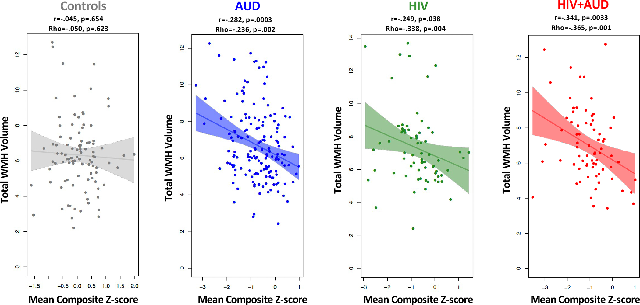
Linear regressions and 95% confidence intervals for significant correlations between mean composite Z-scores and Total WMH volumes at initial study time, indicating that larger WMH volumes correlated with poorer performance in each group. Additional functional-WMH volume correlations appear in Tables 3 and 4.
The virally-suppressed HIV+AUD group achieved significantly better composite scores than the non-suppressed group on Attention/Memory (t=2.92, p=.005), Verbal/Visual Learning (t=1.991, p=.0498), Verbal/Visual Memory (t=2.52, p=.0135), and the mean of the five composite scores (t=2.44, p=.0167). None of the performance scores of the HIV-only subgroups differed significantly by viral suppression.
Group differences in lobar WMH volumes
The regression analysis results, adjusted for sex, race, SES, education, BMI, BDI, and systolic and diastolic blood pressure, on group and group-by-age2 effects of the four lobar volumes, appear in Table 4 (Supplemental Figure 2). In general, the three diagnostic groups had larger regional WMH volumes than controls. Relative to the control group, the AUD group had significantly larger volume in frontal, temporal, and parietal regions; the volume difference was significant in the occipital region in the HIV only group; and the frontal volume difference from controls was marginal for the HIV+AUD group. Both HIV groups exhibited accelerated aging of the frontal WMH volumes relative to controls (Table 4).
Table 4.
Diagnostic difference in lobar WMH volume and interaction with aging.
| Group difference | Group × age2 interaction | |||
|---|---|---|---|---|
|
|
|
|||
| t | p | t | p | |
|
| ||||
| AUD vs. control | ||||
| Frontal | 2.450 | 0.015 | 1.212 | 0.226 |
| Temporal | 2.390 | 0.018 | 0.499 | 0.618 |
| Parietal | 2.501 | 0.013 | 0.066 | 0.947 |
| Occipital | 0.864 | 0.388 | −0.613 | 0.541 |
| HIV vs. control | ||||
| Frontal | 1.254 | 0.212 | 2.185 | 0.030 |
| Temporal | 1.495 | 0.138 | 0.163 | 0.871 |
| Parietal | 1.783 | 0.076 | 1.145 | 0.253 |
| Occipital | 2.024 | 0.045 | 1.596 | 0.112 |
| HIV+AUD vs. control | ||||
| Frontal | 1.963 | 0.051 | 2.377 | 0.018 |
| Temporal | 1.443 | 0.151 | −1.074 | 0.284 |
| Parietal | 0.524 | 0.601 | 0.786 | 0.432 |
| Occipital | −0.943 | 0.347 | −0.430 | 0.667 |
| HIV+AUD vs. HIV | ||||
| Frontal | 0.731 | 0.466 | 1.819 | 0.070 |
| Temporal | −0.192 | 0.848 | −0.390 | 0.697 |
| Parietal | −0.494 | 0.622 | 0.784 | 0.434 |
| Occipital | −1.912 | 0.058 | −1.531 | 0.127 |
Group differences resulting from multiple regression using lmer accounting for sex, race (Asian, Black, other), SES, education, BMI, BDI, systolic and diastolic blood pressure
Bold font indicates p≤.05 without correction for multiple comparisons.
Correlations between cognitive test composite scores and lobar WMH volumes
These exploratory analyses identified the most consistent set of correlations in the AUD group occurred between each composite score and frontal WMH volumes. In the HIV only group, Attention/Working Memory correlated with frontal, temporal, and parietal WMH volumes. The HIV+AUD group had its strongest correlations between Motor Skills and temporal and parietal WMH volumes (Table 5).
Table 5.
Results of lm analyses testing relations between diagnostic and other study variables and bilateral lobar WMH.
| AUD | Frontal WMH | Temporal WMH | Parietal WMH | Occipital WMH | ||||
|---|---|---|---|---|---|---|---|---|
| lm(variable~WMH) | t | p | t | p | t | p | t | p |
|
|
|
|
|
|||||
| Attention/Working Memory | −2.402 | 0.017 | −1.612 | 0.109 | −2.231 | 0.027 | −1.029 | 0.305 |
| Executive Functions | −3.057 | 0.003 | −1.129 | 0.261 | −2.856 | 0.005 | −1.443 | 0.151 |
| Verbal/Visual Learning | −1.609 | 0.110 | −0.390 | 0.697 | −0.031 | 0.976 | −0.931 | 0.353 |
| Verbal/Visual Memory | −2.164 | 0.032 | −0.303 | 0.762 | −0.838 | 0.404 | −1.462 | 0.146 |
| Motor Skills | −2.884 | 0.004 | −2.491 | 0.014 | −2.830 | 0.005 | −0.970 | 0.333 |
| Mean | −3.316 | 0.001 | −1.675 | 0.096 | −2.510 | 0.013 | −1.502 | 0.135 |
|
| ||||||||
| HIV | Frontal WMH | Temporal WMH | Parietal WMH | Occipital WMH | ||||
| lm(variable~WMH) | t | p | t | p | t | p | t | p |
|
|
|
|
|
|||||
| Attention/Working Memory | −2.875 | 0.005 | −2.207 | 0.031 | −3.846 | 0.000 | −1.841 | 0.070 |
| Executive Functions | −2.152 | 0.035 | −1.790 | 0.078 | −1.242 | 0.219 | 0.100 | 0.920 |
| Verbal/Visual Learning | −1.361 | 0.178 | −0.160 | 0.874 | −1.499 | 0.138 | −0.967 | 0.337 |
| Verbal/Visual Memory | −1.330 | 0.188 | −0.347 | 0.729 | −1.357 | 0.179 | −0.448 | 0.655 |
| Motor Skills | −2.131 | 0.037 | −0.721 | 0.473 | −3.067 | 0.003 | −0.958 | 0.341 |
| Mean | −2.532 | 0.014 | −1.337 | 0.186 | −2.663 | 0.010 | −0.881 | 0.381 |
|
| ||||||||
| HIV+AUD | Frontal WMH | Temporal WMH | Parietal WMH | Occipital WMH | ||||
| lm(variable~WMH) | t | p | t | p | t | p | t | p |
|
|
|
|
|
|||||
| Attention/Working Memory | −0.515 | 0.608 | −1.017 | 0.313 | −1.085 | 0.282 | −1.899 | 0.062 |
| Executive Functions | −0.595 | 0.554 | −0.336 | 0.738 | −1.085 | 0.282 | −0.823 | 0.413 |
| Verbal/Visual Learning | −1.927 | 0.058 | −1.805 | 0.075 | −1.581 | 0.118 | −1.635 | 0.107 |
| Verbal/Visual Memory | −1.586 | 0.117 | −1.667 | 0.100 | −1.714 | 0.091 | −1.354 | 0.180 |
| Motor Skills | −0.866 | 0.389 | −3.304 | 0.002 | −2.383 | 0.020 | −1.937 | 0.057 |
| Mean | −1.334 | 0.186 | −2.097 | 0.040 | −1.706 | 0.092 | −2.004 | 0.049 |
Group differences resulting from multiple regression using lm accounting for age and systolic and diastolic blood pressure.
Neuropsychological test composite scores were standardized Z-scores adjusted for age, sex, and education of controls.
Bold font indicates p≤.05 without correction for multiple comparisons; corrected p≤.017.
DISCUSSION
Quantitative analysis of the 1,056 FLAIR datapoints acquired longitudinally in 528 participants from four study groups supported the mainstay of our hypotheses regarding presence and progression of WMH volumes, their moderating factors, and functional outcomes: 1) the HIV, AUD, and HIV+AUD groups each had larger WMH volumes in both periventricular and deep white matter regions than the control group. All groups including controls exhibited accelerated volume increases with aging. Volume acceleration was greater in the two HIV groups than in controls, whereas the AUD acceleration did not differ from that of controls in any region despite larger WMH volumes in the AUD than control groups in periventricular and deep white matter regions. 2) Sex, length of HIV status, and blood pressure were moderating factors of WMH volumes and differed by diagnosis. 3) Functional correlates of WMH volumes occurred most consistently with the composite measure of upper limb Motor Skills.
Acceleration of WMH volumes with normal aging and disease
WMH volumes were larger and expanded faster in older than younger control participants screened to meet health criteria. Extending the pattern of squared fits of WMH volume over age relative to controls, longitudinal examination revealed faster acceleration of WMH progression in the two HIV groups despite successful ART treatment. Although several participants in both HIV groups had unsuppressed viral counts (that is, their counts exceed the 50 copies/mL cut-off), their WMH burden was not greater statistically than their virally-suppressed counterparts. This lack of viral suppression difference comports with other attempts seeking such differences in brain age gaps for WMH(19). Insensitivity to a viral-suppression effect may reflect the variable measured, in this case edematous brain tissue, because in the current study splitting the HIV group by viral load suppression was successful in identifying viral load effect on cognitive performance in the HIV groups. By contrast, a previous study found that viral load did not differentiate the HIV groups in cognitive function but did so in cerebral blood flow, notably in older, treated HIV participants with unsuppressed viral load(but see47, 84), suggesting a role for small vessel disease even in treated HIV infection.
Our study had adequate power to detect an aging effect and age-HIV interactions, not forthcoming in earlier studies. One reported a 2-year follow-up of HIV and control participants age 60 years and older(35); another followed participants for 3 years using Fazekas ratings for WMH quantification(72). A direct comparison of DTI measures of white matter tissue integrity and WMH volume quantification revealed an interaction with age in the WMH volumes but not the DTI metrics(36).
The HIV groups showed a graded effect for WMH volume acceleration in the periventricular area, where the HIV-only participants significantly exceeded the aging effect of the controls, and the HIV+AUD group acceleration was faster than that of the HIV-only group. Despite the compounded HIV+AUD effect, AUD by itself did not show an age acceleration beyond control levels. Nonetheless, the AUD group had greater presence of WMH as noted in some earlier reports(85–87) but not others(88, 89). Further, age acceleration occurred in the AUD group in both deep and periventricular measures, which had been assessed but never before identified(85).
Moderating factors of WMH volume acceleration with aging
Older age of AUD onset, systolic and diastolic blood pressure, and more depressive symptoms were each at least modest predictors of larger WMH volumes in the AUD group. Although the role of hypertension as a correlate of WMH is well established(37, 40, 72, 90), high blood pressure metrics have been found to correlate with WMH volume in HIV in some studies(52) but not others(72). In the current study, only in the HIV group comorbid for AUD were high values of either blood pressure measure modestly related to WMH volumes. The commonality of AUD suggests a salient role for alcohol as a relevant moderator of WMH volume in the HIV group.
Location and shape of WMHs have been proposed to be caused by different pathological mechanisms(91). Multimodal imaging of regional WMH was interpreted to indicate that PVWMHs were attributable to ischemic damage to veins caused by glymphatic dysfunction, whereas DWMH arose from both ischemic-related hypoperfusion and glymphatic dysfunction(41). Such dysfunction would attenutate reabsorption of paraventricular interstitial fluid and result in edematous tissue imaged as hyperintense image signal. Significant volume increases at both WMH sites were detected in our control and diagnostic cohorts, and both sites showed accelerated aging, possibly reflecting cerebral small vessel disease or ischemia, which may spontaneously reverse, measured as WMH volume shrinkage(92), or be amenable to treatment.
Even though female participants in the diagnostic groups had larger total and PVWMH volumes for their head size than the male affected or control groups, the AUD men showed faster acceleration of these volumes than the AUD women and control men and women. By contrast, the comorbid group showed the opposite sex-age interaction in the DWMH, where women with HIV+AUD showed accelerated aging relative to controls. Further, the Black and Asian comorbid participants had a greater WMH burden than controls. Recent studies report that postmenopausal women(93) and middle-age Black individuals(94) in general are at heightened risk of cerebral small vessel disease evidenced by WMH burden.
Functional correlates of WMH volumes
Cerebrovascular risk factors of silent brain infarcts, intracranial large artery stenosis, and compromised white matter tissue integrity are risk factors for poor cognitive performance in populations in general(95). In PLWH, poorer cognitive scores correlated with greater WMH burden despite well-controlled HIV infection(26, 52). Herein, larger WMH volumes consistently predicted upper limb motor skills regardless of diagnosis, also observed by others(35, 55). Further, virally-suppressed PLWH comorbid for AUD achieved higher cognitive and motor scores than their non-suppressed counterparts; this difference did not hold for the HIV only group. Significant correlations between test scores and WMH volumes in any of our three diagnostic groups endured correction for age and blood pressure. Consistent with known functional localization, Attention/Working Memory and Executive Function scores of the AUD group were most strongly correlated with frontal and parietal WMH volumes as were the Attention/Working Memory scores of the HIV only group.
Limitations
Despite successful ART and viral suppression in longitudinal assessment, precise tracking when ART was initiated in the course of the infection and whether individuals suffered rebound effects with intermittent treatment(20, 96) are unknown. These factors have the potential to linger as legacy effects or to trigger inflammatory immune responses(11–13, 26). Although we attempted to account for comorbidities statistically, such concomitants, including tobacco use, high blood pressure, and diabetes, occur with high prevalence in both the HIV and the AUD populations(20) and may have contributed beyond statistical accounting to accelerate WMH volume expansion. Further, low vitamin D may be a risk factor for greater WMH burden(97), whereas physical exercise may enhance CNS health(98); these measures were not available in our cohorts. Despite efforts to maintain contact with all study participants for longitudinal assessment, follow-up was voluntary and yielded an average return of 53% per group and potential attrition biases despite used of mixed cross-sectional/longitudinal modeling (lmer) designed to overcome problems of missing data. These limitations notwithstanding, we note that selection of fully rarified, comorbid-free groups of PLWH or AUD participants re-examined at precise target follow-up times would be unlikely to represent the reality of clinical study or these diseases.
Conclusion
Determination of the composition and constituents of neuroradiologically detectable abnormal tissue may provide insight into mechanisms causing the disruption, which would be an essential step for therapeutic targeting. WMH detection as a noninvasive measure of cerebral small vessel disease is commonly taken as evidence for microvascular rarefaction that occurs with aging and disease(99). Tracking WMH volumes may be especially relevant in treating HIV and AUD given that each is associated with liabilities for inflammatory or hypertensive processes and may display WMH volume reduction in response to treatment (cf., 20). Accelerated aging in HIV hastened by AUD comorbidity highlights a role for alcohol in increasing risk for premature cognitive and motor decline(15, 27–29) and HIV-related dementia. Although aging by time is linear, modifiable factors—such as good nutrition, exercise, and reduction alcohol in consumption—might mitigate its acceleration.
Supplementary Material
KEY RESOURCES TABLE.
| Resource Type | Specific Reagent or Resource | Source or Reference | Identifiers | Additional Information |
|---|---|---|---|---|
| Add additional rows as needed for each resource type | Include species and sex when applicable. | Include name of manufacturer, company, repository, individual, or research lab. Include PMID or DOI for references; use “this paper” if new. | Include catalog numbers, stock numbers, database IDs or accession numbers, and/or RRIDs. RRIDs are highly encouraged; search for RRIDs at https://scicrunch.org/resources. | Include any additional information or notes if necessary. |
| Antibody | NA | NA | NA | NA |
| Bacterial or Viral Strain | NA | NA | NA | NA |
| Biological Sample | NA | NA | NA | NA |
| Cell Line | NA | NA | NA | NA |
| Chemical Compound or Drug | NA | NA | NA | NA |
| Commercial Assay Or Kit | NA | NA | NA | NA |
| Deposited Data; Public Database | Pending upload of data to NIH/NIAAA NDAr | Pending upload of data to NIH/NIAAA NDAr | Pending upload of data to NIH/NIAAA NDAr | Pending upload of data to NIH/NIAAA NDAr |
| Genetic Reagent | NA | NA | NA | NA |
| Organism/Strain | NA | NA | NA | NA |
| Peptide, Recombinant Protein | NA | NA | NA | NA |
| Recombinant DNA | NA | NA | NA | NA |
| Sequence-Based Reagent | NA | NA | NA | NA |
| Software; Algorithm | UBO and other imaging software are open source | UBO and other imaging software are open source | UBO and other imaging software are open source | UBO and other imaging software are open source |
| Transfected Construct | NA | NA | NA | NA |
| Other | NA | NA | NA | NA |
Acknowledgments
This research was funded by grants from the National Institute on Alcohol Abuse and Alcoholism (AA017347 with Adolf Pfefferbaum, Edith V. Sullivan, and Natalie M. Zahr as MPIs; AA010723 with Edith V. Sullivan and Natalie M. Zahr as MPIs; AA005965 with Adolf Pfefferbaum and Natalie M. Zahr as MPIs) and the National Institute of Mental Health (MH113406 with Kilian M. Pohl and Victor Valcour as MPIs).
Footnotes
Disclosures
None of the manuscript authors—Drs. Pfefferbaum, Zhao, Pohl, Sassoon, Zahr, Sullivan—report any biomedical financial interests or potential conflicts of interest.
Publisher's Disclaimer: This is a PDF file of an unedited manuscript that has been accepted for publication. As a service to our customers we are providing this early version of the manuscript. The manuscript will undergo copyediting, typesetting, and review of the resulting proof before it is published in its final form. Please note that during the production process errors may be discovered which could affect the content, and all legal disclaimers that apply to the journal pertain.
REFERENCES
- 1.UNAIDS (2022): Fact Sheet. In: UNAIDS, editor. https://wwwunaidsorg/en/resources/fact-sheet.
- 2.CDC (2020): HIV Surveillance Report. Center For Disease Control And Prevention. [Google Scholar]
- 3.Weber T (2008): Progressive multifocal leukoencephalopathy. Neurol Clin. 26:833–854, x-xi. [DOI] [PubMed] [Google Scholar]
- 4.May MT, Gompels M, Delpech V, Porter K, Orkin C, Kegg S, et al. (2014): Impact on life expectancy of HIV-1 positive individuals of CD4+ cell count and viral load response to antiretroviral therapy. Aids. 28:1193–1202. [DOI] [PMC free article] [PubMed] [Google Scholar]
- 5.Kirwan PD, Chau C, Brown AE, Gill ON, Delpech VC (2016): HIV in the UK 2016 Report. London, UK: Public Health England. [Google Scholar]
- 6.Centers for Disease Control and Prevention (2016): Diagnosis of HIV infection among adults aged 50 years and older in the United States and dependent areas, 2010–2014. Atlanta, GA: Surveillance Supplemental Report. [Google Scholar]
- 7.Whiteman ML, Post MJ, Berger JR, Tate LG, Bell MD, Limonte LP (1993): Progressive multifocal leukoencephalopathy in 47 HIV-seropositive patients: neuroimaging with clinical and pathologic correlation. Radiology. 187:233–240. [DOI] [PubMed] [Google Scholar]
- 8.Jellinger KA, Setinek U, Drlicek M, Böhm G, Steurer A, Lintner F (2000): Neuropathology and general autopsy findings in AIDS during the last 15 years. Acta Neuropathol. 100:213–220. [DOI] [PubMed] [Google Scholar]
- 9.Yu H, Wu H, R S, Kaur A, Pasham SR, Upadhaya R, et al. (2022): Single-center experience on progressive multifocal leukoencephalopathy (PML) cases, neuroimaging relevance, and management at West Virginia University (WVU). J Med Virol. 94:4015–4022. [DOI] [PubMed] [Google Scholar]
- 10.Sarbu N, Shih RY, Jones RV, Horkayne-Szakaly I, Oleaga L, Smirniotopoulos JG (2016): White Matter Diseases with Radiologic-Pathologic Correlation. Radiographics. 36:1426–1447. [DOI] [PubMed] [Google Scholar]
- 11.Venkataramana A, Pardo CA, McArthur JC, Kerr DA, Irani DN, Griffin JW, et al. (2006): Immune reconstitution inflammatory syndrome in the CNS of HIV-infected patients. Neurology. 67:383–388. [DOI] [PubMed] [Google Scholar]
- 12.Hoffmann C, Horst HA, Albrecht H, Schlote W (2003): Progressive multifocal leucoencephalopathy with unusual inflammatory response during antiretroviral treatment. J Neurol Neurosurg Psychiatry. 74:1142–1144. [DOI] [PMC free article] [PubMed] [Google Scholar]
- 13.Cinque P, Bossolasco S, Brambilla AM, Boschini A, Mussini C, Pierotti C, et al. (2003): The effect of highly active antiretroviral therapy-induced immune reconstitution on development and outcome of progressive multifocal leukoencephalopathy: study of 43 cases with review of the literature. J Neurovirol. 9 Suppl 1:73–80. [DOI] [PubMed] [Google Scholar]
- 14.Petersen KJ, Lu T, Wisch J, Roman J, Metcalf N, Cooley SA, et al. (2023): Effects of clinical, comorbid, and social determinants of health on brain ageing in people with and without HIV: a retrospective case-control study. Lancet HIV. [DOI] [PMC free article] [PubMed] [Google Scholar]
- 15.Pfefferbaum A, Zahr NM, Sassoon SA, Kwon D, Pohl KM, Sullivan EV (2018): Accelerated and Premature Aging Characterizing Regional Cortical Volume Loss in Human Immunodeficiency Virus Infection: Contributions From Alcohol, Substance Use, and Hepatitis C Coinfection. Biological psychiatry Cognitive neuroscience and neuroimaging. 3:844–859. [DOI] [PMC free article] [PubMed] [Google Scholar]
- 16.Cole JH, Caan MWA, Underwood J, De Francesco D, van Zoest RA, Wit F, et al. (2018): No Evidence for Accelerated Aging-Related Brain Pathology in Treated Human Immunodeficiency Virus: Longitudinal Neuroimaging Results From the Comorbidity in Relation to AIDS (COBRA) Project. Clinical infectious diseases : an official publication of the Infectious Diseases Society of America. 66:1899–1909. [DOI] [PubMed] [Google Scholar]
- 17.O’Connor C, Leyritana K, Doyle AM, Lewis JJ, Gill R, Salvaña EM (2022): Interactive Mobile Phone HIV Adherence Support for Men Who Have Sex With Men in the Philippines Connect for Life Study: Mixed Methods Approach to Intervention Development and Pilot Testing. JMIR Form Res. 6:e30811. [DOI] [PMC free article] [PubMed] [Google Scholar]
- 18.Samboju V, Cobigo Y, Paul R, Naasan G, Hillis M, Tsuei T, et al. (2021): Cerebrovascular Disease Correlates With Longitudinal Brain Atrophy in Virally Suppressed Older People Living With HIV. J Acquir Immune Defic Syndr. 87:1079–1085. [DOI] [PMC free article] [PubMed] [Google Scholar]
- 19.Petersen KJ, Strain J, Cooley S, Vaida F, Ances BM (2022): Machine Learning Quantifies Accelerated White-Matter Aging in Persons With HIV. J Infect Dis. 226:49–58. [DOI] [PMC free article] [PubMed] [Google Scholar]
- 20.Verheij E, Boyd A, Wit FW, Verboeket SO, Verburgh ML, van der Valk M, et al. (2023): Long-term evolution of comorbidities and their disease burden in individuals with and without HIV as they age: analysis of the prospective AGE(h)IV cohort study. Lancet HIV. 10:e164–e174. [DOI] [PubMed] [Google Scholar]
- 21.Pfefferbaum A, Rohlfing T, Rosenbloom MJ, Chu W, Colrain IM, Sullivan EV (2013): Variation in longitudinal trajectories of regional brain volumes of healthy men and women (ages 10 to 85 years) measured with atlas-based parcellation of MRI. NeuroImage. 65:176–193. [DOI] [PMC free article] [PubMed] [Google Scholar]
- 22.Fazekas F, Chawluk JB, Alavi A, Hurtig HI, Zimmerman RA (1987): MR signal abnormalities at 1.5 T in Alzheimer’s dementia and normal aging. AJR Am J Roentgenol. 149:351–356. [DOI] [PubMed] [Google Scholar]
- 23.Dhana A, DeCarli C, Dhana K, Desai P, Krueger K, Evans DA, et al. (2022): Association of Subjective Memory Complaints With White Matter Hyperintensities and Cognitive Decline Among Older Adults in Chicago, Illinois. JAMA Netw Open. 5:e227512. [DOI] [PMC free article] [PubMed] [Google Scholar]
- 24.Kohanski RA, Deeks SG, Gravekamp C, Halter JB, High K, Hurria A, et al. (2016): Reverse geroscience: how does exposure to early diseases accelerate the age-related decline in health? Ann N Y Acad Sci. 1386:30–44. [DOI] [PubMed] [Google Scholar]
- 25.Duko B, Ayalew M, Ayano G (2019): The prevalence of alcohol use disorders among people living with HIV/AIDS: a systematic review and meta-analysis. Subst Abuse Treat Prev Policy. 14:52. [DOI] [PMC free article] [PubMed] [Google Scholar]
- 26.Alakkas A, Ellis RJ, Watson CW, Umlauf A, Heaton RK, Letendre S, et al. (2019): White matter damage, neuroinflammation, and neuronal integrity in HAND. J Neurovirol. 25:32–41. [DOI] [PMC free article] [PubMed] [Google Scholar]
- 27.Le Berre AP, Fama R, Sassoon SA, Pfefferbaum A, Sullivan EV, Zahr NM (2019): Cognitive and Motor Impairment Severity Related to Signs of Subclinical Wernicke’s Encephalopathy in HIV Infection. J Acquir Immune Defic Syndr. 81:345–354. [DOI] [PMC free article] [PubMed] [Google Scholar]
- 28.Le Berre AP, Fama R, Sassoon SA, Zahr NM, Pfefferbaum A, Sullivan EV (2020): Cognitive impairment severity in relation to signs of subclinical Wernicke’s encephalopathy in HIV and alcoholism comorbidity. AIDS. 34:391–403. [DOI] [PMC free article] [PubMed] [Google Scholar]
- 29.Sullivan EV, Zahr NM, Sassoon SA, Pfefferbaum A (2022): Aging Accelerates Postural Instability in HIV Infection: Contributing Sensory Biomarkers. Journal of neuroimmune pharmacology : the official journal of the Society on NeuroImmune Pharmacology. [DOI] [PMC free article] [PubMed] [Google Scholar]
- 30.Antinori A, Arendt G, Becker JT, Brew BJ, Byrd DA, Cherner M, et al. (2007): Updated research nosology for HIV-associated neurocognitive disorders. Neurology. 69:1789–1799. [DOI] [PMC free article] [PubMed] [Google Scholar]
- 31.Gisslen M, Price RW, Nilsson S (2011): The definition of HIV-associated neurocognitive disorders: are we overestimating the real prevalence? BMC Infect Dis. 11:356. [DOI] [PMC free article] [PubMed] [Google Scholar]
- 32.Spies G, Ahmed-Leitao F, Hoddinott G, Seedat S (2022): Effects of unhealthy alcohol use on brain morphometry and neurocognitive function among people with HIV. J Neurovirol. 28:35–45. [DOI] [PMC free article] [PubMed] [Google Scholar]
- 33.Monnig MA, Gullett JM, Porges EC, Woods AJ, Monti PM, Tashima K, et al. (2021): Associations of alcohol use, HIV infection, and age with brain white matter microstructure. J Neurovirol. 27:936–950. [DOI] [PMC free article] [PubMed] [Google Scholar]
- 34.Pfefferbaum A, Rosenbloom MJ, Adalsteinsson E, Sullivan EV (2007): Diffusion tensor imaging with quantitative fibre tracking in HIV infection and alcoholism comorbidity: synergistic white matter damage. Brain. 130:48–64. [DOI] [PubMed] [Google Scholar]
- 35.Watson C, Busovaca E, Foley JM, Allen IE, Schwarz CG, Jahanshad N, et al. (2017): White matter hyperintensities correlate to cognition and fiber tract integrity in older adults with HIV. J Neurovirol. 23:422–429. [DOI] [PMC free article] [PubMed] [Google Scholar]
- 36.Seider TR, Gongvatana A, Woods AJ, Chen H, Porges EC, Cummings T, et al. (2016): Age exacerbates HIV-associated white matter abnormalities. J Neurovirol. 22:201–212. [DOI] [PMC free article] [PubMed] [Google Scholar]
- 37.Stewart CR, Stringer MS, Shi Y, Thrippleton MJ, Wardlaw JM (2021): Associations Between White Matter Hyperintensity Burden, Cerebral Blood Flow and Transit Time in Small Vessel Disease: An Updated Meta-Analysis. Front Neurol. 12:647848. [DOI] [PMC free article] [PubMed] [Google Scholar]
- 38.Li Q, Yang Y, Reis C, Tao T, Li W, Li X, et al. (2018): Cerebral Small Vessel Disease. Cell Transplant. 27:1711–1722. [DOI] [PMC free article] [PubMed] [Google Scholar]
- 39.Debette S, Markus HS (2010): The clinical importance of white matter hyperintensities on brain magnetic resonance imaging: systematic review and meta-analysis. Bmj. 341:c3666. [DOI] [PMC free article] [PubMed] [Google Scholar]
- 40.Wardlaw JM, Smith EE, Biessels GJ, Cordonnier C, Fazekas F, Frayne R, et al. (2013): Neuroimaging standards for research into small vessel disease and its contribution to ageing and neurodegeneration. Lancet Neurol. 12:822–838. [DOI] [PMC free article] [PubMed] [Google Scholar]
- 41.Cai J, Sun J, Chen H, Chen Y, Zhou Y, Lou M, et al. (2022): Different mechanisms in periventricular and deep white matter hyperintensities in old subjects. Front Aging Neurosci. 14:940538. [DOI] [PMC free article] [PubMed] [Google Scholar]
- 42.Murray KD, Uddin MN, Tivarus ME, Sahin B, Wang HZ, Singh MV, et al. (2021): Increased risk for cerebral small vessel disease is associated with quantitative susceptibility mapping in HIV infected and uninfected individuals. Neuroimage Clin. 32:102786. [DOI] [PMC free article] [PubMed] [Google Scholar]
- 43.Sargurupremraj M, Suzuki H, Jian X, Sarnowski C, Evans TE, Bis JC, et al. (2020): Cerebral small vessel disease genomics and its implications across the lifespan. Nat Commun. 11:6285. [DOI] [PMC free article] [PubMed] [Google Scholar]
- 44.McArthur JC, Kumar AJ, Johnson DW, Selnes OA, Becker JT, Herman C, et al. (1990): Incidental white matter hyperintensities on magnetic resonance imaging in HIV-1 infection. Multicenter AIDS Cohort Study. J Acquir Immune Defic Syndr (1988). 3:252–259. [PubMed] [Google Scholar]
- 45.Manji H, Connolly S, McAllister R, Valentine AR, Kendall BE, Fell M, et al. (1994): Serial MRI of the brain in asymptomatic patients infected with HIV: results from the UCMSM/Medical Research Council neurology cohort. J Neurol Neurosurg Psychiatry. 57:144–149. [DOI] [PMC free article] [PubMed] [Google Scholar]
- 46.Bernard C, Dilharreguy B, Font H, Diop AN, Tine JM, Diakhate IC, et al. (2021): Cerebral alterations in West African HIV and non-HIV adults aged ≥50: An MRI study. Int J Infect Dis. 103:457–463. [DOI] [PMC free article] [PubMed] [Google Scholar]
- 47.Haddow LJ, Sudre CH, Sokolska M, Gilson RC, Williams IG, Golay X, et al. (2019): Magnetic Resonance Imaging of Cerebral Small Vessel Disease in Men Living with HIV and HIV-Negative Men Aged 50 and Above. AIDS Res Hum Retroviruses. 35:453–460. [DOI] [PMC free article] [PubMed] [Google Scholar]
- 48.Law-Ye B, de Truchis P, Peyrassou D, Force G, Carlier RY (2022): Elevation of brain ADC (apparent diffusion coefficient) in HIV-associated neurocognitive disorders and evolution after treatment: A pilot study. J Neurol Sci. 442:120446. [DOI] [PubMed] [Google Scholar]
- 49.Lewis JP, Suchy-Dicey AM, Noonan C, Blue Bird Jernigan V, Umans JG, Domoto-Reilly K, et al. (2021): Associations of Binge Drinking With Vascular Brain Injury and Atrophy in Older American Indians: The Strong Heart Study. J Aging Health. 33:51s–59s. [DOI] [PMC free article] [PubMed] [Google Scholar]
- 50.Schutte R, Smith L, Wannamethee G (2022): Alcohol - The myth of cardiovascular protection. Clin Nutr. 41:348–355. [DOI] [PubMed] [Google Scholar]
- 51.Ambrosius WT, Sink KM, Foy CG, Berlowitz DR, Cheung AK, Cushman WC, et al. (2014): The design and rationale of a multicenter clinical trial comparing two strategies for control of systolic blood pressure: the Systolic Blood Pressure Intervention Trial (SPRINT). Clin Trials. 11:532–546. [DOI] [PMC free article] [PubMed] [Google Scholar]
- 52.Su T, Wit FW, Caan MW, Schouten J, Prins M, Geurtsen GJ, et al. (2016): White matter hyperintensities in relation to cognition in HIV-infected men with sustained suppressed viral load on combination antiretroviral therapy. Aids. 30:2329–2339. [DOI] [PubMed] [Google Scholar]
- 53.Moulignier A, Savatovsky J, Assoumou L, Lescure FX, Lamirel C, Godin O, et al. (2018): Silent Cerebral Small-Vessel Disease Is Twice as Prevalent in Middle-Aged Individuals With Well-Controlled, Combination Antiretroviral Therapy-Treated Human Immunodeficiency Virus (HIV) Than in HIV-Uninfected Individuals. Clinical infectious diseases : an official publication of the Infectious Diseases Society of America. 66:1762–1769. [DOI] [PubMed] [Google Scholar]
- 54.van Zoest RA, Underwood J, De Francesco D, Sabin CA, Cole JH, Wit FW, et al. (2017): Structural Brain Abnormalities in Successfully Treated HIV Infection: Associations With Disease and Cerebrospinal Fluid Biomarkers. J Infect Dis. 217:69–81. [DOI] [PubMed] [Google Scholar]
- 55.Wu M, Fatukasi O, Yang S, Alger J, Barker PB, Hetherington H, et al. (2018): HIV disease and diabetes interact to affect brain white matter hyperintensities and cognition. Aids. 32:1803–1810. [DOI] [PMC free article] [PubMed] [Google Scholar]
- 56.Cannistraro RJ, Badi M, Eidelman BH, Dickson DW, Middlebrooks EH, Meschia JF (2019): CNS small vessel disease: A clinical review. Neurology. 92:1146–1156. [DOI] [PMC free article] [PubMed] [Google Scholar]
- 57.Chui HC, Ramirez Gomez L (2017): Vascular Contributions to Cognitive Impairment in Late Life. Neurol Clin. 35:295–323. [DOI] [PubMed] [Google Scholar]
- 58.Pantoni L, Fierini F, Poggesi A (2015): Impact of cerebral white matter changes on functionality in older adults: An overview of the LADIS Study results and future directions. Geriatr Gerontol Int. 15 Suppl 1:10–16. [DOI] [PubMed] [Google Scholar]
- 59.Sanford R, Strain J, Dadar M, Maranzano J, Bonnet A, Mayo NE, et al. (2019): HIV infection and cerebral small vessel disease are independently associated with brain atrophy and cognitive impairment. Aids. 33:1197–1205. [DOI] [PMC free article] [PubMed] [Google Scholar]
- 60.Schouten J, Su T, Wit FW, Kootstra NA, Caan MW, Geurtsen GJ, et al. (2016): Determinants of reduced cognitive performance in HIV-1-infected middle-aged men on combination antiretroviral therapy. Aids. 30:1027–1038. [DOI] [PubMed] [Google Scholar]
- 61.Bonnet F, Amieva H, Marquant F, Bernard C, Bruyand M, Dauchy FA, et al. (2013): Cognitive disorders in HIV-infected patients: are they HIV-related? Aids. 27:391–400. [DOI] [PubMed] [Google Scholar]
- 62.Dufouil C, Richert L, Thiébaut R, Bruyand M, Amieva H, Dauchy FA, et al. (2015): Diabetes and cognitive decline in a French cohort of patients infected with HIV-1. Neurology. 85:1065–1073. [DOI] [PMC free article] [PubMed] [Google Scholar]
- 63.Dey AK, Stamenova V, Bacopulos A, Jeyakumar N, Turner GR, Black SE, et al. (2019): Cognitive heterogeneity among community-dwelling older adults with cerebral small vessel disease. Neurobiology of aging. 77:183–193. [DOI] [PubMed] [Google Scholar]
- 64.Grosu S, Rospleszcz S, Hartmann F, Habes M, Bamberg F, Schlett CL, et al. (2021): Associated factors of white matter hyperintensity volume: a machine-learning approach. Sci Rep. 11:2325. [DOI] [PMC free article] [PubMed] [Google Scholar]
- 65.Jokinen H, Melkas S, Madureira S, Verdelho A, Ferro JM, Fazekas F, et al. (2016): Cognitive reserve moderates long-term cognitive and functional outcome in cerebral small vessel disease. J Neurol Neurosurg Psychiatry. 87:1296–1302. [DOI] [PubMed] [Google Scholar]
- 66.Januel E, Godin O, Moulignier A, Lescure FX, Savatovsky J, Lamirel C, et al. (2019): Brief Report: Impact of ART Classes on the Increasing Risk of Cerebral Small-Vessel Disease in Middle-Aged, Well-Controlled, cART-Treated, HIV-Infected Individuals. J Acquir Immune Defic Syndr. 81:547–551. [DOI] [PubMed] [Google Scholar]
- 67.Soontornniyomkij V, Umlauf A, Chung SA, Cochran ML, Soontornniyomkij B, Gouaux B, et al. (2014): HIV protease inhibitor exposure predicts cerebral small vessel disease. Aids. 28:1297–1306. [DOI] [PMC free article] [PubMed] [Google Scholar]
- 68.Morgello S, Murray J, Van Der Elst S, Byrd D (2014): HCV, but not HIV, is a risk factor for cerebral small vessel disease. Neurol Neuroimmunol Neuroinflamm. 1:e27. [DOI] [PMC free article] [PubMed] [Google Scholar]
- 69.Gongvatana A, Cohen RA, Correia S, Devlin KN, Miles J, Kang H, et al. (2011): Clinical contributors to cerebral white matter integrity in HIV-infected individuals. J Neurovirol. 17:477–486. [DOI] [PMC free article] [PubMed] [Google Scholar]
- 70.Lescure FX, Moulignier A, Savatovsky J, Amiel C, Carcelain G, Molina JM, et al. (2013): CD8 encephalitis in HIV-infected patients receiving cART: a treatable entity. Clinical infectious diseases : an official publication of the Infectious Diseases Society of America. 57:101–108. [DOI] [PubMed] [Google Scholar]
- 71.Nir TM, Jahanshad N, Busovaca E, Wendelken L, Nicolas K, Thompson PM, et al. (2014): Mapping white matter integrity in elderly people with HIV. Hum Brain Mapp. 35:975–992. [DOI] [PMC free article] [PubMed] [Google Scholar]
- 72.Mina Y, Wu T, Hsieh HC, Hammoud DA, Shah S, Lau CY, et al. (2021): Association of White Matter Hyperintensities With HIV Status and Vascular Risk Factors. Neurology. 96:e1823–e1834. [DOI] [PMC free article] [PubMed] [Google Scholar]
- 73.Deh K, Ponath GD, Molvi Z, Parel GT, Gillen KM, Zhang S, et al. (2018): Magnetic susceptibility increases as diamagnetic molecules breakdown: Myelin digestion during multiple sclerosis lesion formation contributes to increase on QSM. Journal of magnetic resonance imaging : JMRI. 48:1281–1287. [DOI] [PMC free article] [PubMed] [Google Scholar]
- 74.Sullivan EV, Zahr NM, Sassoon SA, Thompson WK, Kwon D, Pohl KM, et al. (2018): The Role of Aging, Drug Dependence, and Hepatitis C Comorbidity in Alcoholism Cortical Compromise. JAMA psychiatry. [DOI] [PMC free article] [PubMed] [Google Scholar]
- 75.First M (2000): Diagnostic and Statistical Manual of Mental Disorders, Fourth Edition, Text Revision (DSM-IV-TR) Washington, DC: American Psychiatric Association. [Google Scholar]
- 76.American Psychiatric Association (2013): Diagnostic and Statistical Manual of Mental Disorder (DSM-V). Washington, D.C.: American Psychiatric Association. [Google Scholar]
- 77.Skinner HA, Sheu WJ (1982): Reliability of alcohol use indices. The Lifetime Drinking History and the MAST. Journal of studies on alcohol. 43:1157–1170. [DOI] [PubMed] [Google Scholar]
- 78.Skinner HA (1982): Development and Validation of a Lifetime Alcohol Consumption Assessment Procedure. Toronto, Canada: Addiction Research Foundation. [Google Scholar]
- 79.Jiang J, Liu T, Zhu W, Koncz R, Liu H, Lee T, et al. (2018): UBO Detector - A cluster-based, fully automated pipeline for extracting white matter hyperintensities. Neuroimage. 174:539–549. [DOI] [PubMed] [Google Scholar]
- 80.Jiang J, Paradise M, Liu T, Armstrong NJ, Zhu W, Kochan NA, et al. (2018): The association of regional white matter lesions with cognition in a community-based cohort of older individuals. Neuroimage Clin. 19:14–21. [DOI] [PMC free article] [PubMed] [Google Scholar]
- 81.Piekarski D, Sullivan EV, Pfefferbaum A, Zahr NM (2022): Poor subjective sleep predicts compromised quality of life but not cognitive impairment in abstinent individuals with Alcohol Use Disorder. Alcohol. 103:37–43. [DOI] [PMC free article] [PubMed] [Google Scholar]
- 82.Zahr NM, Pohl KM, Saranathan M, Sullivan EV, Pfefferbaum A (2019): Hippocampal subfield CA2+3 exhibits accelerated aging in Alcohol Use Disorder: A preliminary study. Neuroimage Clin. 22:101764. [DOI] [PMC free article] [PubMed] [Google Scholar]
- 83.van Rensburg R, Nightingale S, Brey N, Albertyn CH, Kellermann TA, Taljaard JJ, et al. (2022): Pharmacogenetics of the Late-Onset Efavirenz Neurotoxicity Syndrome (LENS). Clinical infectious diseases : an official publication of the Infectious Diseases Society of America. 75:399–405. [DOI] [PubMed] [Google Scholar]
- 84.Petersen KJ, Metcalf N, Cooley S, Tomov D, Vaida F, Paul R, et al. (2021): Accelerated Brain Aging and Cerebral Blood Flow Reduction in Persons With Human Immunodeficiency Virus. Clinical infectious diseases : an official publication of the Infectious Diseases Society of America. 73:1813–1821. [DOI] [PMC free article] [PubMed] [Google Scholar]
- 85.Fein G, Shimotsu R, Di Sclafani V, Barakos J, Harper C (2009): Increased white matter signal hyperintensities in long-term abstinent alcoholics compared with nonalcoholic controls. Alcohol Clin Exp Res. 33:70–78. [DOI] [PMC free article] [PubMed] [Google Scholar]
- 86.Gallucci M, Amicarelli I, Rossi A, Stratta P, Masciocchi C, Zobel BB, et al. (1989): MR imaging of white matter lesions in uncomplicated chronic alcoholism. J Comput Assist Tomogr. 13:395–398. [DOI] [PubMed] [Google Scholar]
- 87.Jernigan TL, Butters N, DiTraglia G, Schafer K, Smith T, Irwin M, et al. (1991): Reduced cerebral grey matter observed in alcoholics using magnetic resonance imaging. Alcohol Clin Exp Res. 15:418–427. [DOI] [PubMed] [Google Scholar]
- 88.Anstey KJ, Jorm AF, Reglade-Meslin C, Maller J, Kumar R, von Sanden C, et al. (2006): Weekly alcohol consumption, brain atrophy, and white matter hyperintensities in a community-based sample aged 60 to 64 years. Psychosom Med. 68:778–785. [DOI] [PubMed] [Google Scholar]
- 89.Pfefferbaum A, Lim KO, Zipursky RB, Mathalon DH, Rosenbloom MJ, Lane B, et al. (1992): Brain gray and white matter volume loss accelerates with aging in chronic alcoholics: a quantitative MRI study. Alcohol Clin Exp Res. 16:1078–1089. [DOI] [PubMed] [Google Scholar]
- 90.Wartolowska KA, Webb AJS (2021): Blood Pressure Determinants of Cerebral White Matter Hyperintensities and Microstructural Injury: UK Biobank Cohort Study. Hypertension. 78:532–539. [DOI] [PMC free article] [PubMed] [Google Scholar]
- 91.Keller JA, Kant IMJ, Slooter AJC, van Montfort SJT, van Buchem MA, van Osch MJP, et al. (2022): Different cardiovascular risk factors are related to distinct white matter hyperintensity MRI phenotypes in older adults. Neuroimage Clin. 35:103131. [DOI] [PMC free article] [PubMed] [Google Scholar]
- 92.Jochems ACC, Arteaga C, Chappell F, Ritakari T, Hooley M, Doubal F, et al. (2022): Longitudinal Changes of White Matter Hyperintensities in Sporadic Small Vessel Disease: A Systematic Review and Meta-analysis. Neurology. 99:e2454–e2463. [DOI] [PMC free article] [PubMed] [Google Scholar]
- 93.Lohner V, Pehlivan G, Sanroma G, Miloschewski A, Schirmer MD, Stocker T, et al. (2022): Relation Between Sex, Menopause, and White Matter Hyperintensities: The Rhineland Study. Neurology. 99:e935–e943. [DOI] [PMC free article] [PubMed] [Google Scholar]
- 94.Turney IC, Lao PJ, Renteria MA, Igwe KC, Berroa J, Rivera A, et al. (2023): Brain Aging Among Racially and Ethnically Diverse Middle-Aged and Older Adults. JAMA Neurol. 80:73–81. [DOI] [PMC free article] [PubMed] [Google Scholar]
- 95.Gutierrez J, Porras TN, Yoo-Jeong M, Khasiyev F, Igwe KC, Laing KK, et al. (2021): Cerebrovascular Contributions to Neurocognitive Disorders in People Living With HIV. J Acquir Immune Defic Syndr. 88:79–85. [DOI] [PMC free article] [PubMed] [Google Scholar]
- 96.Kincer LP, Joseph SB, Gilleece MM, Hauser BM, Sizemore S, Zhou S, et al. (2023): Rebound HIV-1 in cerebrospinal fluid after antiviral therapy interruption is mainly clonally amplified R5 T cell-tropic virus. Nat Microbiol. 8:260–271. [DOI] [PMC free article] [PubMed] [Google Scholar]
- 97.Schramm S, Schliephake L, Himpfen H, Caspers S, Erbel R, Jockel KH, et al. (2021): Vitamin D and white matter hyperintensities: results of the population-based Heinz Nixdorf Recall Study and 1000BRAINS. European journal of neurology. 28:1849–1858. [DOI] [PubMed] [Google Scholar]
- 98.Kilgore CB, Strain JF, Nelson B, Cooley SA, Rosenow A, Glans M, et al. (2022): Cardiorespiratory Fitness Is Associated With Better White Matter Integrity in Persons Living With HIV. J Acquir Immune Defic Syndr. 89:558–565. [DOI] [PMC free article] [PubMed] [Google Scholar]
- 99.van Dinther M, Voorter PH, Jansen JF, Jones EA, van Oostenbrugge RJ, Staals J, et al. (2022): Assessment of microvascular rarefaction in human brain disorders using physiological magnetic resonance imaging. J Cereb Blood Flow Metab. 42:718–737. [DOI] [PMC free article] [PubMed] [Google Scholar]
Associated Data
This section collects any data citations, data availability statements, or supplementary materials included in this article.