


Figure 1.
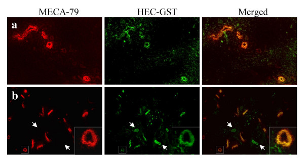
Double immunofluorescent detection of the MECA-79 epitope and GlcNAc6ST-2 in human lymph node and RA synovial tissues. Sections were simultaneously labelled with MECA-79 (red) and anti-GlcNAc6ST-2 (green) antibodies and photographed under appropriate filters. On the right panels, a merged image shows colocalization of both markers (yellow). (a) Lymph node. (b) RA synovium; Two MECA-79 negative and GlcNAc6ST-2 positive vessels are marked by arrows. Original magnification (a) 200×. (b) 150×. Selected areas within squared insets are shown on the lower right corner insets with higher magnification (630×).