



1. Introduction
This chapter reviews recent research on the molecular structural changes induced by the actin-myosin interaction, focusing on spectroscopic probe studies in this laboratory. We ask the following questions: What changes in myosin structure and dynamics are induced by interaction with actin? (addressed in Sect. 2), What changes in actin structure and dynamics are induced by interaction with myosin? (addressed in Sect. 3), and How are these structural changes influenced by the nucleotide-dependent strength of the actin-myosin interaction? This last is particularly important because the weak-to-strong transition (Fig. 1) generates force and movement (Eisenberg and Hill 1985). These questions are illustrated schematically in Fig. 1, which shows the actin-activated myosin ATPase scheme, emphasizing the distinction between weak and strong binding states. Although, in principle, each of the ternary complexes, A.M.N, has both weak and strong structural states (Geeves 1991; Geeves and Lehrer, this Vol.), the brackets in Fig. 1 indicate the predominant state (weak or strong) that exists in each ternary complex.
Fig. 1.
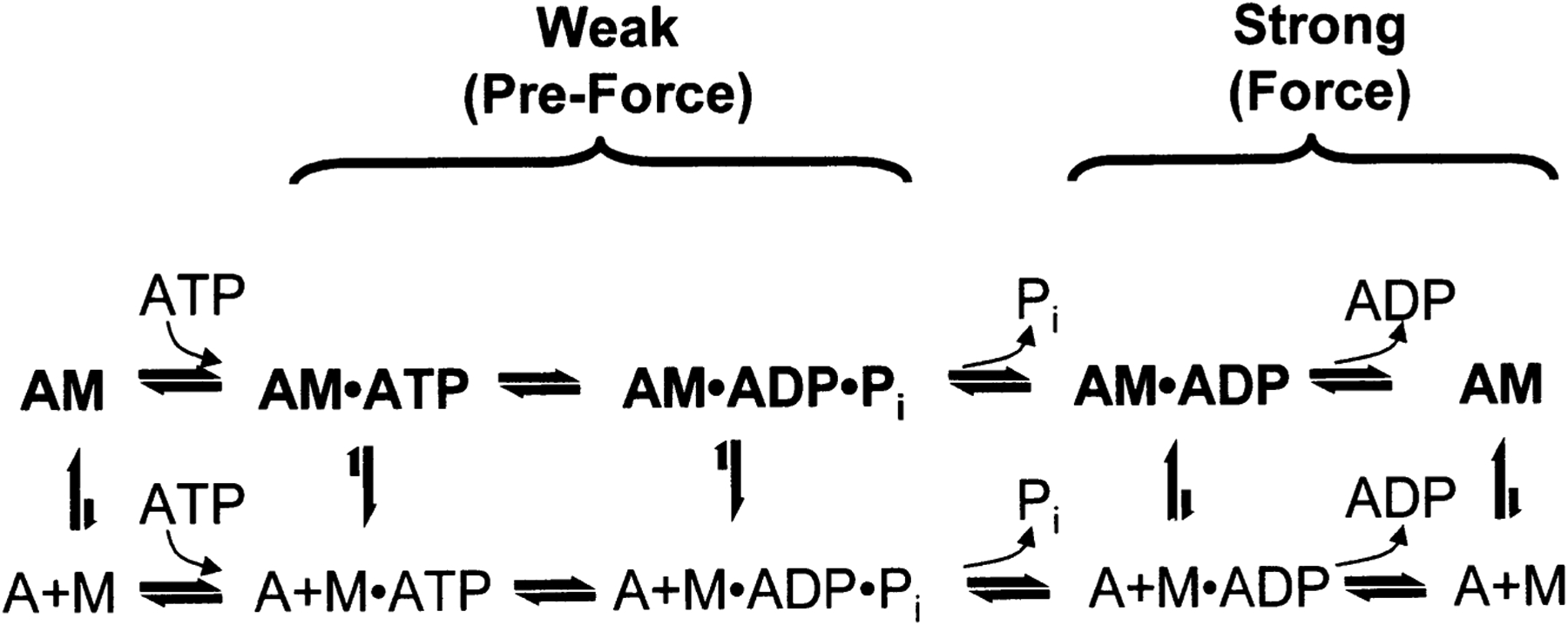
Actomyosin ATPase scheme. The horizontal dimension shows changes in the nucleotide bound to myosin, which determines the force-generating transition from weak (left) to strong (right) states. The vertical dimension shows the transition between the dissociated complexes (A+M.N, bottom) and the bound ternary complexes (A.M.N, top)
In general, we want to know what structural changes occur within the ternary complex A.M.N (top row of Fig. 1), using probes attached specifically to either actin or myosin, in the transition between the weak binding states (bracket on left, N = ATP, ATP-γS, ADP.Pi) and strong binding states (bracket on right, N = ADP or no nucleotide). Although this can, in principle, be observed directly by following the ATPase kinetic cycle transiently from left to right in Fig. 1, it is more common to perform an experiment that focuses on one column of Fig. 1, i.e., on a particular nucleotide (or nucleotide analog) N, mixing A with M.N and asking whether a structural change in the labeled actin or myosin is observed upon formation of the ternary complex A.M.N. The strong-binding ternary complexes are relatively easy to study, but it is difficult to isolate a complex in which most labeled actin monomers are occupied by weakly bound myosin or most labeled myosin heads are bound weakly to actin. This explains why there is much more information on strong-binding structural states than on weak-binding structural states. An alternative approach to studying the structural basis of actomyosin interactions is to apply perturbations (peptides or antibodies or chemical modifications) that are specific for proposed interaction sites on actin or myosin, based on structural modeling (Rayment et al. 1993b; Mendelson and Morris 1997). Although this approach is less direct than studying the actomyosin complex itself, it can provide information about the structural effects of weak interactions.
By using site-directed spectroscopic probes, we are able to probe a specific site on myosin or actin in the presence of the other protein, and in many cases, in the presence of the entire contractile apparatus. The spectroscopic probe techniques discussed here include spin label electron paramagnetic resonance (EPR), which is a high-resolution technique for the analysis of orientational and motional states, time-resolved phosphorescence anisotropy (TPA), which resolves rotational motions within large protein complexes, and fluorescence, which detects nanosecond dynamics, solvent accessibility, and distances between probes. Depending on the probe and the technique, these measurements can reveal information about: (1) global dynamics of actin monomers or myosin head domains, or (2) internal dynamics within the actin monomer or myosin head domain (Fig. 2). Global rotations of actin monomers or myosin head domains correspond to either axial (bending or tilting) motions or azimuthal (torsional or twisting) motions (Fig. 2). Some of these structural effects have been found to be cooperative, i.e., propagated along the filament so that myosin or actin molecules are affected without direct contact.
Fig. 2.
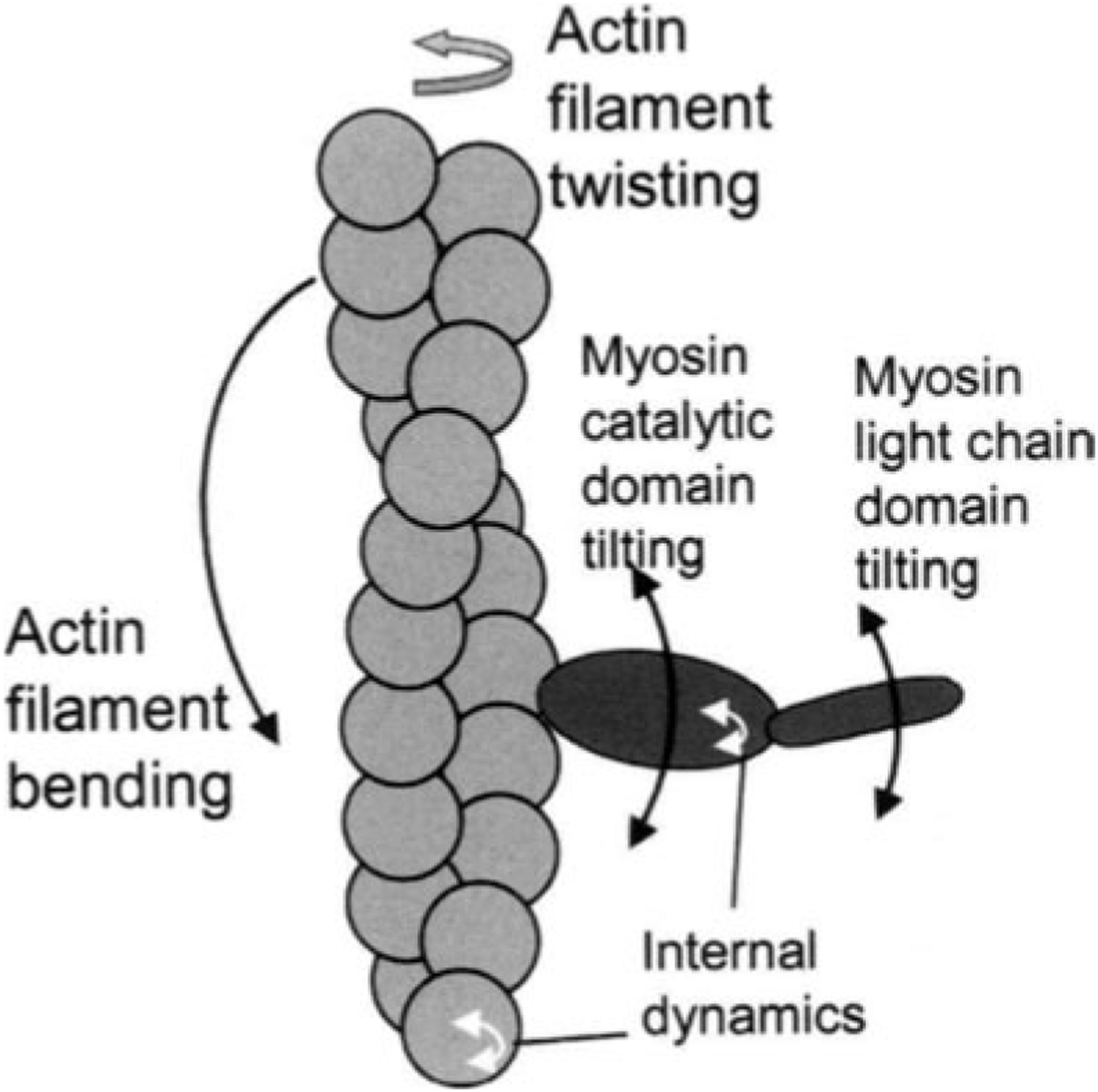
Rotational dynamics of the actomyosin complex showing different modes of motion (global or internal) that are detected by spectroscopic probes. Global rotations of the actin monomer or myosin head domains correspond to either axial (bending or tilting) motions or azimuthal (torsional or twisting) motions. Internal rotations reveal information about local conformational changes that may affect other domains within the molecule
2. Changes in Myosin Structural Dynamics Induced by Actin
The myosin head is functionally divided into the catalytic domain (sometimes designated as the “motor domain”, containing both the ATPase active site and the actin-binding region), and the light chain-domain (LC domain or lever arm). The LC domain contains the essential (ELC) and regulatory light chains (RLC), which wrap around ~8nm of an α-helix in the myosin heavy chain (Rayment et al. 1993a). By specifically attaching probes on Cys 707 (SHl) of the catalytic domain and on cysteines of the RLC, structural dynamics have been probed as a function of the states of the actomyosin ATPase cycle (Fig. 1).
In the classical model for muscle contraction, the myosin head is proposed to rotate as a rigid body from 90° to 45° during muscle contraction (Huxley 1969; Huxley and Simmons 1971). After our EPR data ruled this out (Cooke et al. 1982), models were proposed in which the catalytic domain binds to actin in a fixed rigor-like orientation and the head bends in the middle (Cooke 1986), with the LC domain acting like a lever arm (Rayment et al. 1993b). Our recent spectroscopic data have supported the model of a flexible head, and have led us to propose a model for muscle contraction in which the myosin head undergoes a disorder-to-order transition in both the catalytic domain and LC domain. Specifically, the myosin heads initially attach to actin in a weak and non-stereospecific mode that is characterized by large-scale dynamic disorder, then undergo at least two conformational transitions involving large-scale structural changes within the catalytic domain, and finally attach to actin in a strong binding and stereospecific mode to generate force. The LC domain appears to undergo a similar dynamic disorder-to-order transition (Roopnarine et al. 1998) but within a smaller orientational distribution than the catalytic domain so that the mean angle of the myosin heads is well defined (Baker et al. 1998).
2.1. Global Motions of Catalytic Domain: Disorder-to-Order Transition
We first used a maleimide spin label (MSL) attached to SHI to show that 10–20% of the myosin heads in contracting muscle fibers are distinctly ordered with an angular orientation that is precisely the same as in rigor, the remaining heads being highly disordered as in relaxation (Cooke et al. 1982; Fajer et al. 1990a). Since there was ambiguity about whether the spin labels could directly report an axial rotation of the myosin head, we continued our search for an ideal spin label. We found that an indane-dione spin label (In VSL) bound to SHI had its principal axis oriented almost parallel to the fiber axis so that it could directly detect any axial rotations of the myosin head (Roopnarine and Thomas 1994). Our results with this spin label confirmed that about 20% of the myosin heads in contraction are oriented as in rigor, with the remaining heads being dynamically disordered (Roopnarine and Thomas 1995). Using ATP or ATP-γ-S to trap weak-binding states revealed only dynamic disorder of the catalytic domain, with no evidence for any distinct new orientation of the myosin head either in the weak- or strong-binding states (Fajer et al. 1988, 1990b; Roopnarine and Thomas 1996). Only a very slight reorientation (5–10°), probably corresponding to a conformational change within the catalytic domain, was observed upon addition of ADP to rigor (Fajer et al. 1990b; Tanner et al 1992; Roopnarine and Thomas 1996). These results ruled out the classical model for muscle contraction in which the entire myosin head was proposed to undergo a rigid-body rotation between two large angles (Huxley 1969; Huxley and Simmons 1971). Instead, we proposed a model for contraction in which myosin heads undergo a transition from a disordered-to-ordered state (Berger and Thomas 1994; Thomas et al. 1995; Roopnarine and Thomas 1995).
Using saturation-transfer EPR (ST-EPR), we found that most of the myosin heads in contracting myofibrils or fibers were dynamically disordered in their axial orientation in the microsecond time-scale (Thomas et al. 1980; Barnett and Thomas 1989; Roopnarine and Thomas 1995). Complementary studies with time-resolved phosphorescence anisotropy (TPA) decay using phosphorescent dyes on SHI in muscle fibers confirmed this (Stein et al. 1990). These EPR results suggested that either (1) the catalytic domain of the myosin heads had only one specific orientation of binding to actin (in the rigor-like orientation), and that the myosin heads are only dynamically disordered when detached from actin, or (2) that the myosin head has two modes of binding to actin; in a stereospecific orientation (like the rigor orientation) and another in which the heads are dynamically disordered while attached to actin. Therefore, we conducted ST-EPR experiments on spin-labeled myosin heads in solution or myofibrils under conditions where the fraction of actin-bound heads was known. We determined that actin-bound heads do undergo dynamic microsecond rotational motions during steady state ATPase activity (Svensson and Thomas 1986; Berger et al. 1989; Berger and Thomas 1993). To probe the dynamics of the weak- and strong-binding states we used nucleotide analogs (ATPγS and AMPPNP, respectively). We found that actin-bound heads are rotationally mobile in the weak-binding states, but not in the strong-binding states (Berger and Thomas 1991; Berger and Thomas 1994). This was confirmed by electron microscopy of acto-S1 during steady-state ATP hydrolysis, where it was observed that S1 attached to actin in a variety of conformations in the presence of ATP, but only in one orientation in the presence of ADP (Walker et al. 1994). EM analysis (Walker et al. 1995) and transient EPR (Thomas et al. 1995) both showed that the actin-bound S1 was disordered within 10 ms of adding ATP to the rigor complex.
2.2. Global Motion of Light Chain Domain: Disorder-to-Order Transition, Two Angles
To analyze the orientation of the LC domain, we used spin-labels bound to RLC in scallop muscle, which offers the advantage that light -chain exchange is much easier than in skeletal muscle and that the large functional effect of the RLC (conferring calcium regulation to the muscle) makes it ideal for confirming the functional integrity of labeled LC domain. We found that a spin label, bound to chicken gizzard RLC in scallop muscle, had a single orientation in rigor, but a broad orientational distribution in relaxation, with two distinct and equally populated maxima centered at 38° and 74° with respect to the muscle fiber axis. Upon activation, the same two angles were observed in contraction, but the distribution between these two angles shifted slightly, so that an additional one sixth of them were oriented as in rigor. This was the first direct observation of a distinct angle change in contraction, supporting the model that the LC domain acts as a lever arm in contraction, but also supporting the disorder-to-order transition model (Baker et al. 1998). Subsequent experiments with nucleotide analogs (Brust-Mascher et al. 1999) showed that LC domain reorientation occurs upon strong-binding to actin, but not upon the ATP hydrolysis step.
To measure the microsecond rotational motions of the LC domain, we performed ST-EPR experiments on scallop fibers containing spin-labeled Mercenaria RLC (labeled at Cys 50 with InVSL). We determined that the LC domain is less mobile than the catalytic domain in relaxation but more mobile in rigor, consistent with rotation of one domain relative to the other, and consistent with disorder-to-order transitions in both domains during force generation (Roopnarine et al. 1998). We further tested this by using time-resolved phosphorescence anisotropy (TPA) to measure the rotational dynamics of a phosphorescent probe on the Cysl08 of gizzard RLC on scallop myofibrils. We found that rotational motions of the C-terminus of RLC are consistent with the ST-EPR results of the N-terminus, in that the LC domain is less mobile than the catalytic domain, confirming the segmental flexibility that exists between the catalytic and LC domains (Ramachandran and Thomas 1999).
2.3. Internal Motion of the Myosin Catalytic Domain: Resolve Three Structural States
Are the biochemical states of the myosin ATPase cycle coupled to specific conformational changes within the catalytic domain? We used an iodoacetamide spin label (lASL) bound to SHI to show that conformational changes within the myosin head correspond to specific biochemical states of the ATPase cycle in solution (Barnett and Thomas 1987; Ostap et al. 1993) and in contracting muscle fibers (Ostap et al. 1995; Thomas et al. 1995). The M.ATP state (trapped by ADP.BeFx and ATPγS) showed more internal mobility than in the absence of nucleotide and less than in the transition state M.ADP.Pi (trapped by ADP.AlF4 and MgADP.VO4). Thus, both ATP binding and hydrolysis induce changes in the internal dynamics of the catalytic domain.
A remaining question is: What are the conformational changes that occur within the catalytic domain during its interaction with actin? As in our study of global myosin dynamics (Berger et al. 1989; Berger and Thomas 1991; Berger and Thomas 1994), we answered this question by performing EPR experiments on solutions of IASL-Sl and actin in the presence of various nucleotides, measuring the fraction of S1 bound by centrifugation, and thus calculating the spectrum of the actin-bound ternary complex A.M.N (Grinband et al. 1998). We found that actin greatly restricts the rotational mobility of IASL in strong-binding states (N = no nucleotide or ADP), producing a new conformation that is much more rigid than when myosin is free in solution. In contrast, the effect of actin on the weak-binding states of S1 was simply to perturb the distribution of states slightly, without perturbing the individual myosin conformational states.
3. Changes in Actin Structural Dynamics Induced by Myosin
3.1. Global Dynamics of Actin
The effects of myosin on the global dynamics of actin were first demonstrated by quasi-elastic light scattering (Fujime and Ishiwata 1971), but this technique could not provide site-specific information, nor could it address motions faster than the millisecond time range. Fluorescent probes and spin labels provided site-specific labeling, primarily at Cys 374, but these measurements were limited to the nanosecond time range, which was too fast. In order to obtain direct information about the microsecond dynamics of actin, it was necessary to study spin labels with saturation transfer EPR (Thomas et al. 1979; Ostap and Thomas 1991) or optical probes with time-resolved phosphorescence anisotropy, TPA (Yoshimura et al. 1984; Prochniewicz and Thomas 1999; Prochniewicz and Thomas 1997). These results showed consistently that actin filaments undergo substantial microsecond internal rotation, dominated by torsional motions, and that the strong binding of myosin heads restricts this flexibility cooperatively; i.e., maximal restriction of rotational motion at a ratio of S1 to actin as low as 0.1–0.2 (Thomas et al. 1979; Ostap and Thomas 1991; Prochniewicz and Thomas 1997). Of these studies, the only one to probe directly the difference between weakly and strongly bound states is the EPR study of Ostap and Thomas (1991). To obtain EPR data in weak-binding ternary complexes, these investigators performed EPR on a concentrated solution of S1 and spin-labeled actin (with a maleimide spin label rigidly bound to Cys 374) during the brief steady state following photolysis of caged ATP. They found that the mixture of weak-binding complexes (A.M.ATP and A.M.ADP.Pi) produced just as much restriction of the microsecond flexibility of actin as the strong-binding complexes (A.M and A.M.ADP). These results suggest that the global dynamics of actin do not depend on the weak-to-strong transition.
Phosphorescence (TPA), using erythrosin iodoacetamide attached to eys 374, has provided detailed information about effects of myosin on both the rate and amplitude of actin’s microsecond dynamics (Prochniewicz and Thomas 1997). In the absence of nucleotide (i.e., in a strong-binding state), myosin decreases the rate of actin’s torsional flexibility by a factor of 4 and decreases the amplitude by nearly a factor of 2. As observed previously by ST-EPR (Thomas et al. 1979), actin’s flexibility decreased nonlinearly with S1 binding, indicating that myosin’s effects are propagated cooperatively beyond the monomer to which it binds. Direct analysis of weak-binding states was not attempted in this study, but weak binding was studied indirectly by using antibodies or chemical modifications specific to sites on actin that are proposed to form part of the interface responsible for weak myosin binding (Prochniewicz et al. 1993; Prochniewicz and Thomas 1997). Like strong myosin binding, these perturbations decreased actin flexibility in a cooperative manner, suggesting the propagation of effects from one actin monomer to several others. These results underscore the complex coupling that exists among the actin-myosin interface, global actin rotational dynamics, and the propagation of physical effects along the actin filament.
Other spectroscopic analyses support the propagation of global structural changes along the actin filament. Studies using both electron and optical microscopy revealed an increase in the bending flexibility of the actin filament upon binding of HMM in the presence of ATP (Takebeyashi et al. 1977; Yanagida et al. 1984), which may originate from local conformational changes in actin such as distortion or rotation of protomers and loosening of intermonomer bonds. The dissociation of acto-S1 by AMPPNP induced large undulations in the actin filament as observed using the time-resolved cryo-microscopy. This structural transition appears to originate from the release of structural constraints imposed on actin by binding of myosin heads to two successive monomers along the long-pitch helix (Menetret et al. 1991).
The development of the in vitro motility assay, which is a model of the contraction process, enabled examination of structural changes in individual actin filaments during sliding along myosin heads fixed on a glass surface. Polarized fluorescence from phalloidin-rhodamine actin during sliding indicated myosin-induced deformation of actin filaments (Borejdo and Burlacu 1994). Later fluorescence polarization measurements on sliding filaments labeled with 5-iodoacetamidotetramethylrhodamine led to the conclusion that the sliding actin filament performs an axial rotation with one revolution per 1 μm sliding distance (Sase et al. 1997).
Spectroscopic measurements on contracting glycerinated fibers were made possible by specific labeling of actin with phalloidin-FITC and measurement of polarized fluorescence. The changes in the polarization during development of isometric tension were clearly different from those accompanying transition of the fiber from relaxation to rigor and indicated a possibility that tension development is accompanied by a small axial rotation of the monomers in the actin filament (Prochniewicz et al. 1983). These results indicate that orientational changes in actin are induced by the active interaction of myosin. However, EPR experiments with spin labels attached to oriented actin using either spin-labeled phalloidin in muscle fibers (Naber et al. 1993) or a spin label attached to actin at Cys 374 in flow-oriented actin (Ostap et al. 1992) did not show a significant change in orientation upon the active interaction of myosin and ATP with actin.
3.2. Internal Dynamics of Actin
Fluorescence studies on actin labeled at the C-terminal Cys374 with IAEDANS showed that the rotational mobility of the dye was changing in a non-linear manner following addition of increasing amounts of HMM or S1 (Miki et al. 1982); this change – increase or decrease in mobility – was dependent on the kind of divalent cation (Ca or Mg) tightly bound to actin. Non-linear changes in fluorescence intensity or polarization was further observed when actin was labeled with a fluorescent nucleotide analog E-ADP (Miki et al. 1976). These spectroscopically observed changes were interpreted as the changes in the local environment of the probe or in filament flexibility, and the non-linearity of changes indicated intermonomer cooperativity, where locally induced changes propagate along the larger segments of the filament.
The fluorescence of pyrene-iodoacetamide reacted with Cys 374 on actin has proven to be a valuable probe of actin-myosin interactions (reviewed by Geeves 1991; see Chapter by Geeves and Lehrer, this Vol.). Binding of myosin S1 in the absence of nucleotide (strong binding) decreases the intensity of pyrene fluorescence by 70%, and the change is linear with S1 binding. Although the precise physical basis of this fluorescence change is not clear, it must arise from a change in the local environment of the pyrene label near Cys 374. This label has served as a very useful probe of the kinetics of the actin-myosin interaction. In the presence of saturating ATP or ATP-γ-S (weak binding), myosin has no effect on this fluorescence signal, even under conditions where up to 70% of actin monomers have S1 bound. This result shows clearly that the structural change induced in the C terminus of actin by myosin is much greater when myosin binds strongly to actin than when it binds weakly.
There is an interesting contrast between the effects of myosin on the internal dynamics of actin (measured by pyrene fluorescence; Geeves 1991) and on the global dynamics (measured by ST-EPR; Ostap and Thomas 1991): while the internal dynamics of actin are affected only by strong myosin binding (Geeves 1991), the global dynamics are affected equally by weak and strong binding (Ostap and Thomas 1991).
Attempts to correlate changes in structural and dynamic properties of actin with the changes in its functional interaction with myosin were subsequently pursued in a comparative study on muscle actin and non-muscle actin from yeast (Prochniewicz and Thomas 1999). In this study, fluorescent probes were used to show that muscle and yeast actin have different conformational dynamics near the C terminus, and that strong myosin binding decreases these differences. If the myosin-free state of actin corresponds to the conformation of actin in the weak-binding states (left bracket in Fig. 1), then these results indicate that the transition from weak to strong (right bracket in Fig. 1) involves a larger structural change in yeast actin than in muscle actin. It was suggested that this could be the structural basis explaining why yeast actin is less effective than muscle actin in activating myosin.
4. Summary and Conclusions
Figure 3 summarizes the effects of actomyosin binding on the internal and global dynamics of either protein, as discussed in this chapter. These effects depend primarily on the strength of the interaction; which in turn depends on the state of the nucleotide at the myosin active site. When either no nucleotide or ADP is bound, the interaction is strong and the effect on each protein is maximal. When the nucleotide is ATP or ADP.Pi, or the equivalent nonhydrolyzable analogs, the interaction is weak and the effect on molecular dynamics of each protein is minimal. The weaker effects in weak-binding states are not simply the reflection of lower occupancy of binding sites – the molecular models in Fig. 3 illustrate the effects of the formation of the ternary complex, after correction for the free actin and myosin in the system. Thus EPR on myosin (Berger and Thomas 1991; Thomas et al. 1995) and pyrene fluorescence studies on actin (Geeves 1991) have shown that the formation of a ternary complex has a negligible effect on the internal dynamics of both proteins (left side of Fig. 3, white arrows). As shown by both EPR (Baker et al. 1998; Roopnarine et al. 1998) and phosphorescence (Ramachandran and Thomas 1999), both domains of myosin are dynamically disordered in weak-binding states, and this is essentially unaffected by the formation of the ternary complex (left side of Fig. 3, indicated by disordered myosin domains). The only substantial effect of the formation of the weak interaction that has been reported is the EPR-detected (Ostap and Thomas 1991) restriction of the global dynamics of actin upon weak myosin binding (left column of Fig. 3, gray arrow).
Fig. 3.
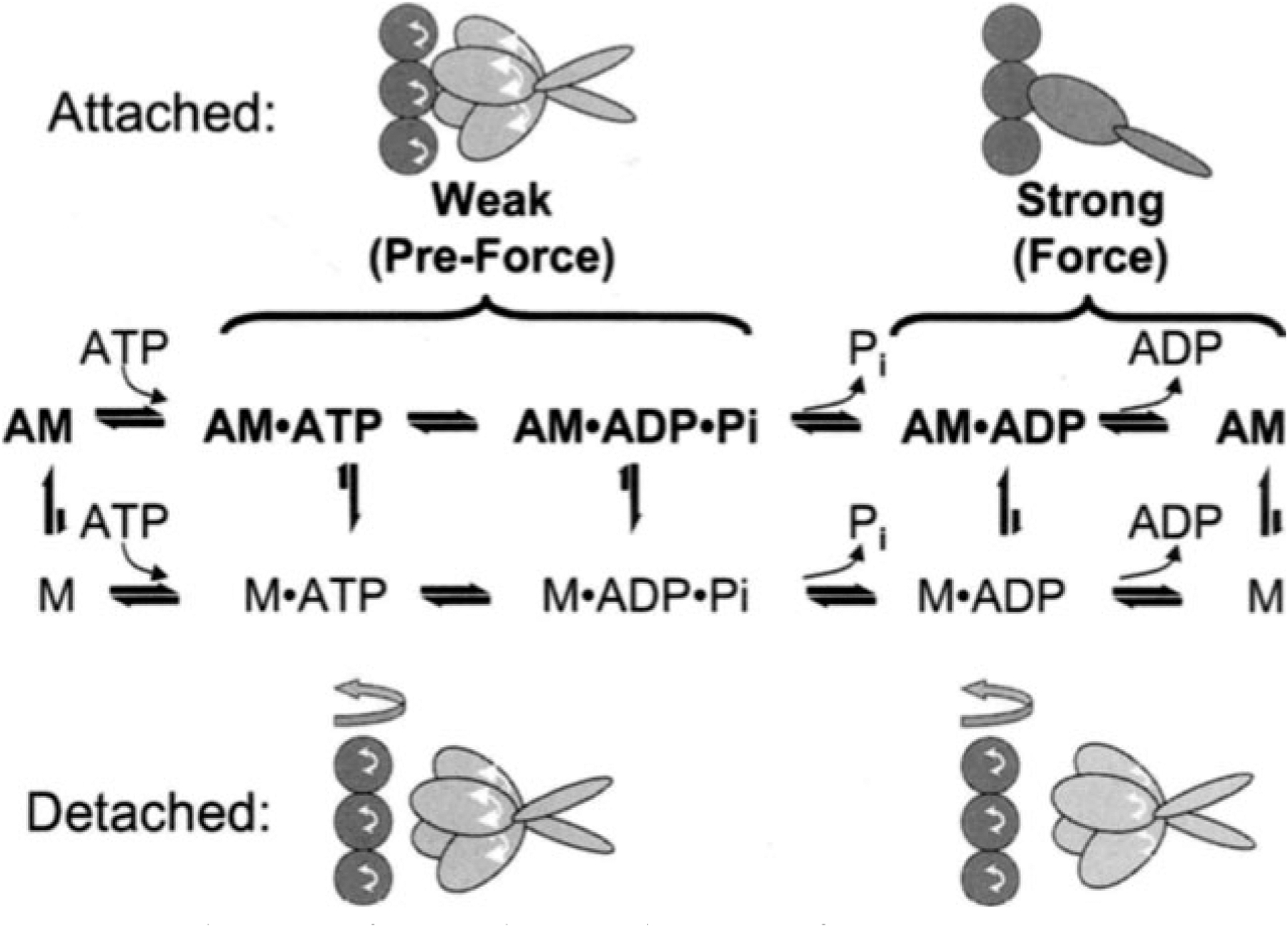
Rotational dynamics of myosin heads and actin as a function of the actomyosin ATPase cycle. In the structural models, the amplitudes of arrows and the disorder of myosin head domains indicate the amplitudes of structural dynamics in each state. Thus the weak states (left) are more dynamic than the strong states (right), and the restriction in dynamics due to actomyosin formation is much greater in strong states than in weak
The effects of strong actomyosin formation are much more dramatic. While substantial rotational dynamics, both internal and global, exist in both myosin and actin in the presence of ADP or the absence of nucleotides, spin label EPR, pyrene fluorescence, and phosphorescence all show dramatic restrictions in these motions upon formation of the strong ternary complex (right column of Fig. 3). One implication of this is that the weak-to-strong transition is accompanied by a disorder-to-order transition in both actin and myosin, and this is itself an excellent candidate for the structural change that produces force (Thomas et al. 1995). Another clear implication is that the crystal structures obtained for isolated myosin and actin are not likely to be reliable representations of structures that exist in ternary complexes of these proteins (Rayment et al. 1993a and 1993b; Dominguez et al. 1998; Houdusse et al. 1999). This is clearly true of the strong-binding states, since the spectroscopic studies indicate consistently that substantial changes occur in both proteins upon strong complex formation. For the weak complexes, the problem is not that complex formation induces large structural changes, but that the structures themselves are dynamically disordered. This is probably why so many different structures have been obtained for myosin S1 with nucleotides bound – each crystal is selecting one of the many different substates represented by the dynamic ensemble. Finally, there is the problem that the structures of actomyosin complexes are probably influenced strongly by their mechanical coupling to muscle protein lattice (Baker at al. 2000).
Thus, even if co-crystals of actin and myosin are obtained in the future, an accurate description of the structural changes involved in force generation will require further experiments using site-directed spectroscopic probes of both actin and myosin, in order to detect the structural dynamics of these ternary complexes under physiological conditions.
References
- Baker JE, Brust-Mascher I, Ramachandran S, LaConte LEW, Thomas DD (1998) A large and distinct rotation of the myosin light chain domain occurs upon muscle contraction. Proc Natl Acad Sci USA 95:2944–2949 [DOI] [PMC free article] [PubMed] [Google Scholar]
- Baker JB, LaConte LEW, Brust-Mascher I, Thomas DD (2000) Mechanochemical coupling in muscle. Biophys J 78:2730–2739 [DOI] [PMC free article] [PubMed] [Google Scholar]
- Barnett VA, Thomas DD (1987) Resolution of conformational states of spin-labeled myosin during steady-state ATP hydrolysis. Biochemistry 26:314–323 [DOI] [PubMed] [Google Scholar]
- Barnett VA, Thomas DD (1989) Microsecond rotational motion of spin-labeled myosin heads during isometric muscle contraction: saturation transfer electron paramagnetic resonance. Biophys J 56:517–523 [DOI] [PMC free article] [PubMed] [Google Scholar]
- Berger CL, Thomas DD (1991) Rotational dynamics of actin-bound intermediates in the myosin ATPase cycle. Biochemistry 30: 11 036–11 045 [DOI] [PubMed] [Google Scholar]
- Berger CL, Thomas DD (1993) Rotational dynamics of actin-bound myosin heads in active myofibrils. Biochemistry 32:3812–3821 [DOI] [PubMed] [Google Scholar]
- Berger CL, Thomas DD (1994) Rotational dynamics of actin-bound intermediates of the myosin ATPase cycle in myofibrils. Biophys J 67:250–261 [DOI] [PMC free article] [PubMed] [Google Scholar]
- Berger CL, Svensson EC, Thomas DD (1989) Photolysis of caged ATP induces microsecond rotation of myosin heads on actin. Proc Natl Acad Sci USA 86:8753–8757 [DOI] [PMC free article] [PubMed] [Google Scholar]
- Borejdo J, Burlacu S (1994) Orientation of actin filaments during motion in vitro motility assay. Biophys J 66:l319–l327 [DOI] [PMC free article] [PubMed] [Google Scholar]
- Brust-Mascher I, LaConte LEW, Baker JE, Thomas DD (1999) Myosin light-chain domain rotates upon muscle activation but not ATP hydrolysis. Biochemistry 38:12607–126l3 [DOI] [PubMed] [Google Scholar]
- Cooke R (1986) The mechanism of muscle contraction. Crit Rev Biochem Mol Bioi 21:53–118 [DOI] [PubMed] [Google Scholar]
- Cooke R, Crowder MS, Thomas DD (1982) Orientation of spin-labels attached to cross-bridges in contracting muscle fibers. Nature 300:776–778 [DOI] [PubMed] [Google Scholar]
- Dominguez R, Freyzon Y, Trybus KM, Cohen C (1998) Crystal structure of a vertebrate smooth muscle myosin motor domain and its complex with the essential light chain: visualization of the pre-power stroke state. Cell 94:559–571 [DOI] [PubMed] [Google Scholar]
- Eisenberg E, Hill T (1985) Muscle contraction and free energy transduction in biological systems. Science 227:999–1006 [DOI] [PubMed] [Google Scholar]
- Fajer PG, Fajer EA, Brunsvold NJ, Thomas DD (1988) Effects of AMPPNP on the orientation and rotational dynamics of spin-labeled myosin heads in muscle fibers. Biophys J 53:513–524 [DOI] [PMC free article] [PubMed] [Google Scholar]
- Fajer PG, Fajer EA, Thomas DD (1990a) Myosin heads have a broad orientational distribution during isometric muscle contraction. Time-resolved EPR studies using caged ATP. Proc Natl Acad Sci USA 87:5538–5542 [DOI] [PMC free article] [PubMed] [Google Scholar]
- Fajer PG,Fajer EA, Matta JJ, Thomas DD (1990b) Effect of ADP on the orientation of spin-labeled myosin heads in muscle fibers: a high resolution study with deuterated spin labels. Biochemistry 29:5865–5871 [DOI] [PubMed] [Google Scholar]
- Fujime S, Ishiwata S (1971) Dynamic light scattering study of muscle F-actin. J Mol Bioi 62: 251–265 [DOI] [PubMed] [Google Scholar]
- Geeves MA (1991) The dynamics of actin and myosin association and the crossbridge model of muscle contraction. Biochem J 274:1–14 [DOI] [PMC free article] [PubMed] [Google Scholar]
- Grinband J, Baker JE, Thomas DD (1998) The conformational state of myosin is affected by actin in the strong binding but not in the weak binding state. Biophys J 74:A263 [Google Scholar]
- Houdusse A, Kalabokis VN, Himmel D, Szent-Gyorgyi AG, Cohen C (1999) Atomic structure of scallop myosin subfragment S1 complexed with MgADP: a novel conformation of the myosin head. Cell 97:459–470 [DOI] [PubMed] [Google Scholar]
- Huxley HE (1969) The mechanism of muscular contraction. Science 114:1356–1366 [PubMed] [Google Scholar]
- Huxley AF, Simmons R (1971) Proposed mechanism of force generation in striated muscle. Nature 233:533–538 [DOI] [PubMed] [Google Scholar]
- Mendelson R, Morris EP (1997) The structure of the acto-myosin subfragment 1 complex: results of searches using data from electron microscopy and X-ray crystallography. Proc Natl Acad Sci USA 94:8533–8538 [DOI] [PMC free article] [PubMed] [Google Scholar]
- Menetret JF, Hofmann W, Schroder RR, Rapp G, Goody RS (1991) Time-resolved cryo-electron microscopic study of the dissociation of actomyosin induced by photolysis of photo labile nucleotides. J Mol Bioi 219:139–144 [DOI] [PubMed] [Google Scholar]
- Miki M, Kouyama T, Mihashi K (1976) Fluorescence study of e-ADP bound to rabbit F-actin: structural change in the adenine subsite of F-actin under the influence of heavy meromyosin FEBS Lett 66:98–101 [Google Scholar]
- Miki M, Wahl P, Auchet JC (1982) Fluorescence anisotropy of labeled F-actin: influence of divalent cations on the interaction between F-actin and myosin heads. Biochemistry 21: 3661–3665 [DOI] [PubMed] [Google Scholar]
- Naber N, Ostap EM, Thomas DD, Cooke R (1993) Orientation and rotational dynamics of spin-labeled phalloidin bound to actin in muscle fibers. Proteins 17:347–354 [DOI] [PubMed] [Google Scholar]
- Ostap EM, Thomas DD (1991) Rotational dynamics of spin-labeled F-actin during activation of myosin S1 ATPase using caged ATP. Biophys J 59:1235–1241 [DOI] [PMC free article] [PubMed] [Google Scholar]
- Ostap EM, Yanagida T, Thomas DD (1992) Orientational distribution of spin-labeled actin oriented by flow. Biophys J 63:966–975 [DOI] [PMC free article] [PubMed] [Google Scholar]
- Ostap EM, White HD, Thomas DD (1993) Transient detection of spin-labeled myosin subfragment 1 conformational states during ATP hydrolysis. Biochemistry 32:6712–6720 [DOI] [PubMed] [Google Scholar]
- Ostap EM, Barnett VA, Thomas DD (1995) Resolution of three structural states of spin-labeled myosin in contracting muscle. Biophys J 69:177–188 [DOI] [PMC free article] [PubMed] [Google Scholar]
- Prochniewicz E, Thomas DD (1997) Perturbations offunctional interactions with myosin induce long range allosteric and cooperative structural changes in actin. Biochemistry 36: 12845–12853 [DOI] [PubMed] [Google Scholar]
- Prochniewicz E, Thomas DD (1999) Differences in structural dynamics of muscle and yeast actin accompany differences in functional interactions with myosin. Biochemistry 38: 14860–14867 [DOI] [PubMed] [Google Scholar]
- Prochniewicz-Nakayama E, Yanagida T, Oosawa F (1983) Studies on conformation of F-actin in muscle fibers in the relaxed state, rigor and during contraction using fluorescent phalloidin. J Cell Bioi 97:1663–1667 [DOI] [PMC free article] [PubMed] [Google Scholar]
- Prochniewicz E, Katayama E, Yanagida T, Thomas DD (1993) Cooperativity in F-actin: chemical modifications of actin monomers affect the functional interactions of myosin with unmodified monomers in the same actin filament. Biophys J 65:113–123 [DOI] [PMC free article] [PubMed] [Google Scholar]
- Ramachandran S, Thomas DD (1999) Rotational dynamics of the regulatory light chain in scallop muscle detected by time-resolved phosphorescence anisotropy. Biochemistry 38:9097–9104 [DOI] [PubMed] [Google Scholar]
- Rayment I, Rypniewski WR, Schmidt-Bäse K, Smith R, Tomchick DR, Benning MM, Winklemann DA, Wesenberg G, Holden HM (1993a) Three-dimensional structure of myosin subfragment-1: a molecular motor. Science 261:50–58 [DOI] [PubMed] [Google Scholar]
- Rayment I, Holden HM, Whittaker M, Yohn CB, Lorenz M, Holmes KC, Milligan RA (1993b) Structure of the actin-myosin complex and its implications for muscle contraction. Science 261:58–65 [DOI] [PubMed] [Google Scholar]
- Roopnarine O, Thomas DD (1994) A spin label that binds to myosin heads in muscle fibers with its principal axis parallel to the fiber axis in rigor. Biophys J 67:1634–1645 [DOI] [PMC free article] [PubMed] [Google Scholar]
- Roopnarine O, Thomas DD (1995) Orientational dynamics of in dane dione spin-labeled myosin head in relaxed and contracting skeletal muscle fibers. Biophys J 68:1461–1471 [DOI] [PMC free article] [PubMed] [Google Scholar]
- Roopnarine O, Thomas DD (1996) Orientational dynamics of intermediate states of indane dione spin-labeled myosin heads in skeletal muscle fibers. Biophys J 70:2795–2806 [DOI] [PMC free article] [PubMed] [Google Scholar]
- Roopnarine O, Szent-Györgyi AG, Thomas DD (1998) Microsecond rotational dynamics of spin-labeled myosin regulatory light chain induced by relaxation and contraction of scallop muscle. Biochemistry 37:14428–14436 [DOI] [PMC free article] [PubMed] [Google Scholar]
- Sase I, Miyata H, Ishiwata S, Kinosita K Jr. (1997) Axial rotation of sliding actin filaments revealed by single-fluorophore imaging. Proc Natl Acad Sci USA 94(11):5646–5650 [DOI] [PMC free article] [PubMed] [Google Scholar]
- Stein RA, Ludescher RD, Dahlberg PS, Bennett RDL, Fajer PG, Thomas DD (1990) Time-resolved rotational dynamics of phosphorescent-labeled myosin heads in contracting muscle fibers. Biochemistry 29: 10023–10031 [DOI] [PubMed] [Google Scholar]
- Svensson EC, Thomas DD (1986) ATP induces microsecond rotational motions of myosin heads crosslinked to actin. Biophys J 50:999–1002 [DOI] [PMC free article] [PubMed] [Google Scholar]
- Takebayashi T, Morita Y, Oosawa F (1977) Electronmicroscopic investigation of the flexibility of F-actin. Biochim Biophys Acta 492:357–363 [DOI] [PubMed] [Google Scholar]
- Tanner JW, Thomas DD, Goldman YE (1992) Transients in orientation of a fluorescent cross-bridge probe following photolysis of caged nucleotides in skeletal muscle fibres. J Mol Bioi 223:185–203 [DOI] [PubMed] [Google Scholar]
- Thomas DD, Seidel JC, Gergely J (1979) Rotational dynamics of F-actin in the submillisecond time range. J Mol Bioi 132:257–273 [DOI] [PubMed] [Google Scholar]
- Thomas DD, Ishiwata S, Seidel JC, Gergely J (1980) Sub-millisecond rotational dynamics of spin-labeled crossbridges in myofibrils. Biophys J 32:873–890 [DOI] [PMC free article] [PubMed] [Google Scholar]
- Thomas DD, Ramachandran S, Roopnarine O, Hayden DW, Ostap EM (1995) The mechanism of force generation in myosin: a disorder-to-order transition, coupled to internal structural changes. Biophys J 68:135s–141s [PMC free article] [PubMed] [Google Scholar]
- Walker M, White H, Belknap B, Trinick J (1994) Electron cryomicroscopy of acto-myosin-Sl during steady-state ATP hydrolysis. Biophys J 66:1563–1572 [DOI] [PMC free article] [PubMed] [Google Scholar]
- Walker M, Trinick J, White H (1995) Millisecond time resolution electron cryo-microscopy of the M-ATP transient kinetic state of the acto-myosin ATPase. Biophys J 68:87S–91S [PMC free article] [PubMed] [Google Scholar]
- Yanagida T, Nakase M, Nishiyama K, Oosawa F (1984) Direct observation of motion of single F-actin filaments in the presence of myosin. Nature 307:58–60 [DOI] [PubMed] [Google Scholar]
- Yoshimura H, Nishio T, Mihashi K (1984) Torsional motion of eosin-labeled F-actin as detected in the time-resolved anisotropy decay of the probe on the sub-millisecond time range. J Mol Bioi 179:453–467 [DOI] [PubMed] [Google Scholar]