



Abstract
The use of CRISPR/Cas9 systems in genome editing has been limited by the inability to efficiently deliver the key editing components to and across tissues and cell membranes, respectively. Spherical nucleic acids (SNAs) are nanostructures that provide privileged access to both but have yet to be explored as a means of facilitating gene editing. Herein, a new class of CRISPR SNAs are designed and evaluated in the context of genome editing. Specifically, Cas9 ProSNAs comprised of Cas9 cores densely modified with DNA on their exteriors and preloaded with single guide (sg) RNA were synthesized and evaluated for their genome editing capabilities in the context of multiple cell lines. The radial orientation of the DNA on the Cas9 protein surface enhances cellular uptake, without the need for electroporation or transfection agents. In addition, the Cas9 proteins defining the cores of the ProSNAs were fused with GALA peptides on their N-termini and nuclear localization signals on their C-termini to facilitate endosomal escape and maximize nuclear localization and editing efficiency, respectively. These constructs were stable against protease digestion under conditions that fully degrade the Cas9 protein, when not transformed into an SNA, and used to achieve genome editing efficiency between 32 and 47%. Taken together, these novel constructs and advances point towards a way of significantly broadening the scope of use and impact of CRISPR-Cas9 genome editing systems.
Graphical Abstract

INTRODUCTION
The CRISPR-Cas9 (clustered regularly interspaced short palindromic repeats/CRISPR-associated protein 9) system provides adaptive immunity in bacteria and has been repurposed into a revolutionary technology for editing and modulating genomes.1-3 As a promising therapeutic agent, this system has the potential to treat a wide range of human diseases, including many forms of cancer, muscular dystrophy and Alzheimer’s disease.4-7 However, the full potential of CRISPR-Cas systems has not yet been realized, in part due to the inability to get the requisite cargo to different tissues, cells, and compartments within cells required to broadly address and treat disease. A primary approach has been to create and deliver plasmids or RNAs that genetically encode the Cas9 protein and single guide RNA (sgRNA) to cells of interest.8-10 Alternatively, direct injection or packaging of Cas9-sgRNA ribonucleoprotein (Cas9 RNP) complexes with heterogenous transfection agents enable cellular uptake and precise control over intracellular protein stoichiometry and editing timeframes.11-13 This latter approach minimizes undesirable off-target editing that can occur due to uncontrollable protein expression levels and overdosing. Consequently multiple cell-permeable systems, such as those based on cationic liposomes or polymers14-16 and inorganic nanoparticles,12, 17-19 have been explored with varying degrees of success due to inefficient cellular delivery as well as toxicity and immunogenicity issues. Direct covalent conjugation20 or noncovalent mixtures11, 21-22 of cell penetrating peptides on Cas9 RNPs may offer promising alternatives; however, variable cell penetrating efficiency and proteolytic instability limit their use in many cases.5 Therefore, a cell-permeable, transfection agent-free, proteolytically-stable, and self-contained genome editing system capable of accessing many tissue types is still needed.
Spherical nucleic acids (SNAs) are formed by chemically modifying nanoparticles with dense layers of single or double stranded DNA or RNA and provide access to many different tissues and over 60 different cell types.23-25 Protein spherical nucleic acids (ProSNAs),24 formed similarly by the conjugation of a nucleic acid shell to a protein, have emerged as promising architectures for intracellular biosensing and therapeutics;26-27 indeed, in many cases, the protein core retains its bioactivity28 and is even more biocompatible and stable than the native protein.26 If one could synthesize SNAs with Cas9 cores and load them with the genome editing nucleic acid cargo, one could create structures that potentially could expand the scope of use for CRISPR-Cas9 genome editing.
Herein, we report a Cas9 ProSNA-sgRNA ribonucleoprotein (Cas9 ProSNA RNP) platform and evaluate its genome editing capabilities in multiple cell lines (Scheme 1). Due to its high genome editing efficiency and specificity, the GeoCas9 (termed Cas9 herein) protein29 was transformed into a ProSNA construct by modifying its surface with a shell of DNA (d(TGG)10) to yield Cas9 ProSNAs for cellular uptake.28 This sequence was chosen because it is known to facilitate cellular uptake in SNA form.24, 30 These structures were designed to maximize endosomal escape by modifying them with three successive N-terminus GALA peptides.31-34 These peptides are glutamate-rich and adopt amphiphilic structures that do not interact with the cell membranes.33-34 In acidic endosomes, the glutamate residues of GALA peptides are protonated to form hydrophobic helices that disrupt endosomal membranes. In addition, Cas9 proteins were modified at the C-terminus with nuclear localization signals (NLSs), which bind to the nuclear pore receptor to facilitate nuclear import. We explored the use of these constructs, in the absence of ancillary transfection agents, with four different cell lines and demonstrated their genome editing efficiencies to be between 32 and 47%.
Scheme 1.
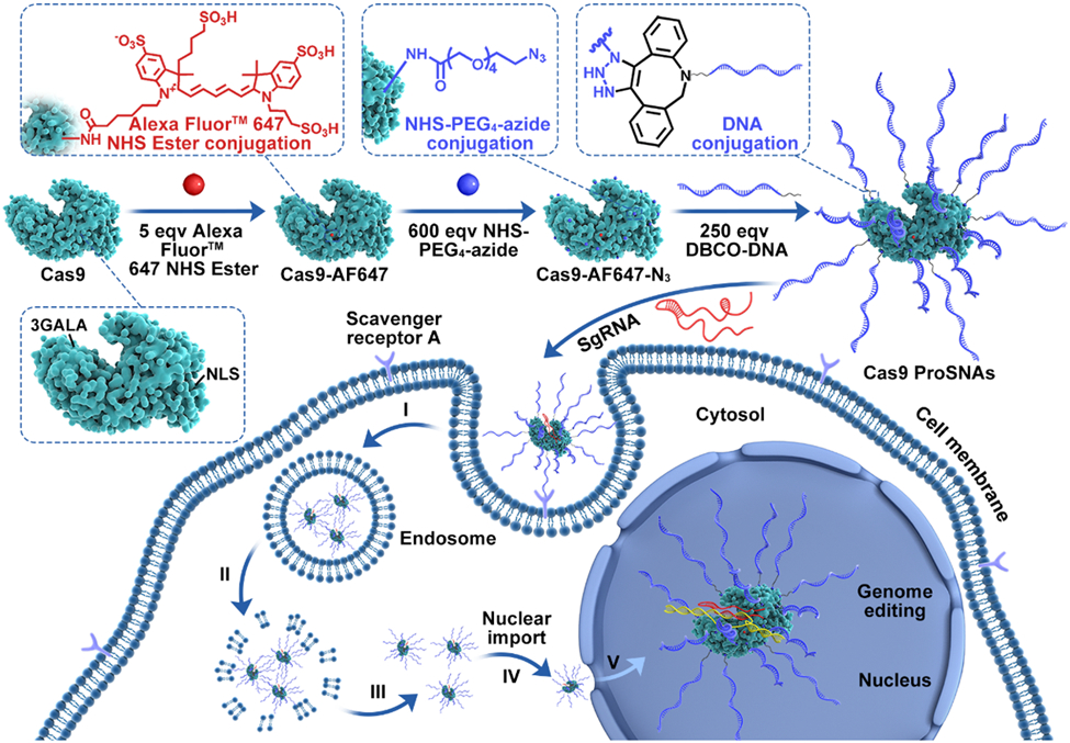
Chemical design of Cas9 ProSNAs and Cas9 ProSNA RNPs that enable delivery of Cas9 protein from the extracellular medium to the nucleus where it engages in genome editing. (I) Interaction with scavenger receptors on the cell surface and delivery via an endosomal pathway. (II) Disruption of endosomal membranes by GALA peptides. (III) Release of Cas9 ProSNA RNPs from the endosome. (IV) Nuclear import directed by nuclear localization signals (NLSs) of Cas9 ProSNA RNPs. (V) Targeting of genomic DNA and introduction of double-strand breaks.
RESULTS AND DISCUSSION
Recombinant Cas9 protein fused with N-terminus GALA peptides and C-terminus NLSs was expressed in Escherichia coli BL21 (DE3) (Figure S1). To prepare Cas9 ProSNAs, the Cas9 protein was first labeled with Alexa Fluor 647 fluorophore (AF647), which allows for the quantification of protein concentrations and fluorescence imaging (Scheme 1, Figures S2-3). The surface lysines were next reacted with N-hydroxy succinimide (NHS) polyethylene glycol polymers (PEG) terminated with azide groups. The terminal azides were then reacted with dibenzocyclooctyne (DBCO)-terminated DNA to yield Cas9 ProSNAs (the d(TGG)10 sequence was chosen due to its formation of a G-quadruplex which facilitates receptor-mediated endocytosis),28,30 and the excess DNA was removed from solution using size-exclusion chromatography (SEC) (Figures S4-6). The Cas9 ProSNAs eluted earlier than the Cas9-AF647-azide, suggesting that Cas9 ProSNAs have larger hydrodynamic radii, which was further confirmed by dynamic light scattering (DLS) (Figure S7). In addition, Cas9 ProSNAs were much less mobile than unmodified Cas9 protein when examined via denaturing polyacrylamide gel electrophoresis (PAGE), indicating that they have a substantially higher molecular weight (Figure S8). Based on the absorbance of DNA and AF647 on Cas9 ProSNAs at 260 nm and 650 nm, respectively, approximately 14 ± 0.5 DNA strands were covalently conjugated per protein (Figure S9). DNA loading can be increased; however, this leads to a decrease in the enzymatic activity of the Cas9 ProSNAs. The experimental spectrum of Cas9 ProSNAs, as measured by circular dichroism (CD), conformed with the calculated spectrum, indicating that negligible changes in protein structure occurred post-DNA conjugation (Figure S10). Moreover, in contrast to the rapid degradation of unmodified Cas9 proteins, Cas9 ProSNAs did not significantly degrade when incubated with trypsin (an enzyme that digests proteins) over the course of 1 h, likely due to the spatial and electrostatic barrier provided by the DNA shell (Figure S11).
SNAs have been found to rapidly enter many cell types without accompanying transfection reagents.23 The cellular uptake was investigated using HaCaT cells (human keratinocytes cells, which can be used for the study of carcinogenesis in human cells35) incubated with 20 nM Cas9 RNPs or Cas9 ProSNA RNPs, and their intracellular delivery was quantified by flow cytometry (Figure 1a and S12). The uptake of Cas9 ProSNA RNPs was significantly higher than that of Cas9 RNPs alone, even after only a 0.5-h incubation period. After 4 h, the fluorescence intensity in cells treated with Cas9 ProSNA RNPs was approximately 45-fold higher than that of cells treated with Cas9 RNPs alone. It is likely that the multivalent binding between ProSNAs and scavenger receptors on the cell surface enabled efficient uptake.36 Next, the cytotoxicity of the Cas9 ProSNA RNPs was assessed in multiple mammalian cell lines owing to their importance in practical applications (i.e., HaCaT; HEK293T, human embryonic kidney 293 cells, which are used widely to produce therapeutic proteins and viruses;37 hBMSCs, human bone marrow stem cells, which are very important for regenerative medicine;38 Raw 264.7, murine macrophage cell line, which is commonly used to study cellular uptake and immune response39), and standard Cell Counting Kit-8 (CCK-8) assays were utilized (Figure 1b). The cell viabilities were not negatively impacted after 24 h of incubation with up to 60 nM Cas9 ProSNA RNPs. The HaCaT cells were further visualized by live/dead staining, and no significant difference in cell viability was observed for the different concentrations of Cas9 ProSNA RNPs that were investigated (Figure S13).
Figure 1:
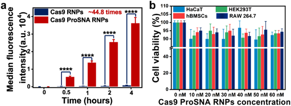
Cellular uptake/biocompatibility assay of Cas9 ProSNA RNPs. (a) Flow cytometry analysis of AF647-labeled Cas9 ProSNA RNPs and Cas9 RNPs that are uptaken by HaCaT cells over 4 h. ****, p < 0.0001. (b) Cell Counting Kit-8 assay quantifying the number of viable cells upon incubation with Cas9 ProSNA RNPs. The error bars represent standard error of the mean with three independent replicates.
Endosomal entrapment remains a major obstacle to cytosolic protein delivery via receptor-mediated endocytosis.11 HaCaT cells were incubated with Cas9 ProSNA RNPs or Cas9 RNPs and visualized using confocal microscopy (Figure 2a and Figure S14). To obtain detailed information regarding the colocalization of Cas9 ProSNA RNPs and endosomes, Z-stack images were taken to calculate the Mander’s overlap coefficient (r). The r score decreased over time (i.e., 2 h (r = 0.73 ± 0.05), 4 h (r = 0.46 ± 0.03), and 8 h (r = 0.27 ± 0.02)), confirming the effective endosomal escape of Cas9 ProSNA RNPs induced by GALA peptides (Figure 2b). We next calculated the nuclear import efficiency as the percentages of nuclei colocalized by Cas9 ProSNA RNPs. The nuclear import efficiency increased over time (i.e., 2 h (13.5 ± 2%), 4 h (44.7 ± 2.4%), 8 h (80.2 ± 3.7%)), highlighting NLS-directed nuclear entry (Figure 2c). As expected, in the absence of DNA functionalization, we did not observe the colocalization of Cas9 RNPs in the nucleus (Figure S14). Taken together, these results showcase the efficient cellular uptake, endosomal escape, and nuclear import of Cas9 ProSNA RNPs.
Figure 2:
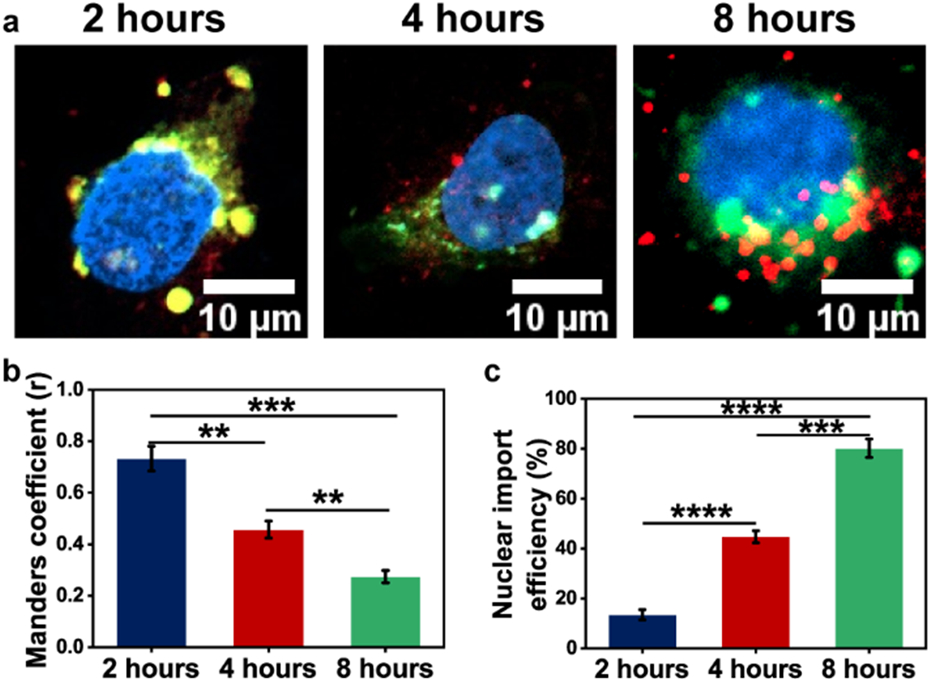
Endosomal escape and nuclear import of Cas9 ProSNA RNPs. (a) Confocal images of HaCaT cells after incubation with 20 nM Cas9 ProSNA RNPs (red), showing endosomes (green) and the cell nuclei (blue) at various time points: 2 h, 4 h, and 8 h. Yellow regions represent colocalization between Cas9 ProSNA RNPs (red) and endosomes (green). Pink regions represent colocalization between Cas9 ProSNA RNPs (red) and the cell nuclei (blue). (b) Mander’s overlap coefficients of Cas9 ProSNA RNPs and the endosomes. (c) Nuclear import efficiency determined by the colocalization of Cas9 ProSNA RNPs within the nuclei. ** , p < 0.01; *** , p < 0.001; **** , p < 0.0001. The error bars represent standard error of the mean with three independent replicates.
Cas9 ProSNA RNP-mediated genomic indel (insertion/deletion) mutations, derived from sgRNA-guided double strand breaks and endogenous non-homologous end joining, were investigated in different cell lines and with different targets. First, GRIN2B was selected as a model target due to its significance in neurodevelopmental disorders and intellectual disabilities.40 Specifically, Cas9 ProSNA RNPs targeting the GRIN2B gene were incubated with HaCaT cells for 3 d, and then a surveyor assay with T7 endonuclease I (T7EI) was performed to detect and quantify the indel efficiency within the target (Figure S15). The surveyor assay revealed an indel frequency of approximately 34.9%, providing further evidence of endosomal escape and subsequent nuclear delivery of Cas9 ProSNA RNPs (Figure 3a). In contrast, the assay did not detect GRIN2B cleavage when Cas9 RNPs were used likely because of their inefficient nuclear delivery (Figure S16). Next, to demonstrate their generalizability to other targets, Cas9 ProSNA RNPs targeting DNase I hypersensitive site were applied to HaCaT cells, resulting in an indel frequency of approximately 38.3% (Figure 3a). hBMSCs cells were also used, and relatively high indel efficiencies were achieved (approximately 39.5% in GRIN2B and 32.6% in DNase I). Furthermore, the gene editing efficiency of Cas9 ProSNA RNPs in difficult-to-transfect cells (RAW 264.7 macrophages) was studied. Compared to the commercial Lipofectamine CRISPRMAX™ Cas9 RNP transfection reagent, Cas9 ProSNA RNPs exhibited a 2.4-fold increase in indel frequency (Figure 3b).
Figure 3:
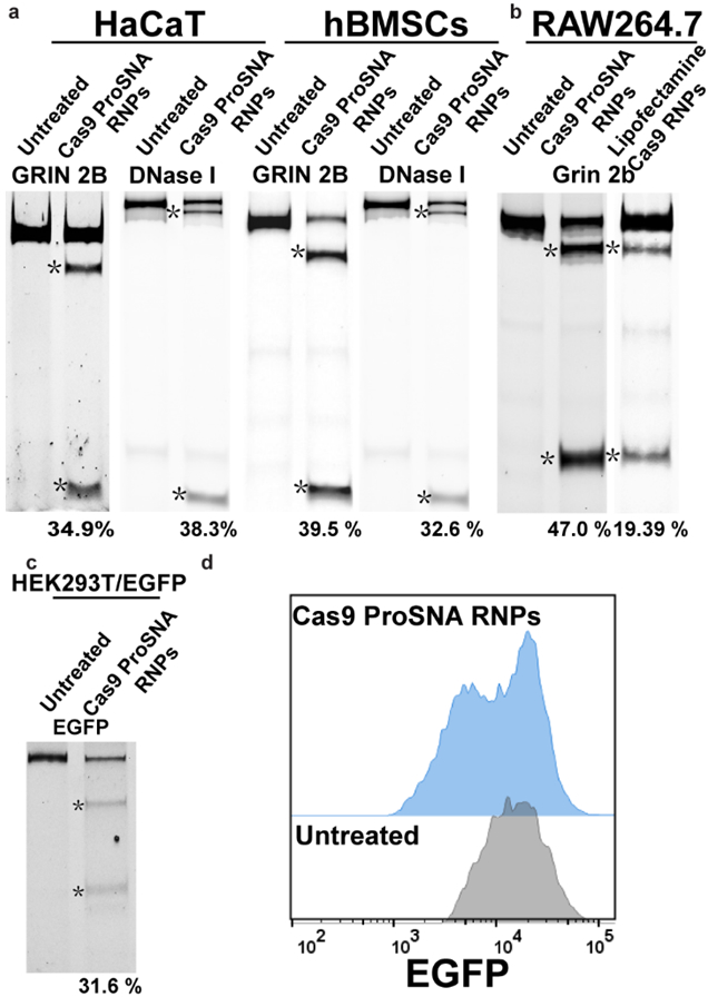
Genome-editing activity of Cas9 ProSNA RNPs in mammalian cells. (a) Surveyor assays in HaCaT and hBMSCs cells. (b) Surveyor assays in RAW 264.7 cells treated with either Cas9 ProSNA RNPs or Cas9 RNPs transfected by commercial Lipofectamine CRISPRMAX™ agent. (c) Surveyor assays of HEK293T/EGFP cells treated with Cas9 ProSNA RNPs targeting the EGFP site. The cleavage products are labeled with asterisks. (d) Flow cytometry histogram of HEK293T/EGFP cells treated with Cas9 ProSNA RNPs.
To evaluate whether the efficient gene editing activity of Cas9 ProSNA RNPs could further induce gene silencing, EGFP sgRNA was used to target the coding region of the expressed EGFP reporter in HEK293T cells (HEK293T/EGFP). After treating HEK293T/EGFP cells with the Cas9 ProSNA RNPs, the corresponding surveyor assay revealed an indel efficiency of 31.6%, indicating that the coding region was successfully targeted and resulted in a frameshift mutation (Figure 3c). The loss of EGFP fluorescence was further confirmed by flow cytometry (Figure 3d and Figure S17) and confocal microscopy (Figure S18). Importantly, the Cas9 RNPs alone did not show detectable genome editing activity in all cell lines with their respective target(s) (Figure S16).
In conclusion, a new chemical construct that can be used for genome editing and gene silencing based on Cas9 ProSNAs was synthesized and developed. Compared to Cas9 RNPs (fused with GALA peptides and NLSs), Cas9 ProSNA RNPs exhibit significantly enhanced cellular internalization, GALA peptide-induced endosomal escape, and NLS-directed nuclear delivery. Importantly, Cas9 ProSNAs are stable against protease digestion, exhibit negligible cellular toxicity, and showed an indel efficiency of up to 47%. In principle, this design can be generalized to other (bio)macromolecular cargos to manipulate cellular functions. The simple and versatile structure of Cas9 ProSNAs provides a direct platform for gene therapy and may inform the future application of a limitless library of protein-based therapies.
Supplementary Material
ACKNOWLEDGEMENT
Research reported in this publication was supported by the National Cancer Institute of the National Institutes of Health under award U54CA199091. The content is solely the responsibility of the authors and does not necessarily represent the official views of the National Institutes of Health. The project was also supported by the Sherman Fairchild Foundation Inc. M.E. was partially supported by the Dr. John N. Nicholson Fellowship and the Alexander S. Onassis Public Benefit Foundation. This work made use of the IMSERC at Northwestern University, which has received support from the Soft and Hybrid Nanotechnology Experimental (SHyNE) Resource (NSF ECCS-1542205), the State of Illinois, and the International Institute for Nanotechnology (IIN).
Footnotes
ASSOCIATED CONTENT
The Supporting Information is available free of charge via the Internet at http://pubs.acs.org.
Methods, Tables S1-S2, and Figure S1-S13 (PDF)
REFERENCES
- 1.Jinek M; Jiang F; Taylor DW; Sternberg SH; Kaya E; Ma E; Anders C; Hauer M; Zhou K; Lin S, et al. , Structures of Cas9 Endonucleases Reveal RNA-Mediated Conformational Activation. Science 2014, 343 (6176), 1247997. [DOI] [PMC free article] [PubMed] [Google Scholar]
- 2.Ran FA; Cong L; Yan WX; Scott DA; Gootenberg JS; Kriz AJ; Zetsche B; Shalem O; Wu X; Makarova KS, et al. , In Vivo Genome Editing using Staphylococcus Aureus Cas9. Nature 2015, 520 (7546), 186–191. [DOI] [PMC free article] [PubMed] [Google Scholar]
- 3.Huang TP; Zhao KT; Miller SM; Gaudelli NM; Oakes BL; Fellmann C; Savage DF; Liu DR, Circularly Permuted and PAM-Modified Cas9 Variants Broaden the Targeting Scope of Base Editors. Nat. Biotechnol 2019, 37 (6), 626–631. [DOI] [PMC free article] [PubMed] [Google Scholar]
- 4.Komor AC; Badran AH; Liu DR, CRISPR-Based Technologies for the Manipulation of Eukaryotic Genomes. Cell 2017, 168 (1-2), 20–36. [DOI] [PMC free article] [PubMed] [Google Scholar]
- 5.Doudna JA, The Promise and Challenge of Therapeutic Genome Editing. Nature 2020, 578 (7794), 229–236. [DOI] [PMC free article] [PubMed] [Google Scholar]
- 6.Doudna JA; Charpentier E, The New Frontier of Genome Engineering with CRISPR-Cas9. Science 2014, 346 (6213), 1258096. [DOI] [PubMed] [Google Scholar]
- 7.Hsu PD; Lander ES; Zhang F, Development and Applications of CRISPR-Cas9 for Genome Engineering. Cell 2014, 157 (6), 1262–1278. [DOI] [PMC free article] [PubMed] [Google Scholar]
- 8.Cong L; Ran FA; Cox D; Lin S; Barretto R; Habib N; Hsu PD; Wu X; Jiang W; Marraffini LA, et al. , Multiplex Genome Engineering Using CRISPR/Cas Systems. Science 2013, 339 (6121), 819–823. [DOI] [PMC free article] [PubMed] [Google Scholar]
- 9.Liu S; Cheng Q; Wei T; Yu X; Johnson LT; Farbiak L; Siegwart DJ, Membrane-Destabilizing Ionizable Phospholipids for Organ-Selective mRNA Delivery and CRISPR-Cas Gene Editing. Nature Materials 2021, 20 (5), 701–710. [DOI] [PMC free article] [PubMed] [Google Scholar]
- 10.Gillmore JD; Gane E; Taubel J; Kao J; Fontana M; Maitland ML; Seitzer J; O'Connell D; Walsh KR; Wood K, et al. , CRISPR-Cas9 In Vivo Gene Editing for Transthyretin Amyloidosis. N. Engl. J. Med 2021, 385 (6), 493–502. [DOI] [PubMed] [Google Scholar]
- 11.Rouet R; Thuma BA; Roy MD; Lintner NG; Rubitski DM; Finley JE; Wisniewska HM; Mendonsa R; Hirsh A; de Onate L, et al. , Receptor-Mediated Delivery of CRISPR-Cas9 Endonuclease for Cell-Type-Specific Gene Editing. J. Am. Chem. Soc 2018, 140 (21), 6596–6603. [DOI] [PMC free article] [PubMed] [Google Scholar]
- 12.Alsaiari SK; Patil S; Alyami M; Alamoudi KO; Aleisa FA; Merzaban JS; Li M; Khashab NM, Endosomal Escape and Delivery of CRISPR/Cas9 Genome Editing Machinery Enabled by Nanoscale Zeolitic Imidazolate Framework. J. Am. Chem. Soc 2018, 140 (1), 143–146. [DOI] [PubMed] [Google Scholar]
- 13.Lin Y; Wagner E; Lachelt U, Non-Viral Delivery of the CRISPR/Cas System: DNA Versus RNA Versus RNP. Biomater. Sci 2022, 10 (5), 1166–1192. [DOI] [PubMed] [Google Scholar]
- 14.Wang M; Zuris JA; Meng F; Rees H; Sun S; Deng P; Han Y; Gao X; Pouli D; Wu Q, et al. , Efficient Delivery of Genome-Editing Proteins using Bioreducible Lipid Nanoparticles. Proc. Natl. Acad. Sci. U.S.A 2016, 113 (11), 2868–2873. [DOI] [PMC free article] [PubMed] [Google Scholar]
- 15.Lyu Y; He S; Li J; Jiang Y; Sun H; Miao Y; Pu K, A Photolabile Semiconducting Polymer Nanotransducer for Near-Infrared Regulation of CRISPR/Cas9 Gene Editing. Angew. Chem. Int. Ed 2019, 58 (50), 18197–18201. [DOI] [PubMed] [Google Scholar]
- 16.Zuris JA; Thompson DB; Shu Y; Guilinger JP; Bessen JL; Hu JH; Maeder ML; Joung JK; Chen ZY; Liu DR, Cationic Lipid-Mediated Delivery of Proteins Enables Efficient Protein-Based Genome Editing in Vitro and in Vivo. Nat. Biotechnol 2015, 33 (1), 73–80. [DOI] [PMC free article] [PubMed] [Google Scholar]
- 17.Mout R; Ray M; Yesilbag Tonga G; Lee YW; Tay T; Sasaki K; Rotello VM, Direct Cytosolic Delivery of CRISPR/Cas9-Ribonucleoprotein for Efficient Gene Editing. ACS Nano 2017, 11 (3), 2452–2458. [DOI] [PMC free article] [PubMed] [Google Scholar]
- 18.Zhou W; Cui H; Ying L; Yu XF, Enhanced Cytosolic Delivery and Release of CRISPR/Cas9 by Black Phosphorus Nanosheets for Genome Editing. Angew. Chem. Int. Ed 2018, 57 (32), 10268–10272. [DOI] [PubMed] [Google Scholar]
- 19.Huang C; Sun Z; Cui H; Pan T; Geng S; Zhou W; Chu PK; Yu XF, InSe Nanosheets for Efficient NIR-II-Responsive Drug Release. ACS Appl. Mater. Interfaces 2019, 11 (31), 27521–27528. [DOI] [PubMed] [Google Scholar]
- 20.Ramakrishna S; Kwaku Dad AB; Beloor J; Gopalappa R; Lee SK; Kim H, Gene Disruption by Cell-Penetrating Peptide-Mediated Delivery of Cas9 Protein and Guide RNA. Genome Research 2014, 24 (6), 1020–1027. [DOI] [PMC free article] [PubMed] [Google Scholar]
- 21.Gustafsson O; Radler J; Roudi S; Lehto T; Hallbrink M; Lehto T; Gupta D; Andaloussi SE; Nordin JZ, Efficient Peptide-Mediated In Vitro Delivery of Cas9 RNP. Pharmaceutics 2021, 13 (6). [DOI] [PMC free article] [PubMed] [Google Scholar]
- 22.Lostalé-Seijo I; Louzao I; Juanes M; Montenegro J, Peptide/Cas9 Nanostructures for Ribonucleoprotein Cell Membrane Transport and Gene Edition. Chemical Science 2017, 8 (12), 7923–7931. [DOI] [PMC free article] [PubMed] [Google Scholar]
- 23.Rosi NL; Giljohann DA; Thaxton CS; Lytton-Jean AK; Han MS; Mirkin CA, Oligonucleotide-Modified Gold Nanoparticles for Intracellular Gene Regulation. Science 2006, 312 (5776), 1027–1030. [DOI] [PubMed] [Google Scholar]
- 24.Kusmierz CD; Bujold KE; Callmann CE; Mirkin CA, Defining the Design Parameters for in Vivo Enzyme Delivery Through Protein Spherical Nucleic Acids. ACS Cent. Sci 2020, 6 (5), 815–822. [DOI] [PMC free article] [PubMed] [Google Scholar]
- 25.Wang S; Chen Y; Wang S; Li P; Mirkin CA; Farha OK, DNA-Functionalized Metal-Organic Framework Nanoparticles for Intracellular Delivery of Proteins. J. Am. Chem. Soc 2019, 141 (6), 2215–2219. [DOI] [PMC free article] [PubMed] [Google Scholar]
- 26.Samanta D; Ebrahimi SB; Kusmierz CD; Cheng HF; Mirkin CA, Protein Spherical Nucleic Acids for Live-Cell Chemical Analysis. J. Am. Chem. Soc 2020, 142 (31), 13350–13355. [DOI] [PMC free article] [PubMed] [Google Scholar]
- 27.Elghanian R; Storhoff JJ; Mucic RC; Letsinger RL; Mirkin CA, Selective Colorimetric Detection of Polynucleotides Based on the Distance-Dependent Optical Properties of Gold Nanoparticles. Science 1997, 277 (5329), 1078–1081. [DOI] [PubMed] [Google Scholar]
- 28.Brodin JD; Sprangers AJ; McMillan JR; Mirkin CA, DNA-Mediated Cellular Delivery of Functional Enzymes. J. Am. Chem. Soc 2015, 137 (47), 14838–14841. [DOI] [PMC free article] [PubMed] [Google Scholar]
- 29.Harrington LB; Paez-Espino D; Staahl BT; Chen JS; Ma E; Kyrpides NC; Doudna JA, A Thermostable Cas9 with Increased Lifetime in Human Plasma. Nat. Commun 2017, 8 (1), 1424. [DOI] [PMC free article] [PubMed] [Google Scholar]
- 30.Narayan SP; Choi CH; Hao L; Calabrese CM; Auyeung E; Zhang C; Goor OJ; Mirkin CA, The Sequence-Specific Cellular Uptake of Spherical Nucleic Acid Nanoparticle Conjugates. Small 2015, 11 (33), 4173–4182. [DOI] [PMC free article] [PubMed] [Google Scholar]
- 31.Kusumoto K; Akita H; Ishitsuka T; Matsumoto Y; Nomoto T; Furukawa R; El-Sayed A; Hatakeyama H; Kajimoto K; Yamada Y, et al. , Lipid Envelope-Type Nanoparticle Incorporating a Multifunctional Peptide for Systemic siRNA Delivery to the Pulmonary Endothelium. ACS Nano 2013, 7 (9), 7534–7541. [DOI] [PubMed] [Google Scholar]
- 32.Abd Elwakil MM; Khalil IA; Elewa YHA; Kusumoto K; Sato Y; Shobaki N; Kon Y; Harashima H, Lung-Endothelium-Targeted Nanoparticles Based on a pH-Sensitive Lipid and the GALA Peptide Enable Robust Gene Silencing and the Regression of Metastatic Lung Cancer. Adv. Funct. Mater 2019, 29 (18), 1807677. [Google Scholar]
- 33.Schach DK; Rock W; Franz J; Bonn M; Parekh SH; Weidner T, Reversible Activation of a Cell-Penetrating Peptide in a Membrane Environment. J. Am. Chem. Soc 2015, 137 (38), 12199–12202. [DOI] [PubMed] [Google Scholar]
- 34.Akishiba M; Takeuchi T; Kawaguchi Y; Sakamoto K; Yu HH; Nakase I; Takatani-Nakase T; Madani F; Graslund A; Futaki S, Cytosolic Antibody Delivery by Lipid-Sensitive Endosomolytic Peptide. Nat. Chem 2017, 9 (8), 751–761. [DOI] [PubMed] [Google Scholar]
- 35.Boukamp P; Petrussevska RT; Breitkreutz D; Hornung J; Markham A; Fusenig NE, Normal Keratinization in a Spontaneously Immortalized Aneuploid Human Keratinocyte Cell Line. J. Cell Biol 1988, 106 (3), 761–771. [DOI] [PMC free article] [PubMed] [Google Scholar]
- 36.Choi CH; Hao L; Narayan SP; Auyeung E; Mirkin CA, Mechanism for the Endocytosis of Spherical Nucleic Acid Nanoparticle Conjugates. Proc. Natl. Acad. Sci. U.S.A 2013, 110 (19), 7625–7630. [DOI] [PMC free article] [PubMed] [Google Scholar]
- 37.Swiech K; Picanco-Castro V; Covas DT, Human Cells: New Platform for Recombinant Therapeutic Protein Production. Protein Expression and Purification 2012, 84 (1), 147–153. [DOI] [PubMed] [Google Scholar]
- 38.Tiwari JN; Seo YK; Yoon T; Lee WG; Cho WJ; Yousuf M; Harzandi AM; Kang DS; Kim KY; Suh PG, et al. , Accelerated Bone Regeneration by Two-Photon Photoactivated Carbon Nitride Nanosheets. ACS Nano 2017, 11 (1), 742–751. [DOI] [PubMed] [Google Scholar]
- 39.Roger T; David J; Glauser MP; Calandra T, MIF Regulates Innate Immune Responses Through Modulation of Toll-Like Receptor 4. Nature 2001, 414 (6866), 920–924. [DOI] [PubMed] [Google Scholar]
- 40.Endele S; Rosenberger G; Geider K; Popp B; Tamer C; Stefanova I; Milh M; Kortüm F; Fritsch A; Pientka FK, et al. , Mutations in GRIN2A and GRIN2B Encoding Regulatory Subunits of NMDA Receptors Cause Variable Neurodevelopmental Phenotypes. Nat. Genet 2010, 42 (11), 1021–1026. [DOI] [PubMed] [Google Scholar]
Associated Data
This section collects any data citations, data availability statements, or supplementary materials included in this article.