



Abstract
Objective:
The aim of this randomized controlled clinical trial was to determine the effect of the omega-3 fatty acid supplementations 300 mg per day for 8 weeks on the serum levels of ACE/ACE2 ratio in Jordanian participants with vitamin D deficiency (VDD).
Methods:
The physical and clinical characteristic of individuals in both intervention and control randomized controlled clinical trial were measured and analyzed. The comparisons between the two groups and the changes in each group before and after taking omega-3 doses were studied through independent t test and paired t test, respectively. Possible factors that have a role in the changes were determined by multivariate stepwise regression. Follow-up period lasted 10 weeks.
Results:
The sample consisted of 82 participants with VDD and a mean age of 37.85 ± 9.85 years. Omega-3 Supplements resulted in a significant decrease in serum ACE levels, ACE/ACE2 ratio and serum 25-hydroxy vitamin D (25OHD). While the change in serum ACE2 levels and serum triglycerides levels were insignificant. Also, a significant increase in serum LDL levels were observed.
Conclusion:
It is possible that taking high doses of omega-3 fatty acid supplementations have positive effects on the heart and circulatory system and could protect from COVID-19 or decrease disease severity, in connection with a decrease in the ACE/ACE 2 ratio. On the other hand, omega-3 supplement may have negative effect on cardiovascular system due to the significant increase in serum LDL levels.
Keywords: omega-3, ACE, ACE2, ACE/ACE2 ratio, non-HDL, vitamin D, 25- hydroxy vitamin D (25OHD)
INTRODUCTION
The Renin Angiotensin Aldosterone system (RAAS) has been focused on for several years because of its crucial role in the physiology and pathophysiology of cardiovascular disease (CVD). It involves also in blood pressure regulation, fluid, and electrolyte balance through action on heart, kidney, and blood vessels.1 Angiotensin converting enzyme (ACE), is a critical regulator of RAAS by converting Angiotensin I (Ang-I) to Angiotensin II (Ang-II), which is the most powerful biologically active product of RAAS.2 Ang-II increases blood pressure stimulates aldosterone secretion, which results in sodium reabsorption and potassium excretion.3 However, ACE2 a second ACE is a negative regulator of RAAS and opposing the effect of ACE in the heart, kidneys, and lungs.4 ACE2 converts Ang-II to Angiotensin, which is a vasodilator, antihypertrophic, antithrombotic peptide.5
It is interesting to note that in almost all the pathological conditions especially in cardiovascular diseases, there is a disturbance in ACE/ACE2 ratio and its usually due to down regulation of ACE2 levels. Indeed, ratio disturbance accompanied by disturbance in RAAS homeostasis.6 For example, ACE2 deficiency has been associated with exacerbation of hypertension (HTN) and ventricular remodeling.7 Also, it has been found that ACE/ACE2 ratio increased in case of moderate to severe chronic heart failure.8
Another leading role of ACE2 was emerged by the evolution of Corona virus disease 2019 (COVID-19).9 Human ACE2 serves as a receptor for Severe Acute Respiratory Syndrome Coronavirus 2 (SARS-CoV-2) entry into target cells by binding of the spike protein to ACE2 and a specific transmembrane serine protease 2 (TMPRSS2) required for the spike (S) protein priming, which also leads to down regulation of ACE2.10 COVID-19 outcome is related to age and the expression of ACE2 decreases with increasing age.11 Interestingly, poor outcomes of COVID-19 have been observed in elderly patients, and those with pre-existing CVD, in whom have already deficiency in ACE2 and increased ACE/ACE2 ratio.12
CVDs are the major cause of death worldwide. For example, in Jordan, 37.6% of deaths were related to CVDs. Moreover, the Jordanian Ministry of Health ranked coronary heart disease as the number one killer among CVDs in Jordan.13
American College of Cardiology and national cardiac societies recommend the intake of 1 gram/day of the two marine omega-3 fatty acids eicosapentaenoic acid (EPA) and docosahexaenoic acid (DHA) for prevention of CVD, treatment post-myocardial infarction and prevention of sudden cardiac death.14 However, randomized controlled trials (RCTs) checking the effect of these supplementations on cardiovascular outcomes are lacking. The aim of this study was to determine the effect of the omega-3 fatty acid supplementations 300 mg per day for 8 weeks on serum levels of ACE/ACE2 ratio in Jordanian participants with vitamin D deficiency (VDD).
MATERIALS AND METHODS
Study setting and participants
This RCT conducted between December 2020 and March 2021 at Applied Science Private University (ASU). Among Jordanian participants in ASU aged (35-55) years were recruited to participate in this study. This RCT was approved by the ASU ethics committee for the protection of human subjects (protocol number 2020-PHA-2). Trial registration: This trial was registered at clinicaltrials.gov as NCT04658433. The RCT was conducted following the Helsinki Declaration. Written informed consent was obtained from all study participants. All participants were informed of the study requirements, benefits, and risks from taking the supplementations and their freedom to withdraw from the study at any time. Eligible participants were enrolled based on a confirmed diagnosis of VDD by internal medicine consultants in Ibn Al-Haytham hospital laboratories. Inclusion criteria included males and females in the age range of 35-55 years without a medical diagnosis of COVID-19. Exclusion criteria included any subject with any chronic disease such as CVD, diabetes, or immune problems, including autoimmune diseases, chronic or severe infections. Also, subjects who take any type of supplement including vitamin D, pregnant, breastfeeding, and females using hormonal contraceptives were excluded.
Intervention
The participants were randomized at baseline into two groups, and the randomization performed by an external statistician and is not involved in the study. In n-3FAs groups, n-3FAs provided by (Jamieson Laboratories, Canada N8W 585). Each soft gel capsule contains: Wild Salmon and Fish oil complex 1000 mg (n-3FAs= 300 mg) providing 180 mg as EPA and 120 mg as DHA. Food and drug administration (FDA) has concluded that dietary dosages of up to 3 grams per day of omega-3 fatty acids from marine sources are generally considered safe. Also, one-gram capsule contains 180 mg EPA and 120 mg DHA generally well tolerated and safe.15 Jordanian participants enrolled in the trial were randomized into two groups as outlined in Figure 1. Group 1 (control group) was participants not received n-3FAs supplementation. Whereas, group 2 (n-3FAs group) was participants treated with 1000 mg wild salmon and fish oil complex (contains 300 mg of n-3FAs) once daily for eight weeks.
Figure 1.
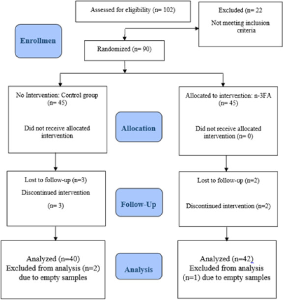
CONSORT flow diagram for the study, indicating the number of subjects screened, recruited, and randomly assigned to the different intervention groups
Anthropometric measurement
Body mass index (BMI), height (Ht), hip circumference (H), body weight (BW), W/H ratio (WHR), waist circumference (W), and Ht/W ratio (HtWR) data were collected using slandered techniques. Before the measurement, the study staff confirmed that the participants should be clothed in light clothing and without shoes.
BW was measured to the closest 0.5 kilos using an electronic scale (Tanita, THD-646). Ht was measured to the closest 0.1 cm using a vertical ruler (stadiometer).
Clinical parameters assays
Fasting venous blood samples at baseline and at ten weeks, using a sterile vacutainer (VACEUTTE® TUBE), needle and needle holder. Blood was collected into a 5 ml serum tube. After collection they were stored at room temperature at least 45-60 minutes then were centrifuged. Blood samples were centrifuged at 4000 rpm for 10 minutes. Aliquots of at least 1 ml of serum were measured into labeled Eppendorf tubes for the measurement of the following clinical measures (Table1). All samples were stored at (-80°C) in Ibn Al-Haytham laboratories, Amman, Jordan. All analysis was undertaken in a quality controlled registered laboratory Ibn Al-Haytham hospital.
Table 1.
Baseline descriptive statistics of the anthropometric and clinical parameters characteristic of the study participants (n=82)
| Parameter | Mean (SD) | Normal range |
|---|---|---|
| Age (years) | 37.85 (9.85) | N/A |
| Wt (kg) | 81.91 (20.65) | N/A |
| Ht (cm) | 168.27 (7.82) | N/A |
| BMI (kg/m2) | 28.61 (6.40) | 18.5-24.9 |
| Waist (cm) | 95.95 (17.39) | NA |
| Hip (cm) | 108.21 (14.17) | N/A |
| WHR | 89.93 (22.55) | N/A |
| ACE (ng/ml) | 246.03 (60.93) | N/A |
| ACE2 (ng/ml) | 1.23 (0.58) | N/A |
| ACE/ACE2 R | 22813.37 (9097.48) | N/A |
| 25OHD (ng/ml) | 21.04 (6.74) | 30-50 |
| FBG (mg/dl) | 86.41 (21.52) | 70-110 |
| TG (mg/dl) | 133.44 (84.37) | Up to 150 |
| LDL (mg/dl) | 100.85 (33.90) | Up to 100 |
| PTH (pg/ml) | 33.33 (8.51) | 9-90 |
| Calcium (mg/dl) | 9.51 (1.64) | 8.6-10.3 |
| PO4 (mg/dl) | 4.20 (0.14) | 2.5-4.5 |
Abbreviations: WHR, waist/hip ratio; Wt., weight; Ht, height; BMI, body mass index; SD, standard deviation; kg, kilograms; cm, centimeter; ACE, angiotensin converting enzyme; ACE/ACE2 R, angiotensin converting enzyme/ angiotensin converting enzyme2 Ratio; 25OHD, 25-hydroxy vitamin D; FBG, fasting blood glucose, TG, triglycerides; LDL, low density lipoprotein; PTH, parathyroid hormone; PO4, phosphate; mg, milligram; ml, milliliter; dl, deciliter; pg, picogram; ng, nanogram; N/A, non-applicable.
Laboratory analysis
Serum ACE assay: serum ACE was measured using Human ELISA (Enzyme Linked Immunosorbent Assay) KIT (ab 263889, abcam, United states). Human ACE ELISA Kit (ab263889) designed for quantitative measurement of ACE protein in serum. It uses Simple Step ELISA technology. Quantitate Human ACE with 0.15 ng/ml sensitivity. Simple Step ELISA technology allows the formation of the antibody-antigen complex in one single step. Samples were added and antibody mix to wells all at once, incubate, wash, and then add final substrate. This approach sandwich ELISA allows the formation of the antibody-analyte sandwich complex in a single step, significantly reducing assay time. Serum ACE2 assay: serum ACE was measured using Human ELISA KIT (ab235649, abcam, United states). Human ACE2 ELISA Kit (ab235649) is a sandwich ELISA designed for the quantitative measurement of native ACE2 protein in serum. It uses Simple Step ELISA technology. Quantitate Human ACE2 with 1052 pg/ml sensitivity. Simple Step ELISA technology allows the formation of the antibody-antigen complex in one single step, reducing assay time to 90 minutes. Samples were added or standards and antibody mix to wells all at once, incubate, wash, and add your final substrate. Serum IL-6 assay serum IL-6 was measured using Human ELISA KIT (ab 46027, abcam, United states). Abcam’s IL-6 Human ELISA kit used for quantitative measurement of IL-6 in Human serum with sensitivity of 2 pg/ml. A monoclonal antibody specific for IL-6 has been coated onto the wells of the microtiter strips provided. IL-6 concentrations are pipetted into these wells. During the first incubation, the samples, and a biotinylated monoclonal antibody specific for IL-6 are simultaneously incubated. After washing, the enzyme Streptavidin-HRP, that binds the biotinylated antibody is added, incubated, and washed. A TMB substrate solution is added which acts on the bound enzyme to induce a colored reaction product. The intensity of this colored product is directly proportional to the concentration of IL-6 present in the samples.
Serum 25OHD assay: 25-hydroxyvitamin D2 and D3 in serum were measured as a standard procedure at the clinical laboratories of Ibn Al-Haytham hospital using chemiluminescent immunoassay technology by LIAISON® 25-hydroxyvitamin D Assay (DiaSorin). Specific antibody to VD was used for coating magnetic particles (solid phase) and VD was linked to an isoluminol derivative. Its lower limit of assay approximately was (4 ng/mL), and its intra- and inter- assay coefficients of variation was 5.0% and 8.2% respectively. The assay has a 100% cross-reactivity with both metabolites of 25OHD namely, 25OHD2 and 25OHD3 and thus measures total serum 25OHD content. Serum Fasting Blood Glucose (FBG): the FBG samples were performed at the laboratory of clinical chemistry, Ibn Al- Haytham hospital on a Roche Cobas C501 analyzer (GLUC3 application, Roche, Mannheim, Germany). LDL Cholesterol: low density lipoprotein (LDL) cholesterol was measured using LDL cholesterol precipitating reagent (BioSystems, M11579i-16). LDL concentrations were calculated from the difference between the serum total cholesterol (TC) and the cholesterol in the supernatant after centrifugation. The concentration was obtained detection limit = 0.45 mg/dl. Triglycerides (TGs): serum TG level was measured using TGs BioSystems kit (M11528i-20) the absorbance was measured at 500 nm. Detection limit for TG was 1.6 mg/dl.
Statistical analysis
Data were analyzed using statistical package for social science version 25 (SPSS, Chicago, IL, USA). Descriptive statistics were used to describe the sample, normally distributed quantitative variables were described using mean and standard deviation, qualitative variables were described using frequency and percentages. Normality of distribution for laboratory measurements was tested using the Kolmogorov–Smirnov test. As the data were normally distributed independent sample t-test was used to investigate if there were any significant differences in the mean values for each parameter between the two groups of the study. To determine any significant differences in each trial group before and after administration of the supplements (n-3FAs) paired t-test was conducted. Stepwise regression analysis was used to evaluate which independent variable influenced serum (ACE, ACE2, ACE/ACE2 Ratio) in n-F3A group at the end of the trial. A p-value of < 0.050 was considered statistically significant.
RESULTS
In stage two randomized distribution of the eligible participants (n=102) were enrolled according to inclusion and exclusion criteria. 102 participants enrolled in this RCT, of which 90 began and 82 participants completed the whole trial (Figure 1). Forty-five percent of the sample were female, 48.3% were in the control group and 51.7% were in the intervention group. Baseline anthropometric and clinical characteristics of the study participants are shown in Table 1. Mean age of the participants was 37.85 ± 9.85 years. Baseline BMI mean for all participants was 28.61 ± 6.40 kg/m2 which indicated that the population of the study sample generally was over-weight.
The mean values of clinical parameters at baseline including parathyroid hormone (PTH), calcium (Ca), phosphate (PO4), TG, LDL for all participants were within normal ranges (Table 1). Baseline mean value for serum 25OHD was 21.04 ± 6.74 ng/ml for all trial participants which mean they were vitamin D deficient. No single participant presented in this study with baseline serum 25OHD higher than 30 ng/m. Mean serum levels for ACE and ACE2 were 246.03 ± 60.93 ng/ml and 1.23 ± 0.58 ng/ml, respectively. Other mean values of serum parameters at baseline were within normal ranges are presented in Table 1.
The comparison of baseline and follow-up anthropometric characteristics of the participants between the study groups
Independent samples t-tests were conducted to determine the significant differences between control and n-F3A groups in anthropometric characteristics. The results showed that there were no significant differences between the two groups in all anthropometric characteristics at the baseline or at the end of the study as shown in Table 2.
Table 2.
Comparison of baseline and follow-up anthropometric characteristics of the participants between the study groups (n=82)
| Parameter | C | n-3FAs | P-value |
|---|---|---|---|
| Mean (SD) | Mean (SD) | ||
| Wt1 | 79.83 (19.43) | 83.85 (21.87) | 0.457 |
| Wt2 | 79.41(19.23) | 83.82 (22.84) | 0.423 |
| Ht1 | 166.62 (7.98) | 169.81(7.46) | 0.116 |
| Ht2 | 166.62 (7.98) | 169.81 (7.46) | 0.116 |
| BMI1 | 28.00 (5.30) | 29.18 (7.31) | 0.480 |
| BMI2 | 27.61 (5.10) | 29.19 (7.42) | 0.345 |
| Waist1 | 95.94 (14.66) | 95.97 (19.85) | 0.995 |
| Waist2 | 94.28 (13.17) | 97.45 (20.24) | 0.478 |
| Hip1 | 106.81 (14.99) | 109.52 (13.47) | 0.465 |
| Hip2 | 108.68 (9.61) | 109.16 (13.32) | 0.874 |
| WHR1 | 92.88 (30.42) | 87.17 (10.94) | 0.331 |
| WHR2 | 86.80 (9.71) | 88.91 (12.33) | 0.467 |
Note: P-value for comparison between groups (Independent Samples T- test); significant at 0.05 significant level. Abbreviations: n-3FA, omega-3 supplementation group; C, control group; SD, standard deviation; WHR, waist/ hip ratio; Wt, weight; Ht, hight; BMI, body mass index; 1, baseline value; 2, follow up value.
Changes in mean ACE, ACE2, and ACE/ACE2 ratio levels
At the end of the trial, paired samples t-test showed a significant difference in the mean ACE. ACE. Follow up levels were significantly reduced from the baseline with a change of about 38.06 ng /ml (269.76 ± 60.53 vs 231.70 ± 62.31 ng/ml, P=0.043) in the n-3FA group. Independent t-test showed a significant difference in mean ACE between n-3FA and control group (231.70 ± 62.31 vs 206.91 ± 60.17 ng/ml, P=0.006). On the other hand, there was no significant change between mean ACE2 of baseline and follow-up among those in n-3FA group (1.19 ± 0.48 vs 1.34 ± 0.36 ng/ml, P =0.453). Also, according to p-value obtained from independent t-test, there were no significant differences in the mean ACE2 between control and n-3FA group at baseline or follow-up end of the study as shown in Table 2. Finally, a significant reduction in mean ACE/ACE2 ratio of the baseline and follow-up was observed in n-3FA group (24908.48 ± 8714.86 vs 17725.19 ± 6147.40, P = 0.0387). Mean ACE/ACE2 ratio was significantly changed in n-3FA group, without significant difference in the control group for the same ratio. Also, no significant differences in mean serum ACE/ACE2 ratio were observed between the two study groups at baseline and at the end of the study.
Changes in the serum level of selected clinical parameters
Serum 25OHD Level
A significant reduction in serum 25OHD level was observed between baseline and the end of the study in n-3FA group with mean (23.48 ± 5.49 vs 16.03 ± 5.64 ng/ml, p <0.001). While in control group, no significant difference was observed in 25OHD level between baseline and at the end of the study with mean (18.42 ± 7.05 vs 17.02±5.52 ng/ml, p=0.052). Also, no significant differences were observed in serum 25OHD level between the two groups at baseline and at the end of the study p-value (>0.05).
TG
No significant differences were observed in serum TG level between the baseline and the end of the study within the control and n-F3A groups. Also, no significant differences were observed in serum TG level between groups at baseline and at the end of the study p-value (>0.05).
LDL
A significant change in serum LDL level were observed between baseline and the end of the study in n-3FA group (86.13 ± 30.91 vs 112.46 ±31.12 mg/dl, p<0.001), as serum LDL was significantly increased after n-3FAs administration. On the other hand, no significant change was observed in serum LDL level in control group between baseline and the end of the study mean (92.59 ± 30.02 vs 104.81 ± 47.24 mg/dl, p=0.061). Also, no significant differences in serum LDL levels were observed between the two groups at baseline and at the end of the study (p>0.05).
Multivariate regression analysis of factors associated with the changes in the levels of selected variables
Multivariate regression analysis demonstrated that 25OHD serum level was predictive of the significant reduction in serum ACE levels observed at 10-week follow-up in n-3FA group (R=0.687, R2=0.472, P=0.010) (Table 3). Other independent predictors of the significant reduction in serum ACE levels were also observed in n-3FAs group as shown in (Table 3). According to the twelfth multiple linear regression model the predictors indicted the following effects on ACE levels: (R=0.989, R2=0.979, F-test=107.345, β1= -1.424, β2= -4.870, β3= 4.714, β4= -1.624, β5= 2.113, β6= -7.831, β7= -1.220, β8= 14.258, β9= -.794, β10= 2.481, β11= 24.676, β12= -.111, P-value= 0.000). This model explained about (97.9%) of the variation in ACE levels, as shown in Table 3. There was a significant effect of the independent variables (WHR, Age, Ratio of (TC/HDL), TG, PTH) on ACE2 according to the fifth multiple linear regression model indicators (R=0.811, R2=0.657, F-test=9.594, β1=0.029, β2=-0.040, β3=0.002, β4=-0.001, β5= 0.013, P-value= 0.000). Also, this model explained about (65.7%) of the variation in ACE2 levels. There was a significant effect of the independent variables (LDL, Non-HDL, TG, Ratio of (TC/HDL), Age, WHR, FBG, Height, IL6) on ACE/ACE2 Ratio according to the ninth multiple linear regression model indicators (R=0.980, R2=0.961, F-test=57.579, β1=-909.930, β2= 862.272, β3= -37.583, β4= -32.844, β5= 261.077, β6= -308.472, β7= 102.070, β8=-249.049, β9= 127.209, P-value= 0.000). This model explained about (96.1%) of the variations in (ACE/ACE2) Ratio levels.
Table 3.
Significant multivariate associations between selected predictors and serum levels of ACE, ACE2, ACE/ACE2 R by stepwise regression at follow up of the trial in n-3FAs study group
| DV | IDV | Coefficient | ||||
|---|---|---|---|---|---|---|
| B | F | R | R2 | P-value | ||
| ACE | 25OHD | -1.424 | 25.873 | 0.687 | 0.472 | 0.010 |
| LDL | -4.870 | 20.039 | 0.767 | 0.589 | <0.01 | |
| Non-HDL | 4.714 | 23.531 | 0.850 | 0.723 | <0.01 | |
| Height | -1.624 | 29.751 | 0.906 | 0.821 | <0.01 | |
| PTH | 2.113 | 28.836 | 0.923 | 0.852 | <0.01 | |
| Weight | -7.831 | 31.195 | 0.941 | 0.886 | <0.01 | |
| HDL | -1.220 | 35.289 | 0.956 | 0.915 | 0.023 | |
| BMI | 14.258 | 41.085 | 0.968 | 0.937 | <0.01 | |
| IL-6 | -0.794 | 46.505 | 0.979 | 0.958 | <0.01 | |
| Hip | 2.481 | 52.596 | 0.982 | 0.965 | <0.01 | |
| ACE2 | 24.676 | 92.790 | 0.987 | 0.974 | 0.001 | |
| TG | -0.111 | 107.345 | 0.989 | 0.979 | <0.01 | |
| ACE2 | WHR | 0.029 | 9.462 | 0.496 | 0.246 | 0.001 |
| Age | -0.040 | 9.876 | 0.643 | 0.414 | < 0.001 | |
| TC/HDL R | 0.002 | 9.197 | 0.711 | 0.505 | 0.007 | |
| TG | -0.001 | 9.483 | 0.770 | 0.593 | 0.007 | |
| PTH | 0.013 | 9.594 | 0.811 | 0.657 | 0.040 | |
| ACE/ACE2 R | LDL | -909.930 | 15.141 | 0.586 | 0.343 | < 0.001 |
| Non-HDL | 862.272 | 19.525 | 0.763 | 0.582 | < 0.001 | |
| TG | -37.583 | 18.393 | 0.819 | 0.671 | < 0.001 | |
| TC/HDL R | -32.844 | 17.902 | 0.857 | 0.734 | 0.003 | |
| Age | 261.077 | 21.559 | 0.901 | 0.812 | 0.003 | |
| WHR | -308.472 | 40.473 | 0.954 | 0.910 | < 0.001 | |
| FBG | 102.070 | 49.448 | 0.968 | 0.938 | < 0.001 | |
| Height | -249.049 | 53.590 | 0.975 | 0.951 | 0.007 | |
| IL-6 | 127.209 | 57.579 | 0.980 | 0.961 | 0.031 | |
Note: R, Pearson linear correlation coefficient; R2, determinant coefficient; B, slope; F, variation between sample mean/variation within the sample; p-value obtained by Multiple liner regression analysis significant at 0.05 significant level Abbreviations: ACE, angiotensin converting enzyme; ACE/ACE2 R, angiotensin converting enzyme/angiotensin converting enzyme2 Ratio; ACE2, angiotensin converting enzyme 2; IDV, independent variable; DV, dependent variable; WHR, waist/hip ratio; Wt, weight; Ht, hight; BMI, body mass index; 25OHD, 25-hydroxy vitamin D; FBG, fasting blood glucose, TG, triglycerides; LDL, low density lipoprotein; PTH, parathyroid hormone; IL-6, interleukin 6; HDL, high density lipoprotein, TC, total cholesterol; R, Ratio.
DISCUSSION
To our knowledge, this is the first RCT which was specifically designed to examine the effect of omega-3 supplementations (300 mg n-3FAs daily dose) on serum ACE/ACE2 ratio level in participants with VDD. The main findings of this trial were that omega-3 supplementation significantly reduced serum level of ACE/ACE2 ratio. Significant reduction in ACE serum level was also observed at the end of the trial. Further, n-3FA supplementation significantly increased serum level of LDL and cause significant decrease in 25OHD serum levels in participants with VDD. Therefore, discussion of the observed results of this trial will be based on association between n-3FAs and ACE/ACE2 ratio and serum lipid profile in participants with and determination of predictors that mediated this association.
There is no RCT to date has asseseed the effect of omega-3 supplementation on serum ACE/ACE2 ratio. Despite there is limited data available but our findings were similar to previous in vitro and animal studies. In vitro study by Vijay Kumar and Das16 that conducted to evaluate the effect of different type of poly unsaturated fatty acids (PUFA) on ACE activity, they use blood sample from both hypertensive and normotensive individuals. This study found that serum ACE activity was significantly higher in patients with HTN compared to normotensive individuals and by the application of 20 microgram/ml of PUFA on isolated ACE resulted with significant reduction in ACE activity. Accordingly, this study suggestd that these PUFA especially EPA and DHA could be used as antihypertensive agent themselves.
The same result was found in animal study conducted on rats (50 diabetic and 10 control) fed with omega-3 and fenugreek terpenes formulation for 8 weeks, they found that plasma ACE activity was increased 45% in diabetic rats compared to control and the administration of omega-3 and fenugreek terpenes formulation for 8 weeks resulted with significant reduction in ACE activity as ACE activity was reduced by 38%, and this activity probably related to omega-3.17
In another animal study conducted on thirty male rats fed with 500mg/kg/day EPA: DHA: 6:1 for old rats for two weeks resulted with significant reduction in ACE and AT1R expression in aortic section.18 The potential role of omega-3 fatty acids in prevention and management of HTN has been mentioned in few review articles, and they hypothesized that omega-3 via suppression of ACE activity and reduce Ang-II formation could be serve as endogenous regulator of ACE activity.19
Another review have mentioned that omega-3 fatty acids suppress aldosterone secretion and this effect is related to the ability of omega-3 to lower ACE activity and reduce Ang-II production.20 Also, another study found that omega-3 administration for 3 weeks in murine models resulted with significant upregulation in ACE2 levels in the kidney of Ang-II dependent HTN model.21 Yet, our result was different, we found no significant change between mean ACE2 of baseline and follow-up among those in n-3FA group.
Another important finding in our study is the significant change in serum LDL level between baseline and the end of the study in n-3FA group (p<0.001), as there is a significant increase in LDL level after n-3FA administration. Our finding was similar to previous RCT conducted on 40 healthy women and men aged 40-65 years treated with 0.7 g DHA/day for three months.22 This RCT resulted in significant increase in serum LDL and total cholesterol by 7.1% and 4.2%, respectively, and this may be related to decrease expression of LDL receptors by DHA. Also, this study found that DHA resulted in increased LDL size and the authors proposed that larger LDL less atherogenic compared to small LDL, which is more exposed to oxidation.22
In another RCT conducted on 59 overweight mildly hyperlipidemic men, received 4-gram purified EPA or DHA for 6 weeks, the results showed that DHA not EPA resulted with significant increase in serum LDL level by 8%, and LDL particle size was also increased by 0.25+ 0.08 nm, and significant reduction in triacylglycerol in DHA and EPA groups by 20% and 18%, respectively.23 On the other hand, Satoh et al. mentioned that 1.8g/day EPA for three months in patients with metabolic syndrome was associated with significant reduction in serum total cholesterol, LDL and TG.24
Also, we observed that serum 25OHD was significantly reduced after n-3FA administration. This finding was similar to previous RCT; 300 mg/day of n-3FAs were given to premenopausal women with VDD and n-3FAs decreased serum 25OHD level significantly after 8 weeks.24 However, a recent systematic review found a total of 10 RCTs that evaluated the influence of omega-3 supplementation on 25-hydroxy vitamin D (25OHD) levels in humans. The review concluded that omega-3 supplementation increased 25OHD levels with dosage ≤ 1000 mg/day for interventional duration > 8 weeks. It seems that the dose-dependent outcomes were taken into consideration.25
Vitamin D has been shown to increases ACE2 and reduce ACE expression and reduce damage in lipopolysaccharide induced lung injury in rats, so 25OHD is important regulator of RAAS, induces ACE2/Ang (17) axis activity and inhibit renin and the ACE/Ang-II/AT1R axis.26 Chronic VDD is associated with increased risk of cardiovascular diseases, diabetes mellitus and HTN and induce lung fibrosis, all as result of activation of RAAS.27
There is growing evidence of presence of correlation between VDD and severity of COVID-19. Mortality rate from COVID-19 was dramatically lower in countries that have greater day light during winter, which consequently results with lower incidence of VDD, as vitamin D consider negative regulator of rennin biosynthesis, vitamin D inhibit rennin, ACE and Ang-II expression and induce ACE2 overexpression.28 While African American population have higher morbidity and mortality rate compared to other populations, and this may link to that most African American have low vitamin D levels.29 In a study conducted to evaluate vitamin D and ACE in COVID-19 patients compared to un infected individuals, the study found that ACE levels were significantly higher in COVID-19 patients and the highest quantity of circulating ACE were observed in patients with vitamin D insufficiency (<30 ng/ml), serum ACE concentration and disease severity were significantly higher in COVID-19 patients that have VDD compared to patient with higher vitamin D levels.27
Lin et al. observed that calcitriol decreased ACE and ACE/ACE2 ratio in diabetic rats and increased ACE2 concentration. In addition, calcitriol suppressed the mitogen-activated protein kinase P38 (MAPK) pathway, which plays a significant role in inflammation, apoptosis, and kidney injury in several glomerular and tubulointerstitial diseases, including diabetic kidney disease. However, the exact mechanism by which calcitriol caused change in ACE and ACE2 levels still unclear but they proposed that it may related to P38 MAPK suppression.30 According to all of these data the vitamin D has critical role in the pathogenesis of COVID-19 and CVD due to its role in the regulation of RAAS, as the HTN is one the most comorbidity associated with severe consequences of COVID-19.31 Stepwise Regression Analysis showed that there was a significant effect of the independent variables (25OHD, LDL, Non-HDL, Height, PTH, Weight, HDL, BMI, IL6, Hip, ACE2, TG) on ACE and this model explained about (97.9%) of the variation in ACE levels.
In Multivariate regression analysis, independent relationship was seen between (LDL, Non-HDL, TG, Ratio of (TC/HDL), age, WHR, FBG, height, IL6) and ACE/ACE2 ratio according to the ninth multiple linear regression model indicators (R=0.980, R2=0.961, F-test=57.579, β1=-909.930, β2= 862.272, β3= -37.583, β4= -32.844, β5= 261.077, β6= -308.472, β7= 102.070, β8=-249.049, β9= 127.209, P-value < 0.001). Also, this model explained about (96.1%) of the variation in (ACE/ACE2) ratio levels.
IL-6 is a proinflammatory cytokine that plays important role in the pathogenesis of several inflammatory diseases, binding of IL-6 to IL-6 receptor, which is transmembrane protein highly expressed in our bodies, result with activation of mitogene activated protein kinase.32 This may justify how IL-6 affects ACE/ACE2 ratio levels, in accordance to our finding, multivariate stepwise regression analysis indicated that IL-6 is a predictor for ACE/ACE2 ratio variation (p=0.031) as there is positive association between IL-6 and ACE/ACE2 ratio. The association between circulatory IL-6 and the development of HTN has been reported. For example, in cross-sectional study conducted on 340 women, the circulatory IL-6 levels were significantly higher in women with HTN compared to women without HTN and increasing systolic and diastolic blood pressure was positevely associated with increasing circulatory levels of IL-6, the baseline level of IL-6 can be used as predictor of cardiovascular event.33 Also there is an association between Ang-II and increased IL-6 levels, as the Ang-II has pro inflammatory properity.33
Beside the contribution of inflammation in the pathogenesis of coronary heart disease, Kunutsor and Laukkanen suggest the presence of association between increased IL-6 levels and risk of HTN.34 A large meta analysis included fourteen prospective cohort studies, two retrospective cohort studies and five nested case-control studies involving 142 640 participants and 20 676 cases, conducted to evaluate the association between inflammation and incident of HTN, found that HTN increased linearly with increasing circulating inflammation markers and high level of IL-6 increase the risk of HTN by 51% and they suppose that IL-6 may increase the risk of HTN by increasing hepatocyte synthesis of C-reactive protein, which could increase the risk of HTN by decreasing expression and activation of the nitric oxide synthase and impairing endothelium dependent vasorelaxation.35
Further, IL-6 plays an important role in the progression of COVID-19, as elevated level of IL-6 was associated with higher fatility rate in COVID-19 patients and serum level of IL-6 can be used as predictor for disease progression.35 Complicated cases of COVID-19 have 2.9 fold higher IL-6 level compared to mild cases.36
CONCLUSION
To the best of our knowledge, this study is the first to show the effect of Omega-3 on serum levels of ACE/ACE2 ratio in participants with VDD. The Omega-3 group showed a significant decrease in serum levels of both ACE/ACE2 ratio and ACE, with significant decrease in serum 25OHD serum levels.
Based on these findings, it seems that omega-3 may have positive effect in prevention of CVDs and reduce susceptibility to COVID-19 and reduce disease severity.
On the other hand, 8 weeks of omega-3 supplementation resulted in significant increase in serum LDL level. So, n-3FAs may accompany with negative effects on lipid profile, which may increase the risk of CVD in individuals with or without VDD.
ACKNOWLEDGMENTS
The authors are grateful to the Applied Science Private University (ASU), Amman, Jordan, for the full financial support granted for this research.
Contributor Information
Sara M. Daboul, MSc. Department of Clinical Pharmacy and Therapeutics, Applied Science Private University, Jordan. m_abusamak@asu.edu.jo
Mohammad Abusamak, MD. Assistant Professor, Department of Surgery, School of Medicine, Al-Balqa Applied University, As-Salt, Jordan, Amman Eye Clinic, Amman, Jordan. mabusamak@bau.edu.jo.
Beisan A. Mohammad, PhD. Assistant Professor, Department of Pharmaceutical Sciences, Fakeeh College for Medical Sciences, Jeddah, Saudi Arabia.bmohammad@fcms.edu.sa
Ahmad R. Alsayed, PhD. Associate Professor, Department of Clinical Pharmacy and Therapeutics, Faculty of Pharmacy, Applied Science Private University, Amman, Jordan. a_alsayed@asu.edu.jo
Maha Habash, PhD. Assistant Professor, Michael Sayegh, Faculty of Pharmacy, Aqaba University of Technology, Aqaba, Jordan. mhabash@aut.edu.jo.
Ibrahim Mosleh, PhD. Professor, Departments of Clinical Laboratories, Jordan University, Amman, Jordan. i.mosleh@ju.edu.jo.
Sami Al-Shakhshir, PhD. Assistant Professor, Michael Sayegh, Faculty of Pharmacy, Aqaba University of Technology, Aqaba, Jordan. sshakhshir@aut.edu.jo.
Reem Issa, PhD. Associate Professor, Department of Pharmaceutical Sciences, Pharmacological and Diagnostic Research Center (PDRC), Faculty of Pharmacy, Al-Ahliyya Amman University, Amman 19328, Jordan. r.issa@ammanu.edu.jo.
Mahmoud Abu-Samak, PhD. Professor, Department of Clinical Pharmacy and Therapeutics, Faculty of Pharmacy, Applied Science Private University, Amman, Jordan. m_abusamak@asu.edu.jo.
References
- 1.1. Mahmudpour M, Roozbeh J, Keshavarz M, et al. COVID-19 cytokine storm:The anger of inflammation. Cytokine. 2020;133(1):155151. https://doi.org/10.1016/j.cyto.2020.155151 [DOI] [PMC free article] [PubMed]; Mahmudpour M, Roozbeh J, Keshavarz M, et al. COVID-19 cytokine storm:The anger of inflammation. Cytokine. 2020;133(1):155151. doi: 10.1016/j.cyto.2020.155151. [DOI] [PMC free article] [PubMed] [Google Scholar]
- 2.2. Chaudhary M. COVID-19 susceptibility:potential of ACE2 polymorphisms. Egyptian Journal of Medical Human Genetics. 2020;21(1):1-8. https://doi.org/10.1186/s43042-020-00099-9 [DOI] [PMC free article] [PubMed]; Chaudhary M. COVID-19 susceptibility:potential of ACE2 polymorphisms. Egyptian Journal of Medical Human Genetics. 2020;21(1):1–8. doi: 10.1186/s43042-020-00099-9. [DOI] [PMC free article] [PubMed] [Google Scholar]
- 3.3. Hess DC, Eldahshan W, Rutkowski E. COVID-19-Related Stroke. Translational Stroke Research. 2020;11(3):322-325. https://doi.org/10.1007/s12975-020-00818-9 [DOI] [PMC free article] [PubMed]; Hess DC, Eldahshan W, Rutkowski E. COVID-19-Related Stroke. Translational Stroke Research. 2020;11(3):322–325. doi: 10.1007/s12975-020-00818-9. [DOI] [PMC free article] [PubMed] [Google Scholar]
- 4.4. Rico-Mesa JS, White A, Anderson AS. Outcomes in Patients with COVID-19 Infection Taking ACEI/ARB. Current Cardiology Reports. 2020;22(5):31. https://doi.org/10.1007/s11886-020-01291-4 [DOI] [PMC free article] [PubMed]; Rico-Mesa JS, White A, Anderson AS. Outcomes in Patients with COVID-19 Infection Taking ACEI/ARB. Current Cardiology Reports. 2020;22(5):31. doi: 10.1007/s11886-020-01291-4. [DOI] [PMC free article] [PubMed] [Google Scholar]
- 5.5. Bosso M, Thanaraj TA, Abu-Farha M, et al. The Two Faces of ACE2:The Role of ACE2 Receptor and Its Polymorphisms in Hypertension and COVID-19. Molecular therapy. Methods &Clinical Development. 2020;18(1):321-327. https://doi.org/10.1016/j.omtm.2020.06.017 [DOI] [PMC free article] [PubMed]; Bosso M, Thanaraj TA, Abu-Farha M, et al. The Two Faces of ACE2:The Role of ACE2 Receptor and Its Polymorphisms in Hypertension and COVID-19. Molecular therapy. Methods &Clinical Development. 2020;18(1):321–327. doi: 10.1016/j.omtm.2020.06.017. [DOI] [PMC free article] [PubMed] [Google Scholar]
- 6.6. Verdecchia P, Cavallini C, Spanevello A, et al. The pivotal link between ACE2 deficiency and SARS-CoV-2 infection. European Journal of Internal Medicine. 2020;76(1):14-20. https://doi.org/10.1016/j.ejim.2020.04.037 [DOI] [PMC free article] [PubMed]; Verdecchia P, Cavallini C, Spanevello A, et al. The pivotal link between ACE2 deficiency and SARS-CoV-2 infection. European Journal of Internal Medicine. 2020;76(1):14–20. doi: 10.1016/j.ejim.2020.04.037. [DOI] [PMC free article] [PubMed] [Google Scholar]
- 7.7. Cole-Jeffrey CT, Liu M, Katovich MJ, et al. ACE2 and Microbiota:Emerging Targets for Cardiopulmonary Disease Therapy. Journal of Cardiovascular Pharmacology. 2015;66(6):540-550. https://doi.org/10.1097/fjc.0000000000000307 [DOI] [PMC free article] [PubMed]; Cole-Jeffrey CT, Liu M, Katovich MJ, et al. ACE2 and Microbiota:Emerging Targets for Cardiopulmonary Disease Therapy. Journal of Cardiovascular Pharmacology. 2015;66(6):540–550. doi: 10.1097/fjc.0000000000000307. [DOI] [PMC free article] [PubMed] [Google Scholar]
- 8.8. Wang J, Li N, Gao F, et al. Balance between angiotensin converting enzyme and angiotensin converting enzyme 2 in patients with chronic heart failure. Journal of the renin-angiotensin-aldosterone system. JRAAS. 2015;16(3):553-558. https://doi.org/10.1177/1470320315576257 [DOI] [PubMed]; Wang J, Li N, Gao F, et al. Balance between angiotensin converting enzyme and angiotensin converting enzyme 2 in patients with chronic heart failure. Journal of the renin-angiotensin-aldosterone system. JRAAS. 2015;16(3):553–558. doi: 10.1177/1470320315576257. [DOI] [PubMed] [Google Scholar]
- 9.9. Gómez J, Albaiceta GM, García-Clemente M, et al. Angiotensin-converting enzymes (ACE, ACE2) gene variants and COVID-19 outcome. Gene. 2020;762:145102. https://doi.org/10.1016/j.gene.2020.145102 [DOI] [PMC free article] [PubMed]; Gómez J, Albaiceta GM, García-Clemente M, et al. Angiotensin-converting enzymes (ACE, ACE2) gene variants and COVID-19 outcome. Gene. 2020;762:145102. doi: 10.1016/j.gene.2020.145102. [DOI] [PMC free article] [PubMed] [Google Scholar]
- 10.10. Danser AHJ, Epstein M, Batlle D. Renin-Angiotensin System Blockers and the COVID-19 Pandemic:At Present There Is No Evidence to Abandon Renin-Angiotensin System Blockers. Hypertension (Dallas, Tex.:1979). 2020;75(6):1382-1385. https://doi.org/10.1161/hypertensionaha.120.15082 [DOI] [PMC free article] [PubMed]; Danser AHJ, Epstein M, Batlle D. Renin-Angiotensin System Blockers and the COVID-19 Pandemic:At Present There Is No Evidence to Abandon Renin-Angiotensin System Blockers. Hypertension (Dallas, Tex.:1979) 2020;75(6):1382–1385. doi: 10.1161/hypertensionaha.120.15082. [DOI] [PMC free article] [PubMed] [Google Scholar]
- 11.11. Cheng H, Wang Y, Wang GQ. Organ-protective effect of angiotensin-converting enzyme 2 and its effect on the prognosis of COVID-19. Journal of Medical Virology. 2020;92(9):726-730. https://doi.org/10.1002/jmv.25937 [DOI] [PMC free article] [PubMed]; Cheng H, Wang Y, Wang GQ. Organ-protective effect of angiotensin-converting enzyme 2 and its effect on the prognosis of COVID-19. Journal of Medical Virology. 2020;92(9):726–730. doi: 10.1002/jmv.25937. [DOI] [PMC free article] [PubMed] [Google Scholar]
- 12.12. Viana SD, Nunes S, Reis F. ACE2 imbalance as a key player for the poor outcomes in COVID-19 patients with age-related comorbidities - Role of gut microbiota dysbiosis. Ageing Research Reviews. 2020;62:101123. https://doi.org/10.1016/j.arr.2020.101123 [DOI] [PMC free article] [PubMed]; Viana SD, Nunes S, Reis F. ACE2 imbalance as a key player for the poor outcomes in COVID-19 patients with age-related comorbidities - Role of gut microbiota dysbiosis. Ageing Research Reviews. 2020;62:101123. doi: 10.1016/j.arr.2020.101123. [DOI] [PMC free article] [PubMed] [Google Scholar]
- 13.13. Alfasfos N, Darawad MW, Nofal B, et al. Knowledge, attitudes, beliefs and perceived risk of acute coronary syndrome among Jordanian patients. 2016;8(15):1830-1844. https://doi.org/10.4236/health.2016.815175; Alfasfos N, Darawad MW, Nofal B, et al. Knowledge, attitudes, beliefs and perceived risk of acute coronary syndrome among Jordanian patients. 2016;8(15):1830–1844. doi: 10.4236/health.2016.815175. [DOI] [Google Scholar]
- 14.14. von Schacky C, Harris WS. Cardiovascular benefits of omega-3 fatty acids. Cardiovascular research. 2007;73(2):310-315. https://doi.org/10.1016/j.cardiores.2006.08.019 [DOI] [PubMed]; von Schacky C, Harris WS. Cardiovascular benefits of omega-3 fatty acids. Cardiovascular research. 2007;73(2):310–315. doi: 10.1016/j.cardiores.2006.08.019. [DOI] [PubMed] [Google Scholar]
- 15.15. Covington MB. Omega-3 fatty acids. American family physician. 2004;70:133-140. https://doi.org/10.1533/9780857098863.1.27 [PubMed]; Covington MB. Omega-3 fatty acids. American family physician. 2004;70:133–140. doi: 10.1533/9780857098863.1.27. [DOI] [PubMed] [Google Scholar]
- 16.16. Kumar KV, Das UN. Effect of cis-unsaturated fatty acids, prostaglandins, and free radicals on angiotensin-converting enzyme activity in vitro. Proceedings of the Society for Experimental Biology and Medicine. Society for Experimental Biology and Medicine (New York, N.Y.). 1997;214(4):374-379. https://doi.org/10.3181/00379727-214-44106 [DOI] [PubMed]; Kumar KV, Das UN. Effect of cis-unsaturated fatty acids, prostaglandins, and free radicals on angiotensin-converting enzyme activity in vitro. Proceedings of the Society for Experimental Biology and Medicine. Society for Experimental Biology and Medicine (New York, N. Y.) 1997;214(4):374–379. doi: 10.3181/00379727-214-44106. [DOI] [PubMed] [Google Scholar]
- 17.17. Hamden K, Keskes H, Belhaj S, et al. Inhibitory potential of omega-3 fatty and fenugreek essential oil on key enzymes of carbohydrate-digestion and hypertension in diabetes rats. Lipids in Health and Disease. 2011;10(1):226. https://doi.org/10.1186/1476-511x-10-226 [DOI] [PMC free article] [PubMed]; Hamden K, Keskes H, Belhaj S, et al. Inhibitory potential of omega-3 fatty and fenugreek essential oil on key enzymes of carbohydrate-digestion and hypertension in diabetes rats. Lipids in Health and Disease. 2011;10(1):226. doi: 10.1186/1476-511x-10-226. [DOI] [PMC free article] [PubMed] [Google Scholar]
- 18.18. Farooq MA, Gaertner S, Amoura L, et al. Intake of omega-3 formulation EPA:DHA 6:1 by old rats for 2 weeks improved endothelium-dependent relaxations and normalized the expression level of ACE/AT1R/NADPH oxidase and the formation of ROS in the mesenteric artery. Biochemical Pharmacology. 2020;173(1):113749. https://doi.org/10.1016/j.bcp.2019.113749 [DOI] [PubMed]; Farooq MA, Gaertner S, Amoura L, et al. Intake of omega-3 formulation EPA:DHA 6:1 by old rats for 2 weeks improved endothelium-dependent relaxations and normalized the expression level of ACE/AT1R/NADPH oxidase and the formation of ROS in the mesenteric artery. Biochemical Pharmacology. 2020;173(1):113749. doi: 10.1016/j.bcp.2019.113749. [DOI] [PubMed] [Google Scholar]
- 19.19. Borghi C, Cicero AF. Omega-3 polyunsaturated fatty acids:Their potential role in blood pressure prevention and management. Heart International. 2006;2(2):98. https://doi.org/10.4081/hi.2006.98 [DOI] [PMC free article] [PubMed]; Borghi C, Cicero AF. Omega-3 polyunsaturated fatty acids:Their potential role in blood pressure prevention and management. Heart International. 2006;2(2):98. doi: 10.4081/hi.2006.98. [DOI] [PMC free article] [PubMed] [Google Scholar]
- 20.20. Cabo J, Alonso R, Mata P. Omega-3 fatty acids and blood pressure. The British Journal of Nutrition. 2012;107 Suppl 2:S195-200. https://doi.org/10.1111/j.1753-4887.1989.tb02754.x [DOI] [PubMed]; Cabo J, Alonso R, Mata P. Omega-3 fatty acids and blood pressure. The British Journal of Nutrition. 2012;107(Suppl 2):S195–200. doi: 10.1111/j.1753-4887.1989.tb02754.x. [DOI] [PubMed] [Google Scholar]
- 21.21. Ulu A, Harris TR, Morisseau C, et al. Anti-inflammatory effects of ω-3 polyunsaturated fatty acids and soluble epoxide hydrolase inhibitors in angiotensin-II-dependent hypertension. Journal of Cardiovascular Pharmacology. 2013;62:285-297. https://doi.org/10.3410/f.718108604.793484466 [DOI] [PMC free article] [PubMed]; Ulu A, Harris TR, Morisseau C, et al. Anti-inflammatory effects of ω-3 polyunsaturated fatty acids and soluble epoxide hydrolase inhibitors in angiotensin-II-dependent hypertension. Journal of Cardiovascular Pharmacology. 2013;62:285–297. doi: 10.3410/f.718108604.793484466. [DOI] [PMC free article] [PubMed] [Google Scholar]
- 22.22. Theobald HE, Chowienczyk PJ, Whittall R, et al. LDL cholesterol-raising effect of low-dose docosahexaenoic acid in middle-aged men and women. The American Journal of Clinical Nutrition. 2004;79(4):558-563. https://doi.org/10.1093/ajcn/79.4.558 [DOI] [PubMed]; Theobald HE, Chowienczyk PJ, Whittall R, et al. LDL cholesterol-raising effect of low-dose docosahexaenoic acid in middle-aged men and women. The American Journal of Clinical Nutrition. 2004;79(4):558–563. doi: 10.1093/ajcn/79.4.558. [DOI] [PubMed] [Google Scholar]
- 23.23. Mori TA, Burke V, Puddey IB, et al. Purified eicosapentaenoic and docosahexaenoic acids have differential effects on serum lipids and lipoproteins, LDL particle size, glucose, and insulin in mildly hyperlipidemic men. The American Journal of Clinical Nutrition. 2000;7195):1085-1094. https://doi.org/10.1093/ajcn/71.5.1085 [DOI] [PubMed]; Mori TA, Burke V, Puddey IB, et al. Purified eicosapentaenoic and docosahexaenoic acids have differential effects on serum lipids and lipoproteins, LDL particle size, glucose, and insulin in mildly hyperlipidemic men. The American Journal of Clinical Nutrition. 2000;7195:1085–1094. doi: 10.1093/ajcn/71.5.1085. [DOI] [PubMed] [Google Scholar]
- 24.24. Satoh N, Shimatsu A, Kotani K, et al. Purified eicosapentaenoic acid reduces small dense LDL, remnant lipoprotein particles, and C-reactive protein in metabolic syndrome. Diabetes Care. 2007;30:144-146. https://doi.org/10.2337/dc06-1179 [DOI] [PubMed]; Satoh N, Shimatsu A, Kotani K, et al. Purified eicosapentaenoic acid reduces small dense LDL, remnant lipoprotein particles, and C-reactive protein in metabolic syndrome. Diabetes Care. 2007;30:144–146. doi: 10.2337/dc06-1179. [DOI] [PubMed] [Google Scholar]
- 25.25. Alhabeeb H, Kord-Varkaneh H, Tan SC, et al. The influence of omega-3 supplementation on vitamin D levels in humans:a systematic review and dose-response meta-analysis of randomized controlled trials. Critical Reviews in Food Science and Nutrition. 2022;62(11):3116-3123. https://doi.org/10.1080/10408398.2020.1↿05 [DOI] [PubMed]; Alhabeeb H, Kord-Varkaneh H, Tan SC, et al. The influence of omega-3 supplementation on vitamin D levels in humans:a systematic review and dose-response meta-analysis of randomized controlled trials. Critical Reviews in Food Science and Nutrition. 2022;62(11):3116–3123. doi: 10.1080/10408398.2020.1863905. [DOI] [PubMed] [Google Scholar]
- 26.26. Xu J, Yang J, Chen J, et al. Vitamin D alleviates lipopolysaccharide?induced acute lung injury via regulation of the renin?angiotensin system. Molecular Medicine Reports. 2017;16(5):7432-7438. https://doi.org/10.3892/mmr.2017.7546 [DOI] [PMC free article] [PubMed]; Xu J, Yang J, Chen J, et al. Vitamin D alleviates lipopolysaccharide-induced acute lung injury via regulation of the renin-angiotensin system. Molecular Medicine Reports. 2017;16(5):7432–7438. doi: 10.3892/mmr.2017.7546. [DOI] [PMC free article] [PubMed] [Google Scholar]
- 27.27. Mardani R, Alamdary A, Mousavi Nasab SD, et al. Association of vitamin D with the modulation of the disease severity in COVID-19. Virus research. 2020;289:198148. https://doi.org/10.1016/j.virusres.2020.198148 [DOI] [PMC free article] [PubMed]; Mardani R, Alamdary A, Mousavi Nasab SD, et al. Association of vitamin D with the modulation of the disease severity in COVID-19. Virus research. 2020;289:198148. doi: 10.1016/j.virusres.2020.198148. [DOI] [PMC free article] [PubMed] [Google Scholar]
- 28.28. Honardoost M, Ghavideldarestani M, Khamseh ME. Role of vitamin D in pathogenesis and severity of COVID-19 infection. Archives of Physiology and Biochemistry. 2020;1-7. https://doi.org/10.1080/13813455.2020.1792505 [DOI] [PubMed]; Honardoost M, Ghavideldarestani M, Khamseh ME. Role of vitamin D in pathogenesis and severity of COVID-19 infection. Archives of Physiology and Biochemistry. 2020:1–7. doi: 10.1080/13813455.2020.1792505. [DOI] [PubMed] [Google Scholar]
- 29.29. Martín Giménez VM, Inserra F, et al. Vitamin D deficiency in African Americans is associated with a high risk of severe disease and mortality by SARS-CoV-2. Journal of Human Hypertension. 2021;35(4):378-380. https://doi.org/10.1038/s41371-020-00398-z [DOI] [PMC free article] [PubMed]; Martín Giménez VM, Inserra F, et al. Vitamin D deficiency in African Americans is associated with a high risk of severe disease and mortality by SARS-CoV-2. Journal of Human Hypertension. 2021;35(4):378–380. doi: 10.1038/s41371-020-00398-z. [DOI] [PMC free article] [PubMed] [Google Scholar]
- 30.30. Lin M, Gao P, Zhao T, et al. Calcitriol regulates angiotensin-converting enzyme and angiotensin converting-enzyme 2 in diabetic kidney disease. Molecular Biology Reports. 2016;43(5):397-406. https://doi.org/10.1007/s11033-016-3971-5 [DOI] [PubMed]; Lin M, Gao P, Zhao T, et al. Calcitriol regulates angiotensin-converting enzyme and angiotensin converting-enzyme 2 in diabetic kidney disease. Molecular Biology Reports. 2016;43(5):397–406. doi: 10.1007/s11033-016-3971-5. [DOI] [PubMed] [Google Scholar]
- 31.31. Cereda E, Bogliolo L, de Stefano L, et al. A brief discussion of the benefit and mechanism of vitamin D supplementation on coronavirus disease 2019. Current Opinion in Clinical Nutrition and Metabolic Care. 2021;24(1):102-107. https://doi. org/10.1097/mco.0000000000000701 [DOI] [PubMed]; Cereda E, Bogliolo L, de Stefano L, et al. A brief discussion of the benefit and mechanism of vitamin D supplementation on coronavirus disease 2019. Current Opinion in Clinical Nutrition and Metabolic Care. 2021;24(1):102–107. doi: 10.1097/mco.0000000000000701. [DOI] [PubMed] [Google Scholar]
- 32.32. Schett G. Physiological effects of modulating the interleukin-6 axis. Rheumatology (Oxford, England). 2018;57(2):ii43-ii50. https://doi.org/10.1093/rheumatology/kex513 [DOI] [PubMed]; Schett G. Physiological effects of modulating the interleukin-6 axis. Rheumatology (Oxford, England) 2018;57(2):ii43–ii50. doi: 10.1093/rheumatology/kex513. [DOI] [PubMed] [Google Scholar]
- 33.33. Bermudez EA, Rifai N, Buring J, et al. Interrelationships among circulating interleukin-6, C-reactive protein, and traditional cardiovascular risk factors in women. Arteriosclerosis, Thrombosis, And Vascular Biology. 2002;22(10):1668-1673. https://doi. org/10.1161/01.atv.0000029781.31325.66 [DOI] [PubMed]; Bermudez EA, Rifai N, Buring J, et al. Interrelationships among circulating interleukin-6, C-reactive protein, and traditional cardiovascular risk factors in women. Arteriosclerosis, Thrombosis, And Vascular Biology. 2002;22(10):1668–1673. doi: 10.1161/01.atv.0000029781.31325.66. [DOI] [PubMed] [Google Scholar]
- 34.34. Kunutsor SK, Laukkanen JA. Should inflammatory pathways be targeted for the prevention and treatment of hypertension?Heart (British Cardiac Society). 2019;105(9):665-667. https://doi.org/10.1136/heartjnl-2018-314625 [DOI] [PubMed]; Kunutsor SK, Laukkanen JA. Should inflammatory pathways be targeted for the prevention and treatment of hypertension? Heart (British Cardiac Society) 2019;105(9):665–667. doi: 10.1136/heartjnl-2018-314625. [DOI] [PubMed] [Google Scholar]
- 35.35. Jayedi A, Rahimi K, Bautista LE, et al. Inflammation markers and risk of developing hypertension:a meta-analysis of cohort studies. Heart (British Cardiac Society). 2019;105(9):686-692. https://doi.org/10.1136/heartjnl-2018-314216 [DOI] [PMC free article] [PubMed]; Jayedi A, Rahimi K, Bautista LE, et al. Inflammation markers and risk of developing hypertension:a meta-analysis of cohort studies. Heart (British Cardiac Society) 2019;105(9):686–692. doi: 10.1136/heartjnl-2018-314216. [DOI] [PMC free article] [PubMed] [Google Scholar]
- 36.36. Hallaj S, Ghorbani A, Mousavi-Aghdas SA, et al. Angiotensin-converting enzyme as a new immunologic target for the new SARS-CoV-2. Immunology and Cell Biology. 2021;99(2):192-205. https://doi.org/10.1111/imcb.12396 [DOI] [PubMed]; Hallaj S, Ghorbani A, Mousavi-Aghdas SA, et al. Angiotensin-converting enzyme as a new immunologic target for the new SARS-CoV-2. Immunology and Cell Biology. 2021;99(2):192–205. doi: 10.1111/imcb.12396. [DOI] [PubMed] [Google Scholar]